Practical infectious diseases
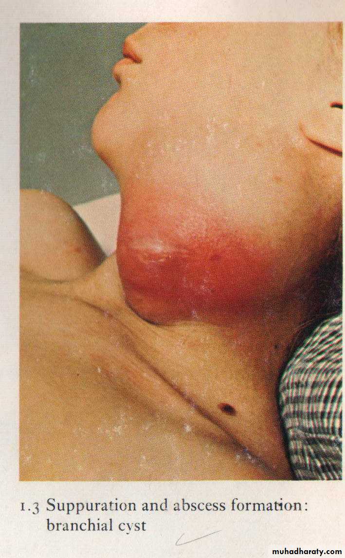
Localization of pus leads to abscess formation which appear here as red congested swelling at the side of the neck. Diagnosis: abscess
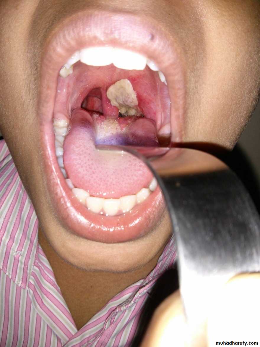
Formation of a superficial, dirty-gray pseudomembrane of diphtheria on the soft palate.
Pharyngeal pseudomembrane. Epithelium is absent; at one side, inflammatory exudate extends to underlying muscle Diagnosis : Diphtheria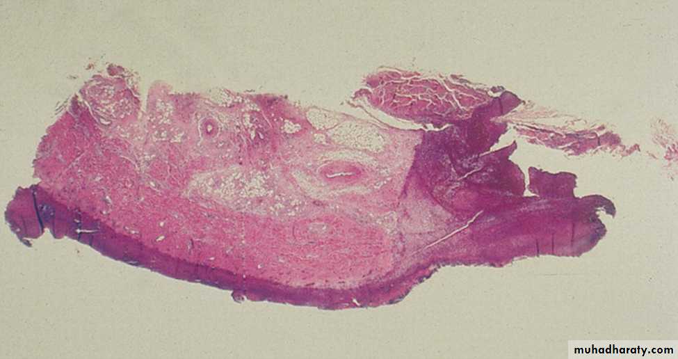
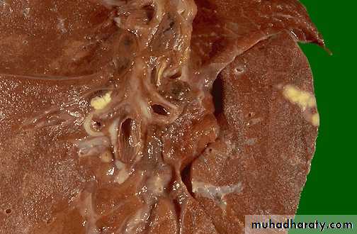
Peripheral sub pleural small whitish yellow lesion (Ghon focus) & hilar lymph node enlargement in primary T.B (cheesy like material) Diagnosis: Ghon’s complex of primary TB..
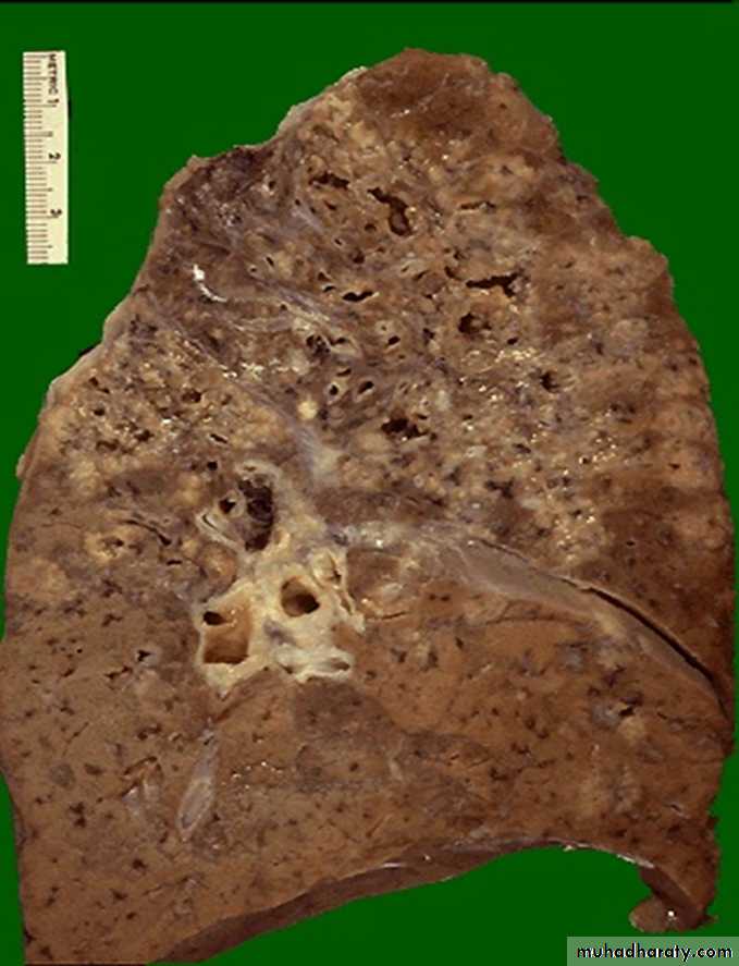
Multiple irregular cavitations & extensive area of necrosis involving the upper lobe of the lung Diagnosis: secondary T.B
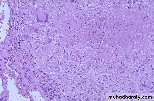
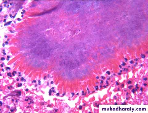
Granuloma with central caseous necrosis surrounded by epithelioid cells & Langhans giant cell (multinucleated giant cells) surrounded by a rim of lymphocytesDiagnosis: T.B lung (caseating granuloma)
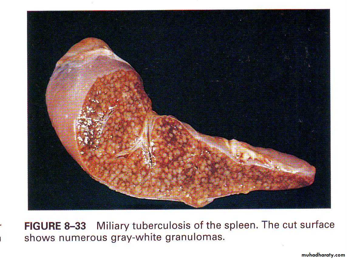
Miliary tuberculosis of the spleen,cut surface show
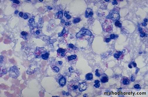
numerous gray-white granulomasThis is an acid fast stain of Mycobacterium tuberculosis Which appear as red rods.
The colony of actinomycosis at high magnification. The micro organism is filamentous & the filaments project as red specular at the periphery of the colony (large arrow) which is surrounded by neutrophils (mycetoma) small arrow.
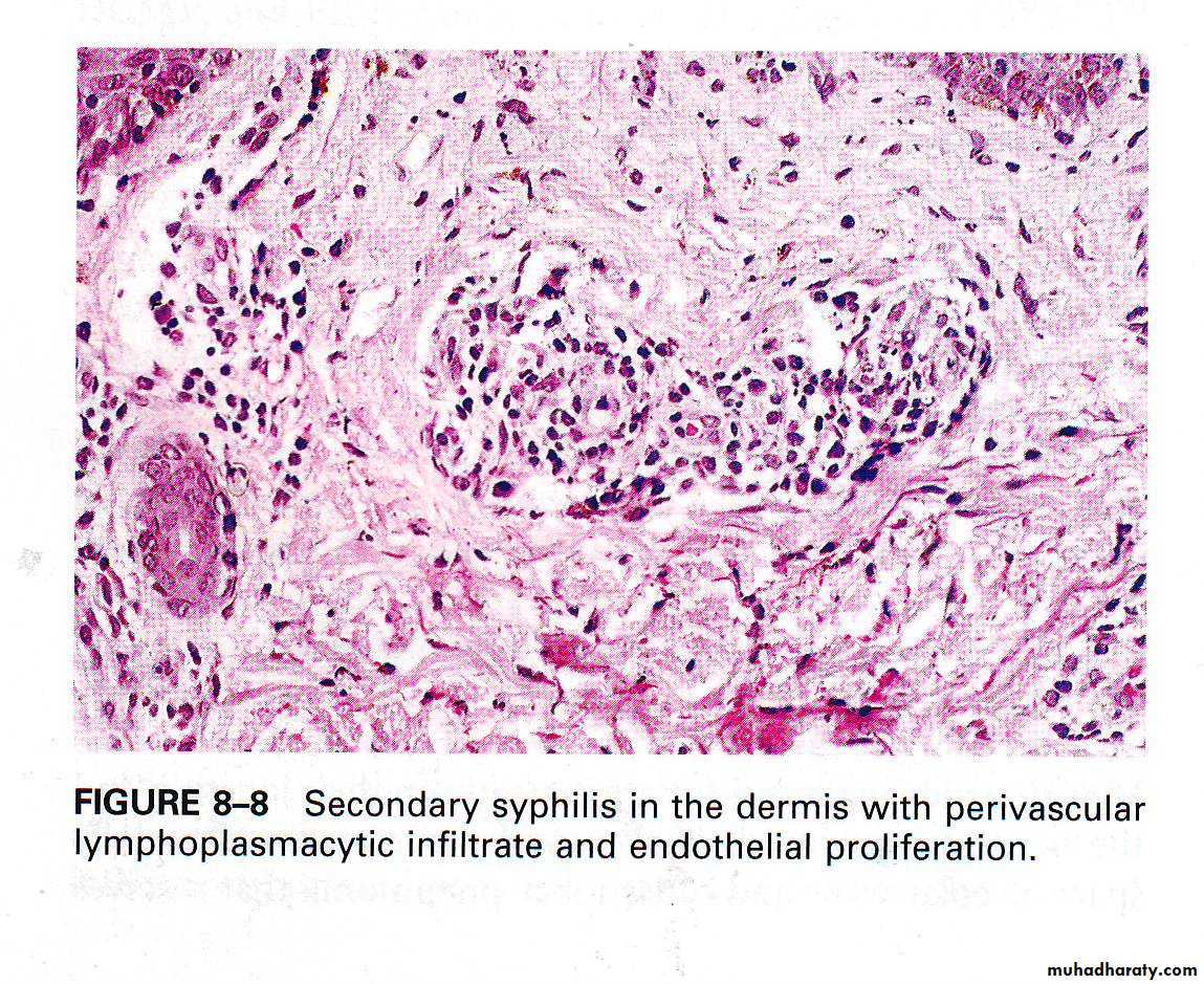
Secondary syphilis, in the dermis with perivascular lymphoplasmacytic infiltrate & endarteritis obliterance.
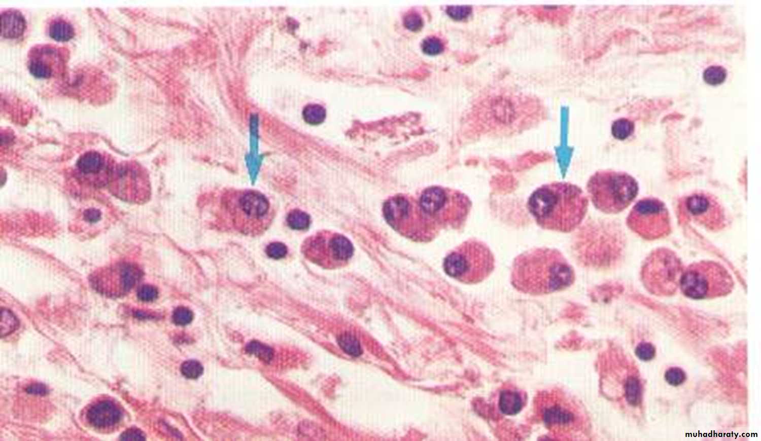
Plasma cells infiltration in syphilis
‘notched incisor Known as Hutchinson's teeth which are characteristic of congenital syphilis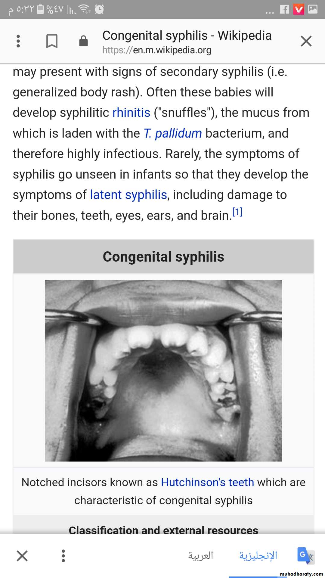
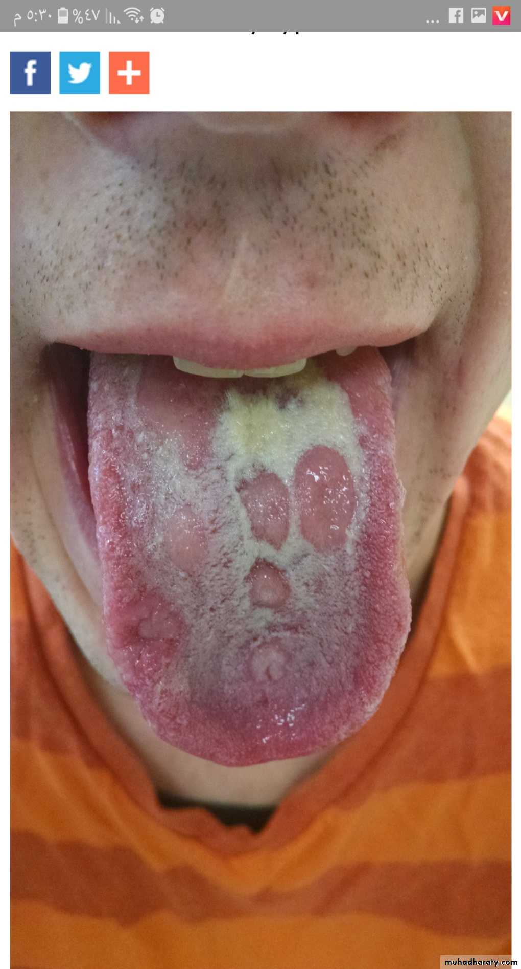
Multiple shallow, lingual
ulcerations in patient withsecondary syphilis
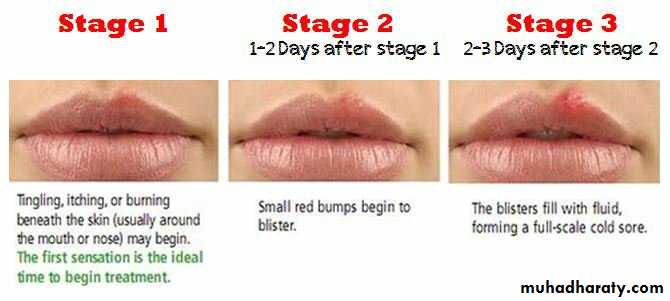
Stages of Herpes Simplex infection (oral sore)
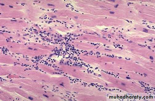
Cardiac muscle fibers show a lymphocytic infiltrate
Diagnosis: Viral myocarditis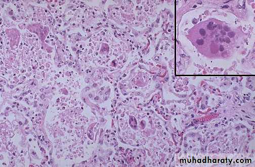
viral infection in the alveolar wall. Showing giant cell originate from epithelial cells with inclusion body of virus within the giant cells
Diagnosis: Viral pneumonia (interstitial pneumonia)
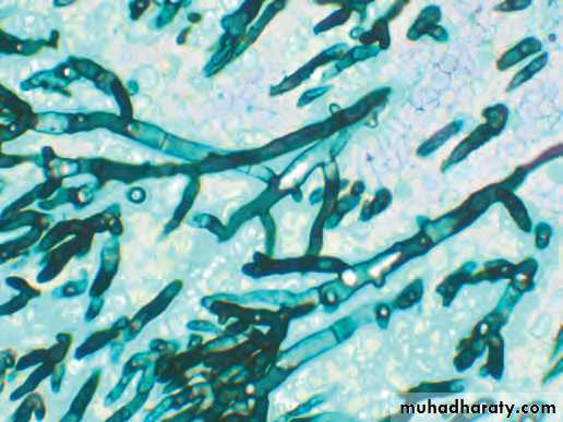
Mold ,Septate hyphae are close-packed with acute-angle branching.
Diagnosis Aspergillus infection, :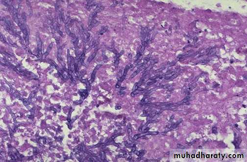
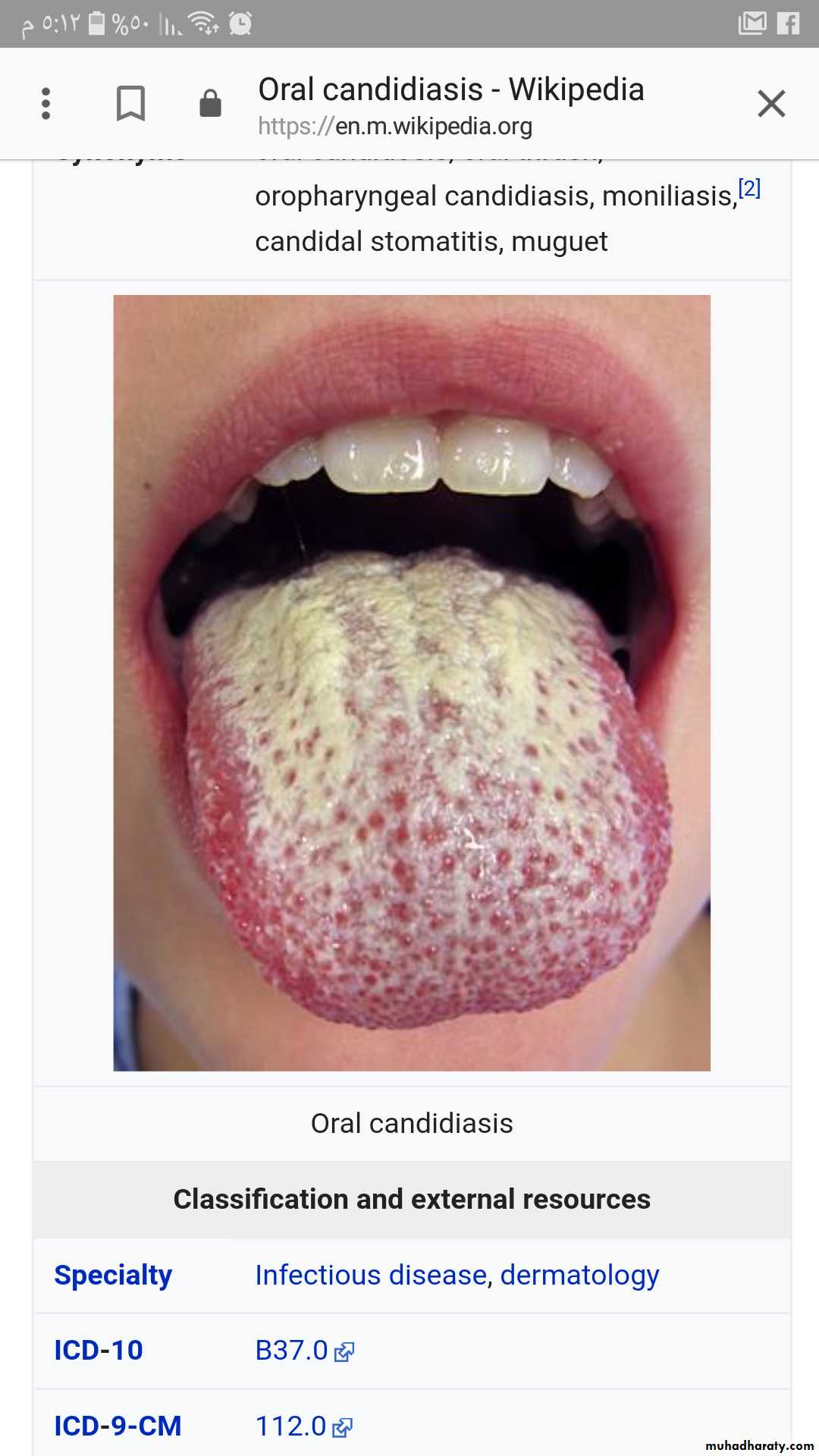
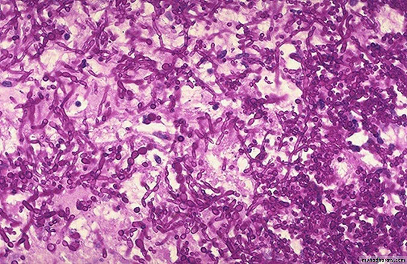
Yeast A numerous budding cells and pseudohyphae characteristic for Candida.
Diagnosis: Candidial infection (fungal infection) lung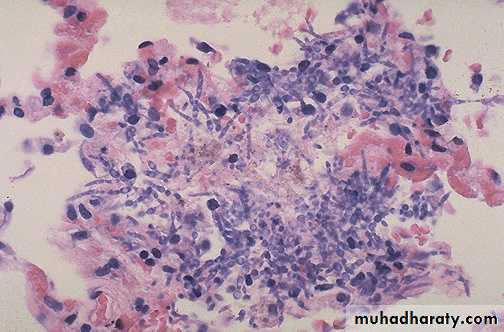
• Oral candidiasis (candidal stomatitis), the fungi creates gray-white, dirty-looking pseudomembranous on the tongue