GENETICSlec. 1
ByLecturer Dr. Zahraa Marwan
Mosul Medical College
Department of pathology
Introduction:
Human diseases in general, whether medical or surgical, can roughly be classified into three categories:1. Those that are genetically determined.
2. Those that are almost entirely environmentally determined.
3. And those to which both nature and nurture contribute.
• Genetics : is the study of single or a few genes and their phenotypic effects.
Genomics: is the study of all the genes in the genome and their interactions.
• Hereditary disorders are derived from one’s parents, are transmitted in the gametes through the generations, and therefore are familial.
• Congenital simply implies “present at birth.”
• Some congenital diseases are not genetic (e.g., congenital syphilis). On the other hand, not all genetic diseases are congenital; the expression of Huntington disease, for example, begins only after the third or fourth decade of life.
Around 20% of pediatric in-patients suffer from disorders of genetic origin.
About 1% of all newborn infants possess a gross chromosomal abnormality.
Approximately 5% of individuals under age 25 develop a serious disease with a significant genetic component.
Classification of Genetic disorders:
1. Classical Genetic Diseases:a. Chromosomal (Cytogenetic) disorders.
b. Single gene (or unifactorial) disorders (Mendelian Disorders).
c. Multifactorial disorders.
2. Non-Classical Diseases "or the single gene disorders with atypical pattern of inheritance":
a. Diseases caused by mutations in mitochondrial genes.
b. Triplet repeat mutations.
c. Uniparental disomy.
d. Genomic imprinting.
e. Gonadal mosaism.
Added to that, is a large group of disorders "malformations" that manifest at birth, called congenital malformations, that many of them are caused by genetic disorders.
1. Classical Genetic Diseases
A. Chromosomal Disorders:Review of what you've already known:
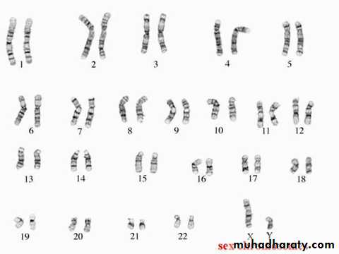
Normal human nucleated cells contain 46 chromosomes arranged in 22 homologous pairs of autosomal chromosomes in addition to one pair of sex chromosomes that could be similar (i.e. XX) or different (i.e. XY). This arrangement into pairs, based on the position of the centromere and on the length of the upper and lower arms of the chromosomes, is known as the Karyotype
It is estimated that about 1 of 200 newborn infants has some form of chromosomal abnormality. The figure is much higher in fetuses that do not survive to term. Cytogenetic disorders may result from alterations in the number or structure of chromosomes and may affect autosomes or sex chromosomes. So, they could either be Numerical or Structural.
Karyotype
A. Numerical abnormalities:
are defined as a gain or loss of one or more whole chromosome(s) (whether an autosome or a sex chromosome) or a whole set of chromosomes.The normal chromosome count is 46 (i.e. 2n = 46) as it is arranged in two sets of chromosomes. An exact multiple of the haploid number (n) is called euploid (normal state).
Any number that is not an exact multiple of the haploid (n) is aneuploid, and it is one of the commonest changes that take place in malignant tissues.
A gain of one or more set of chromosomes is known as polyploidy. This polyploidy may be triploidy when cells have (3n) or tetraploidy when the cells have (4n). Polyploidy generally results in spontaneous abortions.
Haploid=n=23
Diploid=2n=46Euploid is any exact multiple of the haploid number (xn).
Polyploid is 3n (triploid=69) & 4n (tetraploid=92) & generally results in a spontaneous abortion.
Aneuploid is any number that is not an exact multiple of n.
The causes of aneuploidy are:
Non-disjunction
Anaphase lag
Aneuploid
The chief cause of aneuploidy is non-disjunction of the homologous pair of chromosomes at the first meiotic division or failure of sister chromatids to separate during the second meiotic division, leading to the production of two aneuploid cells.When nondisjunction occurs, the gametes formed have either an extra chromosome (n+1) or one less chromosome (n-1).
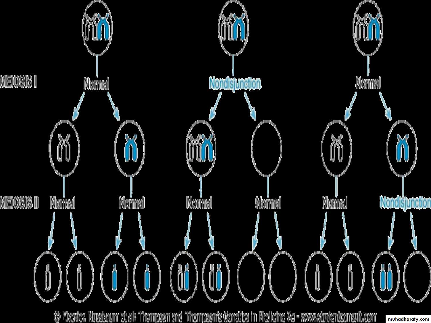
Aneuploid
Fertilization of such gametes by normal gamete will result in two types of zygotes: trisomic with extra chromosome (2n+1) (e.g., Down s), or monosomic (2n-1) (e.g., Turner s).Monosomy involving an autosome is incompatible with life, whereas monosomy involving sex chromosomes & trisomy's of certain autosomes is compatible with life.
Mosaicism
Is a term used to describe the presence of two or more cell populations in the same individual (normal cells and monosomy or trisomy cells)Mosaicism affecting sex chromosomes is common.
While autosomal variety is much less frequent.B. Structural abnormalities:
• In this case, the cell has a normal number of 46 chromosomes but they are morphologically or structurally abnormal.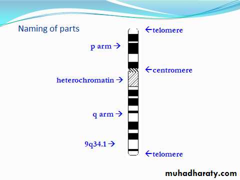
• Each chromosome has 2 arms p (short). and q (long).
• Each arm is divided into 2 or more regions by prominent bands.
• The regions are numbered from the centromere outward. Each region is further subdivided into bands and sub-bands and these are ordered numerically as well.
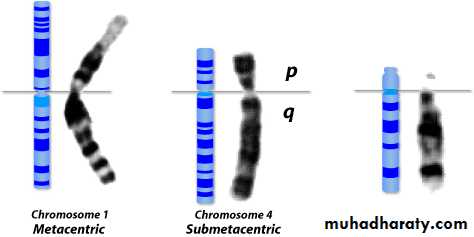
In metacentric chromosomes, the centromere lies near the center of the chromosome.Submetacentric & very Submetacentric chromosomes, have a centromere that is off-center, so that one chromosome arm is longer than the other. In acrocentric chromosomes, the centromere resides very near one end.
p
q
Arm
Region
Band
Subband
2
1
1
2
2
1
1
1
2
3
4
3
2
1
2
1
5
4
3
2
1
1
2
3
1
2
3
1
2, 3
4
1
2
3
17
q
1
1
.
2
Chromosome 17
Defining Chromosomal LocationB. Structural abnormalities:
Structural abnormalities usually result from chromosomal breakage followed by loss or rearrangement of material.They are of different types as follows:
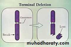
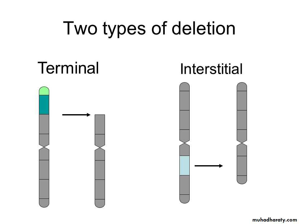
1. DELETION: involves loss of a piece of a chromosome that could be either:
i. Terminal, in which, there is one break in the chromosome and the portion distal to this break is lost. This lost piece could be carrying important genes, and its loss results in signs and symptoms related to the lost genes products
B. Structural abnormalities:
ii. Interstitial, where the piece of a chromosome between two breaks is lost resulting in a chromosome that is shorter than the original with the same consequences.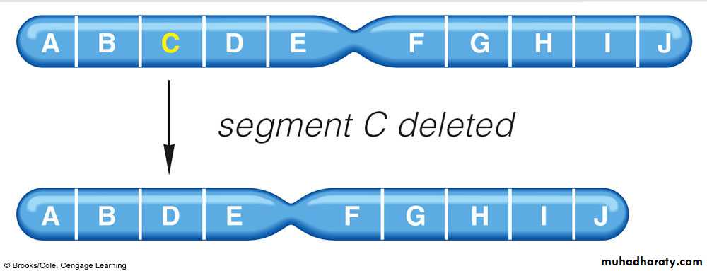
• Ring chromosome is a variant of a deletion, after loss of segments from each end of the chromosome, the arms unite to form a ring, because of the adhesive nature of the exposed DNA, will stick together forming a ring or a circle chromosome.
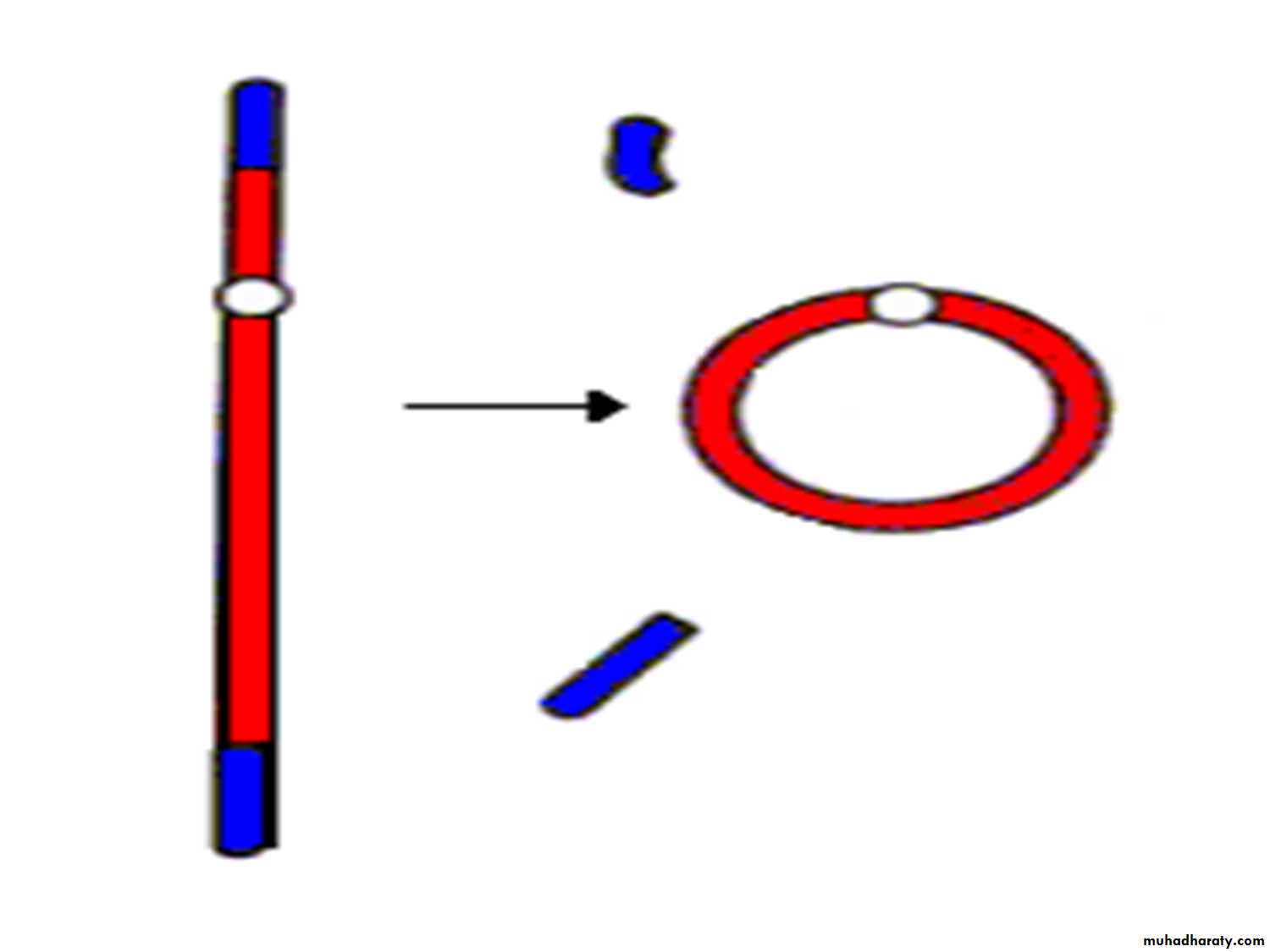
Origin of ring chromosome
reparationdeletion of terminal segments
ring chromosome
r(X)
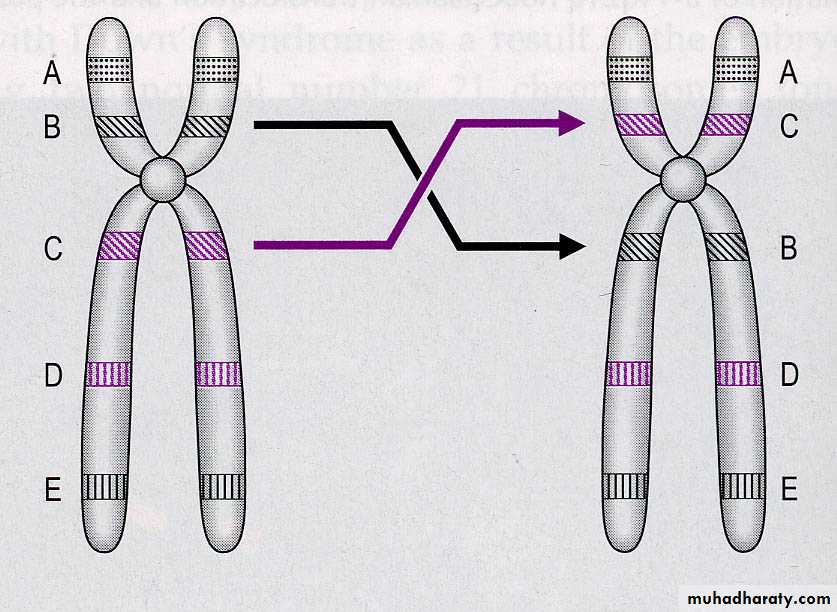
2-TRANSLOCATION: (t)• which involves transfer or exchange of a part of one chromosome to another.
• Two types of translocation are identified:
1- Regular balanced (reciprocal) translocation that takes place between any two chromosomes [other than the acrocentric group of chromosomes], this term used when the entire broken fragments are exchanged (without loss). So, it is not harmful.
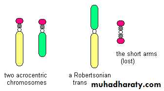
• 2- Another type of translocation called Robertsonian (centric fusion) type that takes place between two acrocentric chromosomes, either one from each group or two chromosomes of the same group but different number or the pair of a homologue where the two acrocentric lose their short arm (due to the breaks occur close to the centromere), Transfer of the segments leads to one very large chromosome & an extremely small one which will be lost and the carrier will have 45 chromosomes. Such loss is compatible with survival.
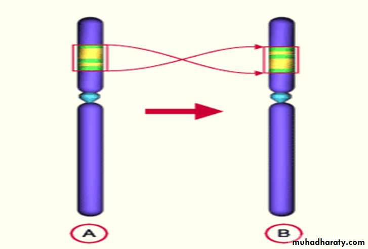
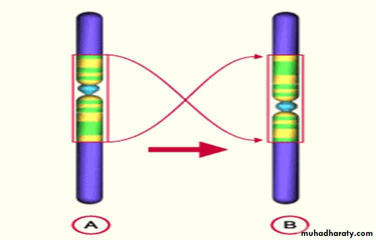
• 3. ISOCHROMOSOMES:
This abnormality results from aberrant division of the centromere (which is the last part of the chromosome that divides in the mitosis to separate the two sister chromatids into individual chromosomes).This aberrant division takes place in a horizontal way rather than the perpendicular (vertical) natural way.
So, the resulting two chromosomes are imbalanced, one formed entirely of two short arms and the other of two long arms.
Each of them is called an isochromosome.
Origin of isochromosomes
i(Xp)i(Xq)
Normal separation in anaphase
Abnormal division – origin of isochromosomes Xp and Xq
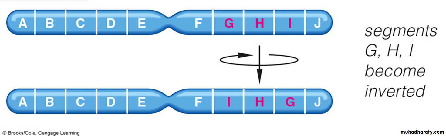
• 4. Inversion:
• occurs when there are two interstitial breaks in the chromosome, and the segment reunite after a complete turnaround (the piece between the two breaks will rotate 180o and is fixed back again in its rotated position).
• There are 2 types, pericentric and paracentric. It is often compatible with normal development
When the breaks involved the short arm or the long arm; the inversion is called paracentric (the breaks are on one side of the centromere), or the breaks may involve both arms and the inversion is called pericentric (the centromere is involved in the inversion), which is more severe.
Inversions
Pericentric Paracentric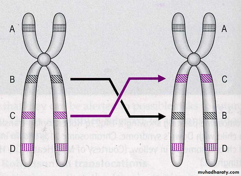
Inversions
Translocation
Deletion
InversionIsochromosome
Ringchromosome
Chromosome Structure Abnormalities
Features of chromosomal disordersChromosomal disorders may be associated with

Absence (deletion or monosomy)
Excess (trisomy) or
Abnormal rearrangement (translocation, inversion)
Loss of chromosomal material produces more severe defects than gain.
Excess chromosomal material may result from complete chromosome (as in trisomy) or part of a chromosome (as in robertsonian translocation).Imbalances of sex chromosome (excess or loss) are tolerated much better than similar changes of autosomes.
Sex chromosomal disorders often produce subtle abnormalities that are sometimes not detected at birth. Infertility which is a common manifestation cannot be diagnosed until adolescence.
Most of chromosomal disorders result from de novo changes (i.e. parents are normal & the risk of recurrence in siblings is low). An important exception is the translocation form of Down syndrome.
I. Disorders of Autosomal Chromosomes:
Trisomy (21, 18, 13) (Down’s syndrome, Edward's syndrome, Patau syndrome)Deletion (Cri du chat syndrome which involves chromosome 5).
Clinical examples of Some Cytogenetic AbnormalitiesTrisomy 21 (Down’s syndrome)
It is the most common chromosomal disorder.Cytogenetic types of Down’s syndrome:
1- Regular Down’s:
which constitutes to 95% of cases of born Down’s syndrome. It usually results from non-disjunction.
In 95%, the extra chromosome is maternal in origin.
The parents are normal in all respects.
Maternal age has a strong influence on the incidence which is 1 in 1550 live births in women < 20, in contrast to 1 in 25 live births in women > 45 years.
No effect of paternal age.
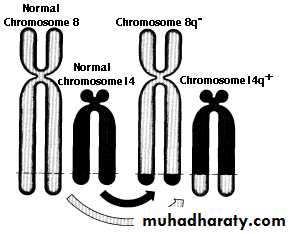
2. Translocation Down’s, they constitute to 3% of cases of born Down's syndrome (the extra chromosomal material is present not as a trisomy but as a translocation of the long arm of chromosome 21 to chromosome 22 or 14, 15). Such cases are usually familial.3. Isochromosome ‘21’ , Isochromosome of the long arm of chromosome 21, may take place resulting in Down's syndrome (rare).
4. Mosaics, approximately 1% of trisomy 21 are mosaics, usually having a mixture of 47 & 46 chromosome cells. Symptoms in such cases are variable & milder, depending on the proportion of abnormal cells.
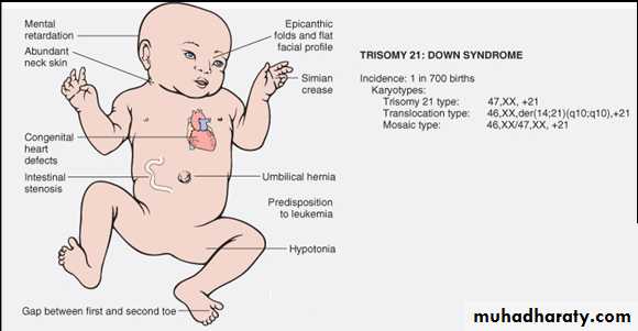
Clinical Features of Down Syndrome
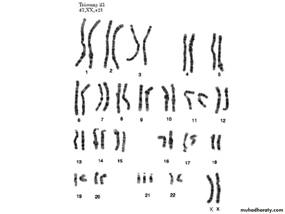
Karyotype of a girl with Down’s syndrome. Notice the 47 total count, the 3 copies of chromosome 21 and the 2 X chromosomes.Autosomal trisomy of chromosome 21 (Down’s syndrome)
II. Abnormalities of Sex Chromosomes:
A number of abnormal karyotypes involving sex chromosomes, ranging from 45X (Turner syndrome) to 47XXY, or 48XXXY, or 49XXXXY (Klinefelter syndrome), are compatible with life.On the other hand males who are phenotypically normal may have two or even three Y- chromosomes.