Lec 1 in Nervous System by Dr Mohammad Ahmad Abdulla
1T
VAULT OF THE SKULL
3T
The internal surface of the vault shows the coronal, sagittal, and lambdoid sutures. In the
midline is a shallow sagittal groove that lodges the
0T3T
superior sagittal sinus.
0T3T
On each side of the
groove are several small pits, called
0T3T
granular pits,
0T3T
which lodge
0T3T
the lateral lacunae
0T3T
and
0T3T
arachnoid granulations.
0T3T
Several narrow grooves are present for the anterior and posterior
divisions of the
0T3T
middle meningeal vessels
0T3T
as they pass up the side of the skull to the vault.
1T
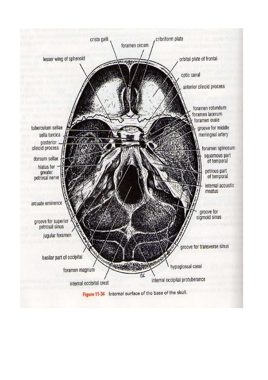
BASE OF THE SKULL
3T
The interior of the base of the skull is divided into three cranial fossae: anterior, middle,
and posterior. The anterior cranial fossa is separated from the middle cranial fossa by the lesser
wing of the sphenoid, and the middle cranial fossa is separated from the posterior cranial fossa
by the petrous part of the temporal bone.
1T
Anterior Cranial Fossa
3T
The anterior cranial fossa lodges the frontal lobes
0T3T
of
0T2T
the
2T3T
cerebral hemispheres. It is bounded
anteriorly by the
2T3T
inner
2T3T
surface of the frontal bone, and in the midline is a crest
2T3T
or
2T3T
the
attachment of the
0T3T
falx cerebri.
0T3T
Its posterior boundary
3T4T
is the
3T4T
sharp lesser wing of the sphenoid,
which articulates
3T4T
laterally
3T4T
with the frontal bone and meets the anteroinferior angle of the
parietal bone, or the pterion. The medial
0T3T
end
0T4T
of
3T4T
the lesser wing of the sphenoid forms the
0T3T
anterior
0T2T
clinoid
2T5T
0T5T
process
0T3T
on each side, which gives attachment to the
3T5T
tentorium
0T5T
cerebelli.
0T3T
The
median part of the anterior cranial
3T5T
3T5T
fossa is limited posteriorly by the groove for the optic
chiasma.
3T
The floor of the fossa is formed by the ridged
0T3T
orbital
0T2T
plates of the frontal bone laterally and
by the
0T2T
cribriform plate
0T2T
of the ethmoid medially. The
0T2T
crista galli is
0T2T
a sharp upward projection
of the ethmoid bone in the midline for the attachment of the falx cerebri. Between the crista
galli and the crest of the frontal bone is a small aperture, the
0T2T
foramen cecum,
0T2T
for the
transmission of a small vein from the nasal mucosa to the superior sagittal sinus. Alongside the
crista galli is a narrow slit in the cribriform plate for the passage of the
0T2T
anterior ethmoidal
nerve
0T2T
into the nasal cavity. The upper surface of the cribriform plate supports the
0T2T
olfactory
bulbs,
0T2T
and the small perforations in the cribriform plate are for the
0T2T
olfactory nerves.
1T
Middle Cranial Fossa
2T
The middle cranial fossa consists of a small median part and expanded lateral parts. The
median raised part is formed by the body of the sphenoid, and the expanded lateral parts form
concavities on either side, which lodge the
0T2T
temporal lobes
0T2T
of the
0T2T
cerebral hemispheres.
2T
It is bounded anteriorly by the lesser wings of the sphenoid and posteriorly by the superior
borders of the petrous parts of the temporal bones. Laterally lie the squamous parts of the
temporal bones, the greater wings of the sphenoid, and the parietal bones.
2T
The floor of each lateral part of the middle cranial fossa is formed by the greater wing of the
sphenoid and the squamous and petrous parts of the temporal bone.
2T
The sphenoid bone resembles a bat having a centrally placed body with
0T2T
greater
0T2T
and
0T2T
lesser
wings
0T2T
that are outstretched on each side. The body of the sphenoid contains the
0T2T
sphenoid air
sinuses,
0T2T
which are lined with mucous membrane and communicate with the nasal cavity; they
serve as voice resonators.
0T
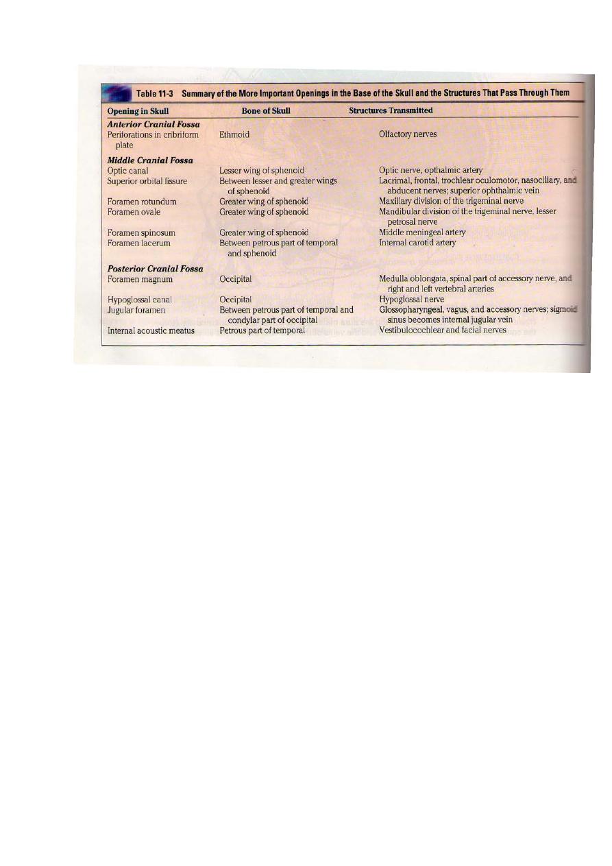
Anteriorly, the
0T6T
optic canal
0T6T
transmits the optic nerve and the ophthalmic artery, a branch of
the internal carotid artery, to the orbit. The
0T6T
superior orbital fissure,
0T6T
which is a slitlike opening

between the lesser and greater wings of the sphenoid, transmits the lacrimal, frontal,
trochlear, oculomotor, nasociliary, and abducent nerves, together with the superior
ophthalmic vein. The sphenoparietal venous sinus runs medially along the posterior border of
the lesser wing of the sphenoid and drains into the cavernous sinus.
0T
The
0T6T
foramen rotundum,
0T6T
which is situated behind the medial end of the superior orbital
fissure, perforates the greater wing of the sphenoid and transmits the maxillary nerve from the
trigeminal ganglion to the pterygopalatine fossa.
0T
The
0T6T
foramen ovale
0T6T
lies posterolateral to the foramen rotundum. It perforates the greater
wing of the sphenoid and transmits the large sensory root and small motor root of the
mandibular nerve to the infratemporal fossa; the lesser petrosal nerve also passes through it.
0T
The small
0T6T
foramen spinosum
0T6T
lies posterolateral to the foramen ovale and also perforates
the greater wing of the sphenoid. The foramen transmits the middle meningeal artery from
the infratemporal fossa into the cranial cavity. The artery then runs forward and laterally in a
groove on the upper surface of the squamous part of the temporal bone and the greater wing of
the sphenoid. After a short distance the artery divides into anterior and posterior branches. The
anterior branch passes forward and upward to the anteroinferior angle of the parietal bone.
Here, the bone is deeply grooved or tunneled by the artery for a short distance before it runs
backward and upward on the parietal bone. It is at this site that the artery may be damaged after
a blow to the side of the head. The posterior branch passes backward and upward across the
squamous part of the temporal bone to reach the parietal bone.
0T
The large and irregularly shaped
0T6T
foramen lacerum
0T6T
lies between the apex of the petrous
part of the temporal bone and the sphenoid bone. The inferior opening of the foramen lacerum
in life is filled by cartilage and fibrous tissue, and only small blood vessels pass through this
tissue from the cranial cavity to the neck.
0T
The
0T6T
carotid canal
0T6T
opens into the side of the foramen lacerum above the closed inferior
opening. The internal carotid artery enters the foramen through the carotid canal and
immediately turns upward to reach the side of the body of the sphenoid bone. Here, the artery
turns forward in the cavernous sinus to reach the region of the anterior clinoid process. At this
point, the internal carotid artery turns vertically upward, medial to the anterior clinoid process,
and emerges from the cavernous sinus.
0T
Lateral to the foramen lacerum is an impression on the apex of the petrous part of the
temporal bone for the
0T6T
trigeminal ganglion.
0T6T
On the anterior surface of the petrous bone are two
grooves for nerves; the largest medial groove is for the
0T6T
greater petrosal nerve,
0T6T
a branch of the
facial nerve; the smaller lateral groove is for the
0T6T
lesser petrosal nerve,
0T6T
a branch of the
tympanic plexus. The greater petrosal nerve enters the foramen lacerum
0T1T
deep to the trigeminal
0T1T
ganglion and joins the
0T1T
deep petrosal nerve
0T1T
(sympathetic fibers from around the internal
carotid artery), to form the
0T1T
nerve of the pterygoid canal.
0T1T
The lesser petrosal nerve passes
forward to the foramen ovale.
0T
The abducent nerve bends sharply forward across the apex of the petrous bone, medial to
the trigeminal ganglion. Here, it leaves the posterior cranial fossa and enters the cavernous
sinus.
0T
The
0T1T
arcuate eminence
0T1T
is a rounded eminence found on the anterior surface of the petrous
bone and is caused by the underlying
0T1T
superior semicircular canal.
0T
The
0T1T
tegmen tympani,
0T1T
a thin plate of bone, is a forward extension of the petrous part of the
temporal bone and adjoins the squamous part of the bone (Fig. 11-34). From behind forward, it
forms the roof of the mastoid antrum, the tympanic cavity, and the auditory tube. This thin
plate of bone is the only major barrier that separates infection in the tympanic cavity from the
temporal lobe of the cerebral hemisphere .
0T
The median part of the middle cranial fossa is formed by the body of the sphenoid bone. In
front is the
0T1T
sulcus chiasmatis,
0T1T
which is related to the optic chiasma and leads laterally to the
0T1T
optic canal
0T1T
on each side. Posterior to the sulcus is an elevation, the
0T1T
tuberculum sellae.
0T1T
Behind

the elevation is a deep depression, the
0T1T
sella turcica,
0T1T
which lodges the
0T1T
hypophysis cerebri.
0T1T
The
sella turcica is bounded posteriorly by a square plate of bone called the
0T1T
dorsum sellae.
0T1T
The
superior angles of the dorsum sellae have two tubercles, called the
0T1T
posterior clinoid pro-
cesses,
0T1T
which give attachment to the fixed margin of the tentorium cerebelli.
0T
The cavernous sinus is directly related to the side of the body of the sphenoid. It carries in
its lateral wall the third and fourth cranial nerves and the ophthalmic and maxillary divisions of
the fifth cranial nerve. The internal carotid artery and the sixth cranial nerve pass forward
through the sinus.
2T
Posterior Cranial Fossa
0T
The posterior cranial fossa is deep and lodges the parts of the hindbrain, namely, the
0T1T
cerebellum, pons,
0T1T
and
0T1T
medulla oblongata.
0T1T
Anteriorly the fossa is bounded by the superior
border of the petrous part of the temporal bone, and posteriorly it is bounded by the internal
surface of the squamous part of the occipital bone. The floor of the posterior fossa is formed by
the basilar, condylar, and squamous parts of the occipital bone and the mastoid part of the
temporal bone.
0T
The roof of the fossa is formed by a fold of dura, the
0T1T
tentorium cerebelli,
0T1T
which intervenes
between the cerebellum below and the occipital lobes of the cerebral hemispheres above.
0T
The
0T1T
foramen magnum
0T1T
occupies the central area of the floor and transmits the medulla
oblongata and its surrounding meninges, the ascending spinal parts of the accessory nerves,
and the two vertebral arteries.
0T
The
0T1T
hypoglossal canal
0T1T
is situated above the anterolateral boundary of the foramen
magnum and transmits the
0T1T
hypoglossal nerve.
0T
The
0T1T
jugular foramen
0T1T
lies between the lower border of the petrous part of the temporal
bone and the condylar part of the occipital bone. It transmits the following structures from
before backward: the
0T1T
inferior petrosal sinus;
0T1T
the
0T1T
ninth, tenth,
0T1T
and
0T1T
eleventh cranial nerves;
0T1T
and the large
0T1T
sigmoid sinus.
0T1T
The inferior petrosal sinus descends in the groove on the lower
border of the petrous part of the temporal bone to reach the foramen. The sigmoid sinus turns
down through the foramen to become the
0T1T
internal jugular vein.
0T
The
0T1T
internal acoustic meatus
0T1T
pierces the posterior surface of the petrous part of the
temporal bone. It transmits the vestibulocochlear nerve and the motor and sensory roots of the
facial nerve.
0T
The
0T1T
internal occipital crest
0T1T
runs upward in the midline posteriorly from the foramen
magnum to the
0T1T
internal occipital protuberance;
0T1T
to it is attached the small
0T1T
falx cere-belli
0T1T
over
the
0T1T
occipital sinus.
0T
On each side of the internal occipital protuberance is a wide groove for the
0T1T
transverse
sinus
0T1T
. This groove sweeps around on either side, on the internal surface of the occipital bone,
to reach the posteroinferior angle or corner of the parietal bone. The groove now passes onto
the mastoid part of the temporal bone, and here the transverse sinus becomes the
0T1T
sigmoid
sinus.
0T1T
The
0T1T
superior petrosal sinus
0T1T
runs backward along the upper border of the petrous bone
in a narrow groove and drains into the sigmoid sinus. As the sigmoid sinus descends to the
jugular foramen, it deeply grooves the back of the petrous bone and the mastoid part of the
temporal bone. Here, it lies directly posterior to the mastoid antrum.
1T
Parts of the Brain
3T
The brain is that part of the central nervous system that lies inside the cranial cavity. It is
continuous with the spinal cord through the foramen magnum
3T
. It is composed of the following
parts:-
1- Forebrain (consists of Cerebrum and Diencephalon)
2- Midbrain
3- Hindbrain (consists of Pons, Medulla oblongata and Cerebellum)
1T
CEREBRUM
3T
The
2T3T
cerebrum
2T3T
is the largest part of the brain and consists of two
2T3T
cerebral hemispheres
2T3T
connected by a mass of white matter called the
2T3T
corpus callosum
2T3T
. Each hemisphere extends
from the frontal to the occipital bones, above the anterior and middle cranial fossae, and,
posteriorly, above the tentorium cerebelli. The hemispheres are separated by a deep cleft, the
2T3T
longitudinal fissure,
2T3T
into which projects the
2T3T
falx cerebri
2T3T
.
The surface layer of each hemisphere is called the
2T
cortex
2T
and is composed of
2T
gray matter
2T
.
The cerebral cortex is thrown into folds, or
2T
gyri,
2T
separated by fissures, or
2T
sulci.
2T
By this means
the surface area of the cortex is greatly increased. Several of the large sulci conveniently
subdivide the surface of each hemisphere into
2T
lobes.
2T
The lobes are named for the bones of the
cranium under which they lie.
The
2T
frontal lobe
2T
is situated in front of the
2T
central sulcus
2T
and above the
2T
lateral sulcus.
2T
The
2T
parietal lobe
2T
is situated behind the central sulcus and above the lateral sulcus. The

2T
occipital lobe
2T
lies below the
2T
parieto-occipital sulcus.
2T
Below the lateral sulcus is situated the
2T
temporal lobe.
The
2T
precentral gyrus
2T
lies immediately anterior to the central sulcus and is known as the
2T
motor area
2T
. The large motor nerve cells in this area control voluntary movements on the
opposite side of the body. Most nerve fibers cross over to the opposite side in the medulla
oblongata as they descend to the spinal cord.
In the motor area the body is represented in an inverted position, with the nerve cells
controlling the movements of the feet located in the upper part and those controlling the
movements of the face and hands in the lower part.
The
2T
postcentral gyrus
2T
lies immediately posterior to the central sulcus and is known as the
2T
sensory area
2T
. The small nerve cells in this area receive and interpret sensations of pain,
temperature, touch, and pressure from the opposite side of the body.
The
2T
superior temporal gyrus
2T
lies immediately below the lateral sulcus
0T
.
0T
The middle of this
gyrus is concerned with the reception and interpretation of sound and is known as the
2T
auditory
area.
2T
Broca's area,
2T
or the
2T
motor speech area,
2T
lies just above the lateral sulcus. It controls the
movements employed in speech. It is dominant in the left hemisphere in right-handed persons
and in the right hemisphere in left-handed persons.
The
2T
visual area
2T
is situated on the posterior pole and medial aspect of the cerebral
hemisphere in the region of the
2T
calcarine sulcus
2T
. It is the receiving area for visual impressions.
The cavity present within each cerebral hemisphere is called the
2T
lateral ventricle.
2T
The
lateral ventricles communicate with the third ventricle through the
2T
interventricular foramina
1T
DIENCEPHALON
1T
The diencephalon is almost completely hidden from the surface of the brain. It consists of a
dorsal
2T
thalamus
2T
and a ventral
2T
hypothalamus.
2T
The thalamus is a large mass of gray matter that
lies on either side of the third ventricle. It is the great relay station on the afferent sensory
pathway to the cerebral cortex.
The hypothalamus forms the lower part of the lateral wall and floor of the third ventricle.
The following structures are found in the floor of the third ventricle from before backward: the
2T
optic chiasma
2T
, the
2T
tuber cinereum
2T
and the
2T
infundibulum,
2T
the
2T
mammillary bodies,
2T
and the
2T
posterior perforated substance.
1T
MIDBRAIN
The midbrain is the narrow part of the brain that passes through the tentorial notch and
connects the forebrain to the hindbrain.
The midbrain comprises two lateral halves called the
2T
cerebral peduncles;
2T
each of these is
divided into an anterior part, the
2T
crus cerebri,
2T
and a posterior part, the
2T
tegmentum,
2T
by a
pigmented band of gray matter, the
2T
substantia nigra
2T
. The narrow cavity of the midbrain is the
2T
cerebral aqueduct,
2T
which connects the third and fourth ventricles. The
2T
tectum
2T
is the part of the
midbrain posterior to the cerebral aqueduct; it has four small surface swellings, namely, the
2T
two superior
2T
and
2T
two inferior colliculi.
2T
The colliculi are deeply placed between the cere-
bellum and the cerebral hemispheres.
The
2T
pineal body
2T
is a small glandular structure that lies between the superior colliculi. It is
attached by a stalk to the region of the posterior wall of the third ventricle. The pineal
commonly calcifies in middle age, and thus it can be visualized on radiographs.

1T
HINDBRAIN
The
2T
pons
2T
is situated on the anterior surface of the cerebellum below the midbrain and
above the medulla oblongata
0T
. It is composed mainly of nerve fibers, which connect the two
halves of the cerebellum. It also contains ascending and descending fibers connecting the
forebrain, the midbrain, and the spinal cord. Some of the nerve cells within the pons serve as
relay stations, whereas others form cranial nerve nuclei.
0T
The
0T1T
medulla oblongata
0T1T
is conical in shape and connects the pons above to the spinal cord
below. A
0T1T
median fissure
0T1T
is present on the anterior surface of medulla, and on each side of this
is a swelling called
0T1T
pyramid
0T1T
. The pyramids are composed of
0T1T
bundles
0T1T
of nerve fibers that
originate in large nerve cells in the
0T
precentral gyrus of the cerebral cortex. The pyramids taper
below, and here most of the descending fibers cross over to the opposite side, forming the
0T
decussation of the pyramids.
Posterior to the pyramids are the
0T
olives,
0T
which are oval elevations, produced by the
underlying
0T
olivary nuclei
0T
. Behind the olives are the
0T
inferior cerebellar peduncles,
0T
which
connect the medulla to the cerebellum.
On the posterior surface of the inferior part of the medulla oblongata are the
0T
gracile
0T
and
0T
cuneate tubercles,
0T
produced by the medially placed underlying
0T
nucleus gracilis
0T
and the
laterally placed underlying
0T
nucleus cuneatus.
The
0T
cerebellum
0T
lies within the posterior cranial fossa
2T
beneath the tentorium cerebelli
2T
.
1T
It is
situated
1T
posterior to the pons and medulla oblongata. It is consists of of two hemispheres
connected by a median portion,
0T
the vermis.
0T
The cerebellum is connected to the midbrain
0T
by
0T
the
0T
superior cerebellar peduncles,
0T
to the pons by
0T
the middle cerebellar peduncles,
0T
and to the
medulla by
0T
the inferior cerebellar peduncles.
The surface layer of each cerebellar hemisphere,
0T
called
0T
the
0T
cortex,
0T
is composed of gray
matter. The cerebellar cortex is thrown into folds, or
0T
folia,
0T
separated by closely se transverse
fissures. Certain masses of gray matter are fount in the interior of the cerebellum, embedded in
the
0T
white
0T
matter; the largest of these is known as the
0T
dentate nucleus.
The cerebellum plays an important role in the control
1T
of
1T
muscle tone and the
coordination of muscle movement
5T
of muscle movements on the same side of the body.
1T
The cavity of the hindbrain is the fourth ventricle. This is bounded in front by the pons and
the medulla oblongata and behind by the
0T1T
superior
0T1T
and
0T1T
inferior medullary vela
0T1T
and the
cerebellum. The fourth ventricle is connected above to the third ventricle by the cerebral aque-
duct, and below it is continuous with the central canal of the spinal cord. It communicates with
the subarachnoid space through three openings in the lower part of the roof: a median and two
lateral openings.