Practical Neoplasia
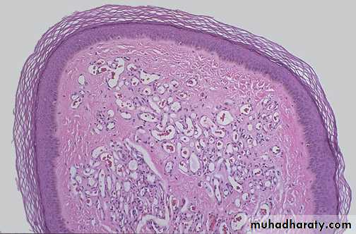
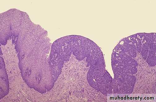
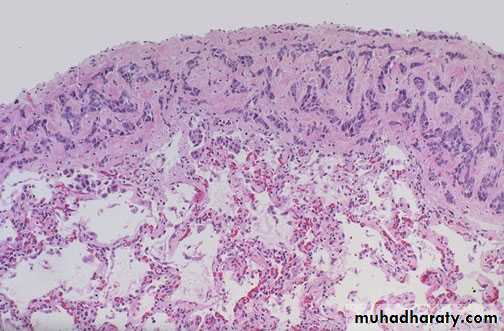
Small well circumscribed cauliflower like growth, whitish in color, occasionally sessile.Diagnosis: squamous papilloma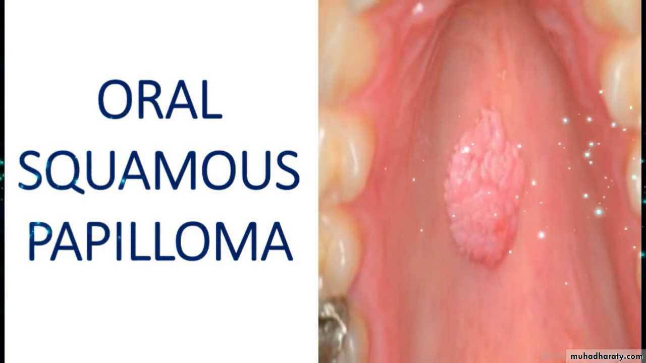
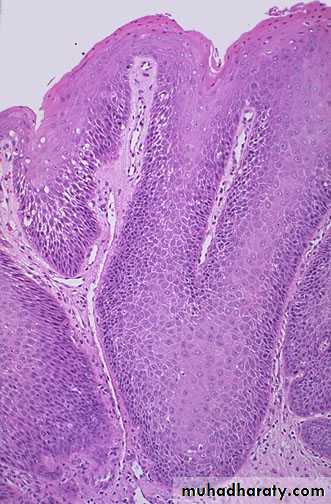
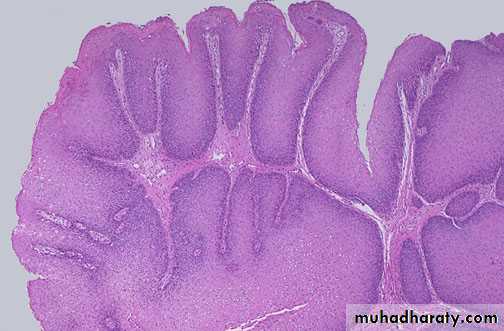
Microscopically: finger like projections, lined by several layers of benign looking squamous cells , with central fibrovascular coreDiagnosis: squamous cell papilloma
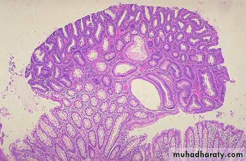
Slide on the left: grossly, multiple nodules of different sizes attached to colonic mucosa by stalkSlide on right: microscopically, nodule composed of proliferating glandular structure some with cystic spaces (crowded, disorganized glands, hyperchromatic and some with cystic spaces) , the polyp is connected to the mucosa by stalk.Diagnosis : adenomatous polyp
Microscopically: there is proliferation of normal-looking, colloid-filled thyroid follicles.Diagnosis: Thyroid adenoma.
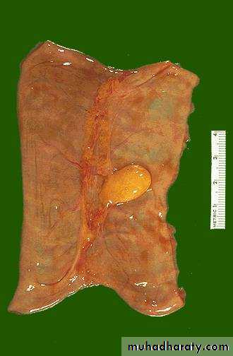
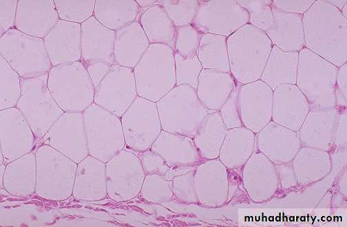
Slide on left: grossly, well circumscribed mass of homogenous yellowish color with lobulated smooth surface on serosal surface of small intestine
Slide on right : Microscopically: proliferation of mature, benign looking fat cells (lipocytes) and there is a capsule in the lower part of the slide.
Diagnosis: lipoma
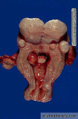
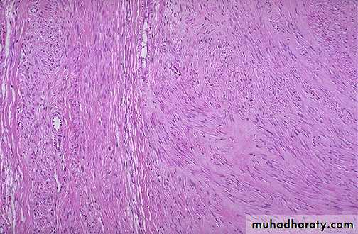
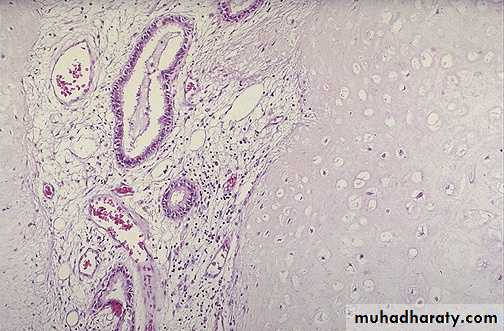
Slide on left: incised uterus showing multiple masses of different sizes rounded regular smooth surface on the mural submucosal & subserosal surfaces of the uterus.slide on right: microscopically, Interlacing bundles of proliferating benign looking spindle cells (smooth muscle cells) with formation of pseudocapsule Diagnosis : lieomyoma of uterus
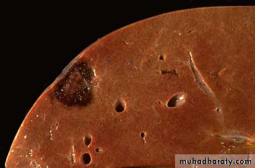
Grossly: section in the liver showing just beneath the capsule there is a well circumscribed mass, dark red in color.Diagnosis:Hemangioma in the liver.
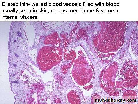
Microscopically: Multiple dilated thin walled blood vessels filled with blood.Diagnosis: Cavernous haemangioma
Microscopically , dermal proliferation of small blood vessels (capillary size) lined by endothelial cells and containing RBCs.Diagnosis : capillary hemangioma
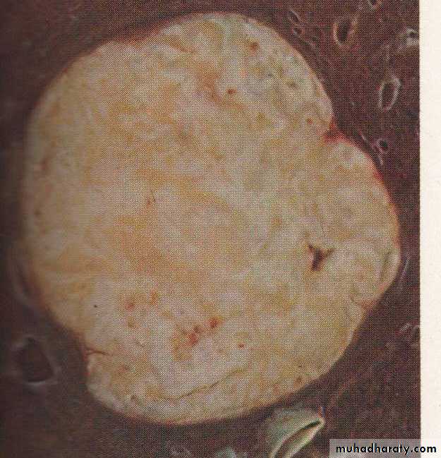
Grossly:Well-defined rounded mass surrounded by fibrotic capsule. Microscopically consists of proliferation of ducts and fibroblastic stroma and the mass surrounded by capsule.
Diagnosis: Fibroadenoma of breast (mixed tumor)
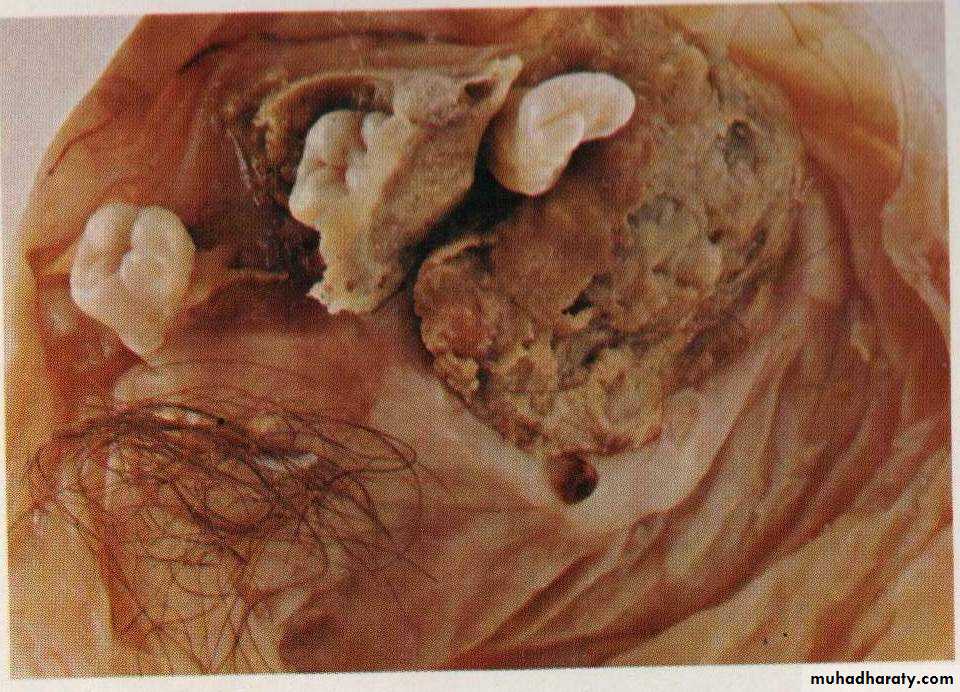
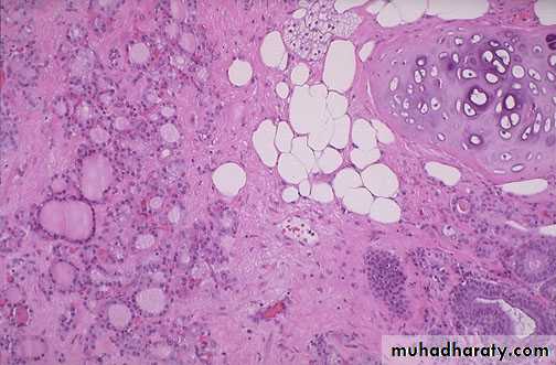
Grossly: cystic mass contains skin, hairs, teeth and cartilage.
Diagnosis: Mature teratoma.Microscopically: A mass consist of mature normal looking cartilage, adipose tissue, intestinal glands, sebaceous glands and thyroid tissue.Diagnosis : mature teratoma
Ectopic pancreas on the wall of jejunum
Diagnosis: choristoma (presence of a normal tissue in an unexpected location)Grossly: solitary mass, whitish to yellowish in cut section and well circumscribed. (relatively common lesion that is usually discovered as an incidental, (rounded radio-opacity (coin lesion) on a routine chest x-ray).Diagnosis: Hamartoma (lung)
Microscopically :mass consists of benign cartilage, fibrovascular stroma and scattered bronchial gland lined by ciliated columnar epithelium, all tissues are mature and normalDiagnosis: Hamartoma (lung)
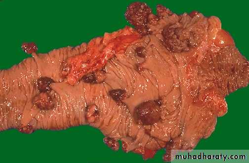
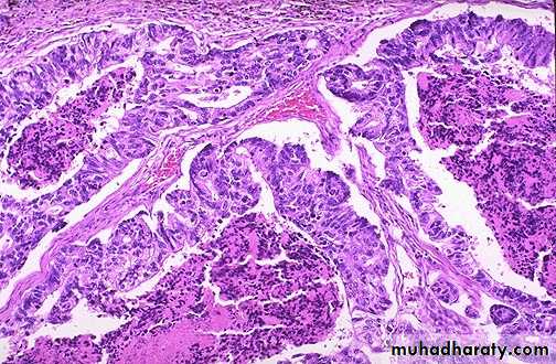
Grossly: a large exophytic growth (mass) with irregular margins arising from the luminal surface of the colon causing partial obstruction.Diagnosis: adenocarcinoma of the colon,

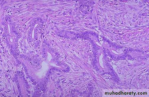
Microscopical slide shows multiple glands lined by malignant cells (increased nuclear/cytoplasmic ratios, mitosis and hyperchromatism). There is a desmoplastic stromal reaction to the infiltrating glands.Diagnosis: Well differentiated Adenocarcinoma of the stomach
Microscopically: malignant epithelial cells (demonstrate mitosis, high nuclear/cytoplasmic ratios & hyperchromatism) forming a gland like structure, but the glands are irregular and branching).Diagnosis: A moderately differentiated adenocarcinoma of colon.
Multiple neoplastic like glands have crowded nuclei with hyperchromatism and pleomorphism & filled with necrotic debris.Diagnosis:Poorly differentiated adenocarcinoma of the colon
Highly cellular tumor showing hyperchromatism, cellular and nuclear pleomorphism & the prominent cell in the center field has an abnormal (tripolar) mitosis.
Diagnosis: Anaplastic tumor
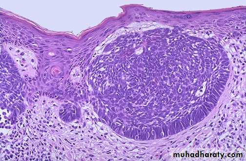
large rounded, rodent ulcer with necrotic base at the lateral angle of left eye .diagnosis:Basal cell carcinoma
Microscopically, dermal islands of small ,rounded to oval dark basophilic (hyperchromatic) cells with scant cytoplasm resemble basal keratinocytes ,the cells at the periphery are elongated and arranged in palisading pattern. .Diagnosis : basal cell carcinoma
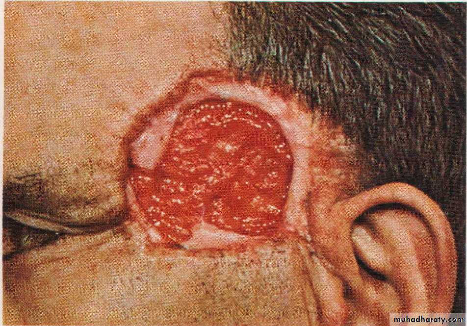
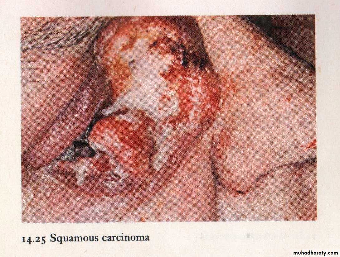
There is ulcer in the inner angle of right eye near the nose destructed the eye , this ulcer is large irregular margins with area of hemorrhage & necrosisdiagnosis: squamous cell carcinoma
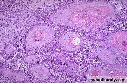
Microscopically: multiple cell nests with keratin pearls , the cell nest composed of malignant squamous cells ( pleomorphic, hyperchromatic nuclei & high N/C ratio)diagnosis: well differentiated squamous cell carcinoma
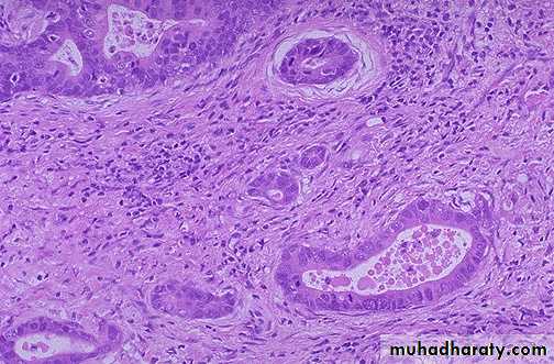
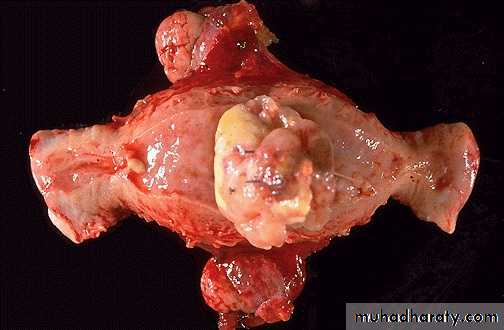
Grossly: There is large irregular mass arise from the fundus of the uterus with area of hemorrhage & necrosis.diagnosis:Leiomyosarcoma
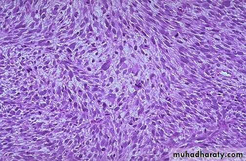
Highly cellular tumor showing proliferation spindle cells(smooth muscle cells), the cells are showing pleomorphism,hyperchromatic nuclei, high mitotic figure with abnormal mitosis).diagnosis:Leiomyosarcoma
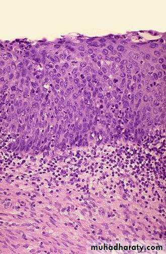
Microscopically: slide of cervix shows part of stratified squamous epithelium with disorderly arranged cells, hyperchromatic nuclei and mitosis but still within the basement membraneDiagnosis: Carcinoma In situ of cervix.
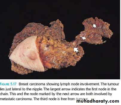
Large ill-defined mass lies just lateral to the nipple with the regional lymph nodes enlargement. The largest arrow indicates the first node in the chain (sentinel node).
Diagnosis: Breast carcinoma with lymph node involvement
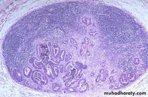
Multiple irregular gland like structure lined by malignant cells infiltrating the lymph node. Diagnosis: Metastatic adenocarcinoma in a lymph node. (lymphatic spread)
Lung tissue showing a large vessel containing malignant cells which forming a glands like structure.Diagnosis: Metastatic adenocarcinoma to the lung (Heamatogenous spread to the lung )
Microscopically: Sheets & cords of malignant cells over the pleural surface of the lungDiagnosis: pleural spread of breast carcinoma(transcoelomic spread)
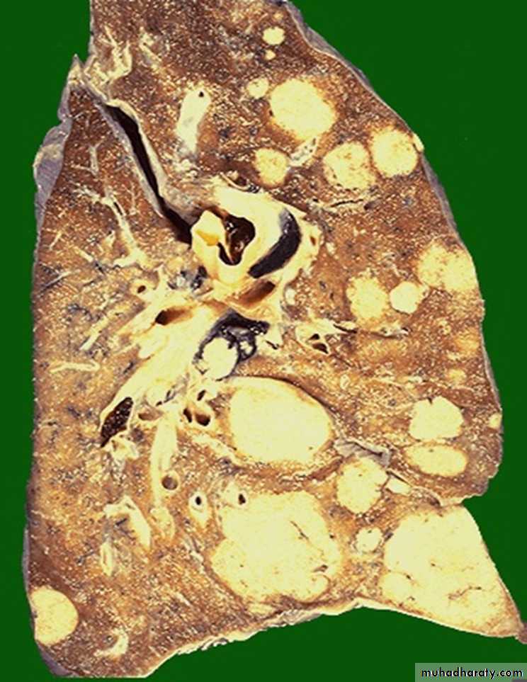
Grossly: section of the lung showing multiple different sizes whitish to yellowish nodules distributed within the lung (Cannonball appearance)Diagnosis: secondary in the lung