Surgical management of non-odontogenic Tumors
Classification:non-odontogenic tumors may be classified according to their presumed tissue of origin into;I- benign mesenchymalII- VascularIII- hematopoietic- reticuloendothelialIV- Neurogenic
I- Benign mesenchymal
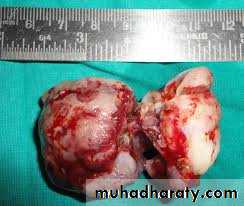
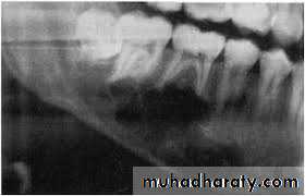
1- Giant- cell lesionsa- Giant -cell reparative granuloma
- Mandible more than maxilla
- Females more than males (ratio 2: 1)
- Usually in children and young adults
- Mostly located in premolar area
- No symptoms, it may be discovered by radiograph
- Appear either as unilocular or multilocular configuration,
Cortical expansion, occasionally with perforation
- Teeth are typically displaced rather than eroded.
Treatment-:
conservatively by local curettage. It has a tendency to recur and many operator now freeze the bony walls of the cavity with liquid nitrogen after curettage in an attempt to prevent this recurrence.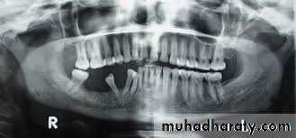
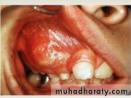
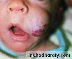
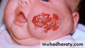
b- Brown tumor of hyperparathyroidism
Hyperparathyroidism may be primary due to over production of parathyroid hormone, (adenoma of parathyroid). Alternatively, it may be secondary to the conditions that reduce calcium level in the blood such (chronic renal disease), causing increased production of parathormon helping calcium to be mobilized from the bony skeleton to raise is blood level and this will affect bones In the jaws cyst-like spaces may develop and are known as brown tumor- Mandible is more affected
- Expansion of the outer and / or the inner cortical plates of bone,- Soft tissue of the brown tumor may grow through the upper border of the alveolar process and appear as dark red colored mass under mucosa
- Lesion is non - painful but may be tender to palpation,
- Radiographically; well circumscribed area of radiolucency with a less defined lamina dura surrounding.
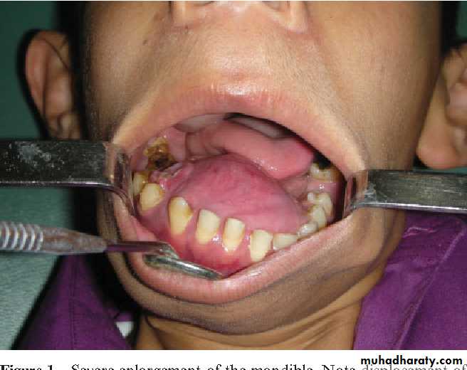
2- Fibrous dysplasia-Affect children- Replacement of normal bone by immature bone with extensive vascular fibrouselements.- Maxilla is more affected- painless enlargement of the maxilla on the affected side , buccal and palatalaspect of the bone may be enlarged and expanded and this may cause spacingbetween the teeth seen as a facial asymmetry of the cheek bone and nasolabial fold.
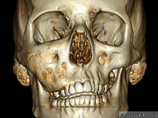
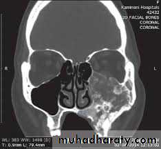
• Diagnosis; usually made on three grounds:- Age- Clinical features- Radiographic appearance of the affected bone; typically show a granular appearance (ground glass)
Treatment: Usually self- limiting and tend to burn out on completion of skeletal growth, and unless there is unacceptable disfigurement most surgeons prefer to postpone until growth is complete. The surgery is normally in the form of re-contouring of the excess bulk of bone, in more gross cases surgery may beneeded at earlier stages, but may require to be repeated when maturity is reached.
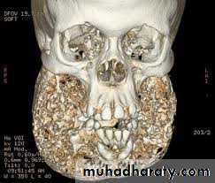
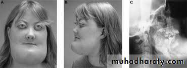
3-Cherubim
Form of fibrous dysplasia.familial, carried by a dominant gene.
mandible is affected more than maxilla,
slow painless swelling which lead to an increase in width and fullness of theangle of the mandible bilaterally
The bone is immature with areas of vascular fibrous tissue
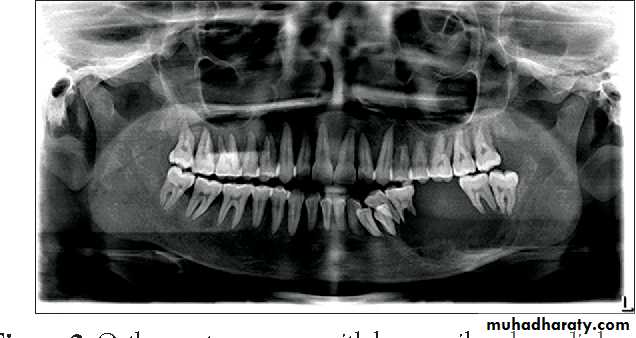
Deciduous teeth exfoliate prematurely, there will be multiple missing andimpacted permanent teeth.
- Radiographically; bilateral multilocular cyst -like radiolucency, teeth seem to be floating within the tumor and may be malformed.- Treatment: surgical contouring is indicated when expansion is sever, leading to functional jaw or airway disturbances, or when the disease causes social problems in the older child. If possible, intervention is delayed until growth has completed.
II- Vascularl- Hemangioma- True tumor of endothelium- Most common benign tumor of the childhood- Usually not present at birth- Exhibits a period of rapid postnatal growth and typically involutes slowly until age 5-7 years- Central hemangioma of the facial bones are rare, most have occurred in the mandible where it extended deeply to involve the oral tissue- The gum may be involved becoming congested, edematous and swollen, False pocketing
Treatment: - Should be avoided unless there have been repeated episodes of hemorrhage. The lesion should then be excised together with its base of normal a, the vessel which supply it may need to be tied off.- Because the involved bone is very vascular, resection may result insignificant blood loss.- IDN is sacrificed if involved Immediate bone and /or nerve grafting should be considered.
2- Neurilemoma- Tumor derived from Schwann cells,- Patient usually experiences pain, swelling and often paresthesia,-The mandible is most commonly involved- Radiographically it appears as a unilocular or multilocular radiolucencywith well defined border.- Treatment: - - Enucleation, recurrence is rare.
Thank
Youfor
listening