
Stage
rd
3
Lung volumes
Physiology (lab)
Omer
1
MdRa
Lung volumes
• Lung volumes and lung capacities refer to the volume of air associated
with different phases of the respiratory cycle.
• Lung volumes are directly measured;
• Lung capacities can be determined from lung volumes.
• The average total lung capacity of an adult human male is about
but only a small amount of this capacity is used during
normal breathing.
• The average human respiratory rate is 30-60 breaths /min. at
birth,
decreasing to 12-20 breaths /min. in adults.
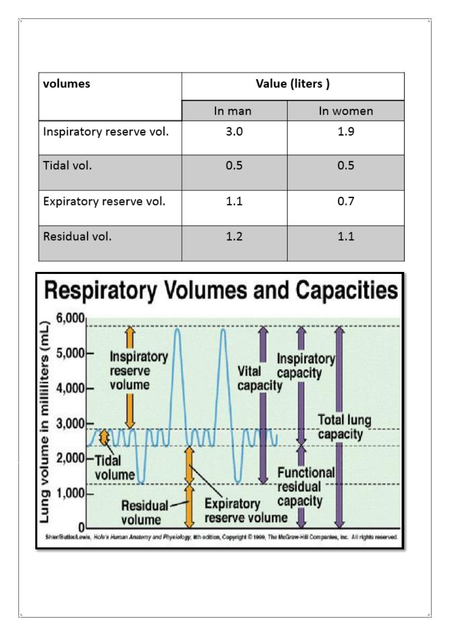
• tidal volume: The amount of air that moves into the lungs with each
inspiration (or the amount that moves out with each expiration).
• inspiratory reserve volume: The air inspired with a maximal inspiratory
effort in excess of the tidal volume.
• expiratory reserve volume: The volume expelled by an active expiratory
effort after passive expiration .
• respiratory minute volume: The amount of air inspired /min..
• maximal voluntary ventilation (MVV) is the largest volume of gas that
can be moved into and out of the lungs in 1 min by voluntary effort=125
to 170 L/min.
• respiratory minute volume: The amount of air inspired /min..
• maximal voluntary ventilation (MVV) is the largest volume of gas that
can be moved into and out of the lungs in 1 min by voluntary effort=125
to 170 L/min.
• FEV
1
:The fraction of the vital capacity expired during the first second of
a forced expiration.
• The FEV
1
to FVC ratio (FEV
1
/FVC) is a useful tool in the diagnosis of
airway disease
Stage
rd
3
Lung volumes
Physiology (lab)
Omer
2
MdRa
Average lung volumes in healthy adults

Stage
rd
3
Lung volumes
Physiology (lab)
Omer
3
MdRa
What Is Obstructive Lung Disease?
• respiratory disease characterized by airway obstruction .
• People with obstructive lung disease have shortness of breath due to
difficulty exhaling all the air from the lungs.
• Because of damage to the lungs or narrowing of the airways inside the
lungs, exhaled air comes out more slowly than normal.
• Obstructive lung disease makes it harder to breathe, especially during
increased activity or exertion.
• As the rate of breathing increases, there is less time to expire all the air
out before the next inhalation.
• The most common causes of obstructive lung disease are:
• Chronic obstructive pulmonary disease (COPD),
• Asthma
• Bronchiectasis
What Is Restrictive Lung Disease?
• are a category of extrapulmonary, pleural, or parenchymal respiratory
diseases that restrict lung expansion,resulting in a ↓lung volume, an
↑work of breathing, and inadequate ventilation and/or oxygenation.
• People with restrictive lung disease cannot fully fill their lungs with air.
• Restrictive lung disease most often results from a condition causing
stiffness in the lungs themselves ( loss of elasticity).
• In other cases, stiffness of the chest wall, weak muscles, or damaged
nerves may cause the restriction in lung expansion.
• Example: emphysema
some autoimmune disease

Stage
rd
3
Lung volumes
Physiology (lab)
Omer
4
MdRa
Physiology
• Basic concepts of normal pulmonary physiology that are involved in
pulmonary function testing include mechanics
• 1-(airflows and lung volumes),
• 2-the ventilation-perfusion interrelationship,
• 3-diffusion and gas exchange,
• 4-and respiratory muscles strength
Diagnosis
• In restrictive lung disease, both forced expiratory volume in one second
(FEV1) and forced vital capacity (FVC) are reduced, however, the decline
in FVC is more than that of FEV1, resulting in a higher than
80% FEV1/FVC ratio.
• In obstructive lung disease however, FEV1 is reduced while FVC remains
stable, producing a lower FEV1/FVC ratio.
• One definition requires a total lung capacity which is 80% or less of the
expected value
Pulmonary Function Tests
• Pulmonary function tests are a group of tests that measure how well
your lungs work.
• This includes how well you are able to breathe and how well your lungs
are able to supply oxygen to the rest of your body.
Types of Lung Function Tests
• 1-Breathing Tests
• Spirometry
• Spirometry measures how much air you breathe in and out and how
fast you blow it out.
• This is measured two ways: peak expiratory flow rate (PEFR) and
forced expiratory volume in 1 second (FEV1).
Stage
rd
3
Lung volumes
Physiology (lab)
Omer
5
MdRa
• During the test, you will take a deep breath in. Then, you'll blow as
hard as you can into a tube connected to a small machine.
• The machine is called a spirometer.

Stage
rd
3
Lung volumes
Physiology (lab)
Omer
6
MdRa
• 2-Lung Volume Measurement
• 3-Lung Diffusion Capacity
• 4-Lung diffusion testing; also called diffusing capacity and diffusing
capacity of the lung for carbon monoxide,
Pulmonary function tests can be used to help diagnose:
• asthma
• allergies
• chronic bronchitis
• respiratory infections
• emphysema
• lung fibrosis
• bronchiectasis (a lung condition in which the airways in the lungs are
stretched and widened)
• chronic obstructive pulmonary disease (COPD)
• asbestosis (a condition caused by exposure to asbestos)
• Sarcoidosis inflammation in the lungs, liver, lymph nodes, eyes, skin,
or other tissues
• scleroderma, a disease of the connective tissue
• pulmonary tumor
• lung cancer
How to Prepare for Pulmonary Function Tests
• 1- stop any medication weather bronchodilator or any other type.
• 2-do not eat a large meal before testing (avoid food and drinks that
contain caffeine).
• 3-do not make hard exercise at least 3 hours before test.
• 4-No smoking for at least 2 hours before test time.

Stage
rd
3
Lung volumes
Physiology (lab)
Omer
7
MdRa
Risks of pulmonary function tests
• Individuals with certain conditions should not take a pulmonary
function test, as it can cause problems. These conditions include:
• -a recent heart attack
• -heart disease
• -recent eye surgery
• -recent chest or abdominal surgery
• -respiratory infections