• 11-11-2018
Medical Parasitology3rd class
Toxoplasmosis• Dr. Abdul Rahman Dahham, PhD
• Department of Microbiology
• College of Medicine
• Nineveh University
1
Toxoplasmosis
Toxoplasmosis is a zoonotic disease caused by the protozoal parasite Toxoplasma gondii. Felids are the definitive hosts for T. gondii, but encysted parasites can survive for very long periods, the parasitic infection are very common in humans and other warm-blooded animals, with approximately a third of the world’s human population estimated to have been exposed to the parasite. Toxoplasmosis can be asymptomatic (no clinical symptoms) or can have more severe consequences such as congenital birth defects, eye disease, or potentially fatal toxoplasmic encephalitis in immuno-compromised individuals.2
All non-feline hosts are intermediate hosts. The word Toxoplasma originated from the Greek world toxon , which mean "bow " and plasmid mean "form". The original Greek meaning is the one used for the word Toxoplasma, meaning "bow" shaped organisms.
3
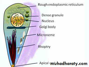
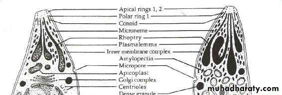
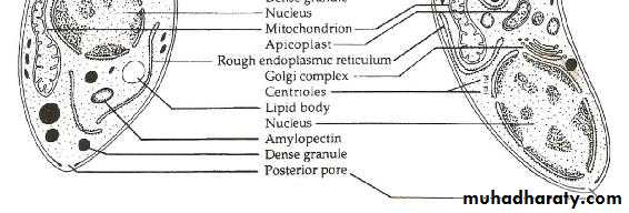
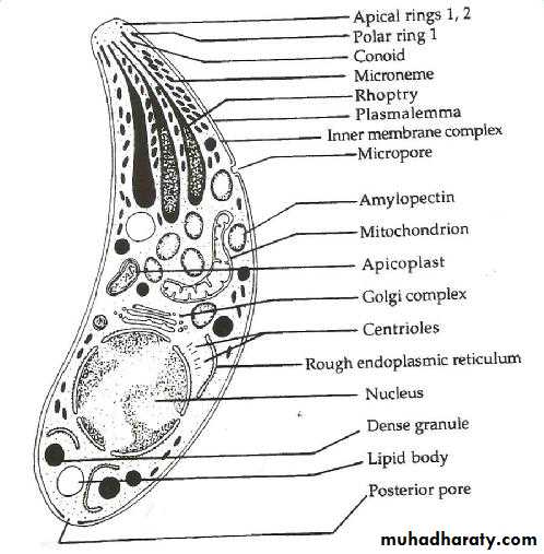
diagram of T. gondii

Epidemiology and geographical distribution
The protozoan parasite T. gondii infects more than a billion people worldwide specially middle east (Iraq).The prevalence data showed that toxoplasmosis is the one of the most common of human infections in the world.
Infection is more common in warm climates and at lower altitudes than in cold climates and mountainous regions.
High prevalence of infection in some European countries has been related to a preference for eating raw or under cooked meat.
A high prevalence in central America has been related to the frequency of stray cats in a climate favoring survival of Oocystes.
4
the organism morphology
Toxoplasmosis is caused by Toxoplasma gondii, an obligate intracellular protozoan parasite in the order Coccidia and phylum Apicomplexa.The infective stages of the parasite can take three different forms:- tachyzoites (trophozoite), bradyzoites (tissue cyst), and oocyst (sporozoites).
Most species of mammals and birds are susceptible and may serve as an intermediate host.
5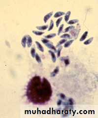
• TACHYZOITES
• TISSUE CYSTS• BRADYZOIT
• OOCYSTS
Oocysts
Tachyzoites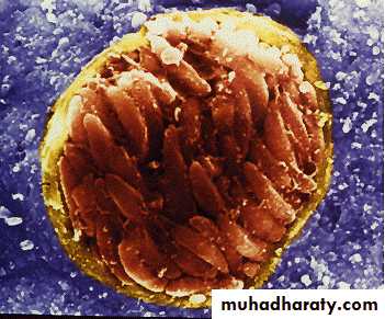
Bradyzoites
TISSUE CYSTSFollowing sporulation in the environment, oocysts containing sporozoites which are infective, and give rise to tachyzoites when ingested by an intermediate host.
The tachyzoites and bradyzoites represent stages in asexual multiplication (Schizogony). While the oocyst is formed by sexual reproduction (gametogony or Sporogony).
All three forms occur in the domestic cat and other felines which is the definitive host and which support both schizogony and gametogony.
7
Tachyziote
The term tachyzoite (tachos = speed in Greek), to describe the stage that rapidly multiplied in any cell of the intermediate host and in non-intestinal epithelial cells of the definitive host.The tachy is crescent – shaped, with one end pointed and the other end rounded. The nucleus is ovoid and situated near the blunt end of the parasite. It can invade any nucleated cell and replicate within cytoplasmic vacuoles by a processes called endodygony or internal budding – daughter trophoziote being formed.
8
The rapid replication and release of tachyzoites from host cells and dissemination around the body via the bloodstream and infect a variety of tissues, causes tissue damage and trigger a strong inflammatory response and therefore is responsible for the clinical manifestations of disease.
The term tachyziote replaces the previously used term trophoziote (trophicos = feeding in Greek). Tachyziote have also been termed endodyzoites or endozoites.
9
10
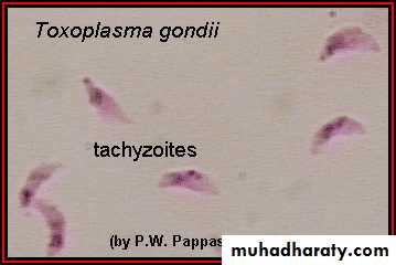
Tachyzoites of Toxoplasma gondii.
Note the characteristic crescent shape.
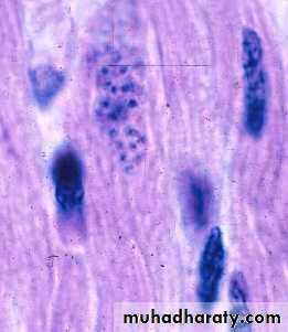
the tachyziote (left) and a bradyziote (right) of
T. gondii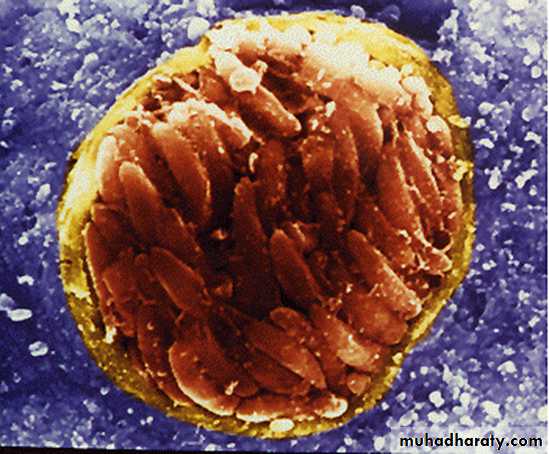
Bradyzoite – A slower reproducing form, contained in tissue
BradyzoitesThe tissue cysts is formed during the chronic phase of the infection and can be found in the muscle and various other tissues and organs, include the Brain the term bradyzoites (Brady = slow in Greek), to describe the organism multiplying slowly within a tissue cysts.
Bradyzoites are not responsible for acute clinical disease; and can persist for the life of the host without causing a host inflammatory response.
11
Tissue cyst vary in size, young tissue cyst may be as small as 5 μm in diameter and contain only two bradyzoite while older ones may contain hundreds of organisms.
Bradyziote differ structurally only slightly from tachyziote. They have situated toward the posterior end whereas the nucleus in tachyzoite is more centrally located
12
the tissue cyst of T.gondii in mouse brain
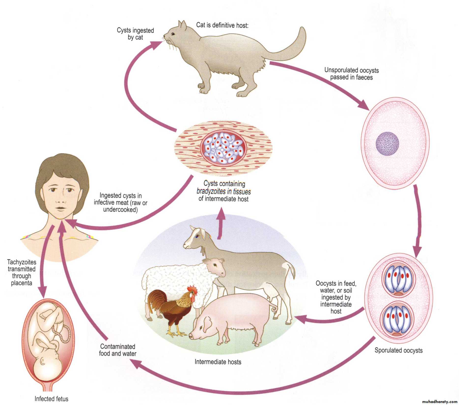
OocystsOocysts develop only in definitive hosts – in the intestine of cats and other felines when cats get infected by ingestion of either tissue cysts or Oocysts, the parasite develop in the intestinal epithelial cells, where both Schizogony and gametogony take place.
Male and female gametocytes develop and after Fertilization, the zygote gets surrounded by a thin, but extremely resistance wall.
13
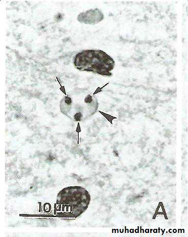
This is Oocyst, which is sepherical or ovoid, about 10 to 12 μm in size and contains a sporoblast. Cats can able to shed millions of oocysts per day in the faeces for about two weeks during the primary infection.
The freshly passed Oocyst is not infectious. It becomes infections only after development in soil or in water for a few days. During this state of sporulation, the sporoblast divides in two sporcysts and four sporozoite is the infective from.
14
It is very resistance to environmental conditions and can remain infective in soil for about a year. When the infective Oocyst is ingested it releases sporozoites in the intestine, which initiate infection.
15
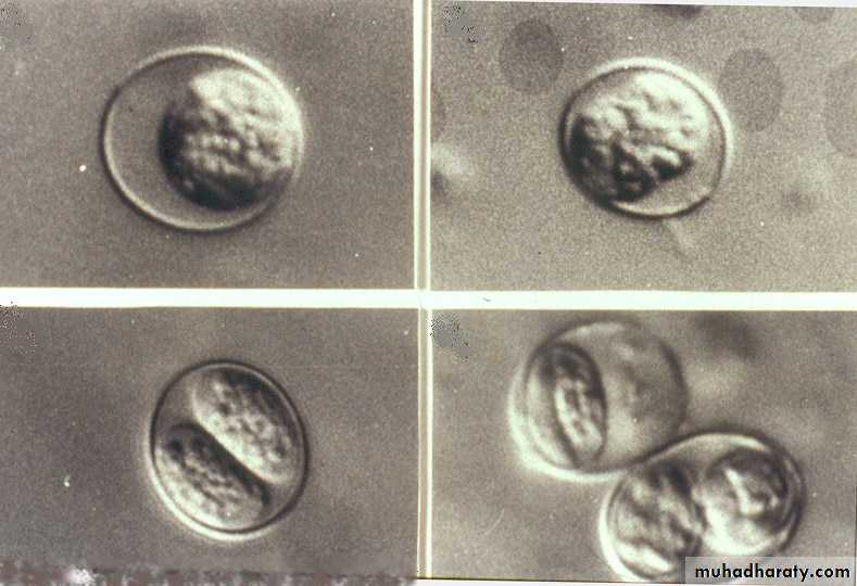
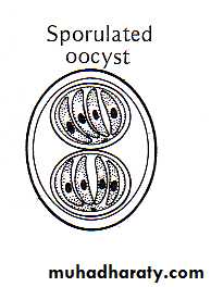
Sporulated oocysts
Un-sporulated oocystsTwo sporocysts
Un-sporulated oocysts are sub-spherical to spherical
Sporulated oocysts are ellipsoidal16
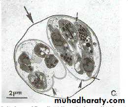
the Oocyst of T.gondii
the sporozoite of T.gondii
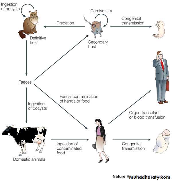
Disease TransmissionsA- Congenital transmitted (Trans-placental transmission which often leads to severe and life long disabilities in the infected infant).
B- Acquired transmitted
Ingestion of Oocyts excreted in the faces of infected cats that contaminated dust, soil and litter box material.
Consumption of tissue cysts under cooked or uncooked meat and certain organs of infected eggs.
Transfusion of blood or blood products.
Organ transplants like kidney transplant and liver transplant
17
18
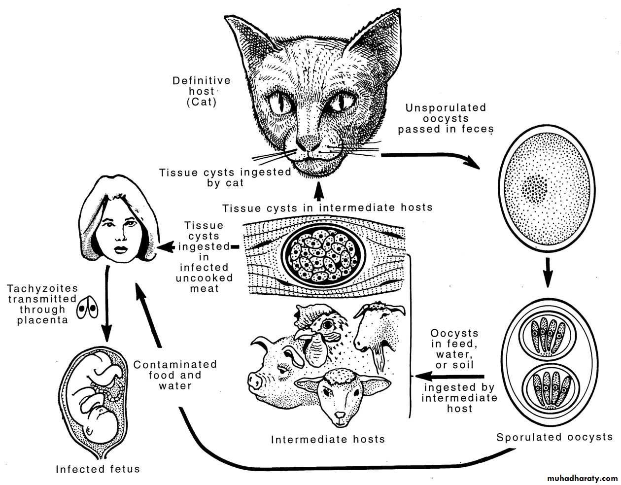
The life cycle of Toxoplasma gondii
Figure 23.13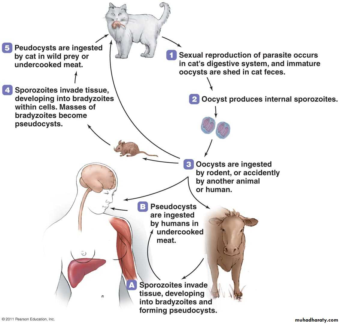
Life Cycle
The life cycle of T. gondii consists of two stages Asexual and sexual:Asexual stage takes place in the intermediate hosts, which are mammals (human) and or bird (During this stage rapid intracellular growth of the parasite as tachyziote takes place).
The tachyziotes can infect and multiply in almost any nucleated mammalian or avian cell, following accumulation tachyziotes are secreted into the blood stream, and spread in the body, leading to development of an acute disease (parasitemia).
20
The normal immune response and transformation of the tachyziote into cyst formation bradyziote limit the acute stage and establish a chronic infection.
Bradyziotes differ from tachyziotes in slow multiplication rate, and in the distinct set of proteins they express. The cysts are formed mainly in neural and muscular tissues especially brain, skeletal and cardiac muscles and can persist, inactivated, in the body for a very long time. In the immunocompromised patient the release of bradyziotes from the cyst, may cause acute encephalitis.
21
• The sexual stage takes place in intestine of the definitive host (domestic cat). When bradyziotes or Oocysts are ingested by a feline, The tissue cyst which contains bradyzoites travel to the intestine via digestive tract, released in the intestine, infect cells and become trophozoites.
• Fertilization occurs in the intestine, and immature oocyst are passed in the cat’s feces. The oocyst contaminate water, food and soil are ingested by intermediate host.
22
The cat can produce million of un-sporulated Oocysts in the feces over a period of 3 – 18 days, depending on the stage of T. gondii ingested. Under mild environmental conditions Oocysts may sporulate within a 3 week period.
23
24
25
• • 25
Symptoms of diseaseMost human infections with T. gondii are asymptomatic, but infection may result in severe clinical disease and on occasion be fatal.
Congenital infections occur in about 1-5 per 1000 pregnancies of which 5-10% result in miscarriage, 8-10% result in serious brain and eye damage to the fetus, 10-13% of the babies will have visual handicaps.
26
Congenital toxoplasmosis occurs when a woman becomes infected with T. gondii during pregnancy. Tachyzoites circulating in the mother’s bloodstream can invade and multiply in the placenta and subsequently infect the foetus.
Transmission of the parasite in utero can cause congenital defects or spontaneous abortion. These congenital defects can include ocular toxoplasmosis, hydrocephalus (big head), mental retardation and intracranial calcifications.
27
In immuno-competent adults, toxoplasmosis, may produce flu-like symptoms, sometimes associated with lymphadenopathy. Enlarged lymph nodes are the most commonly observed signs of human toxoplasmosis.
In immuno-compromised individuals, infection results in generalized parasitemia involvement of brain, liver lung and other organs, This leads to symptoms that affect the central nervous system, including headache, altered mental status, seizures, hemiparesis (muscle weakness on one side of the body), ataxia and/or facial weakness and often death.
28
Toxoplasmic retinochoroiditis (inflammation of the retina and choroid) can be associated with congenital or postnatally acquired disease as a result of acute infection or reactivation of a latent infection.
29
• Diagnosis of Toxoplasmosis:
• The demonstration of the T. gondii organism in blood, body fluids, or tissue.• Detection of T. gondii antigen in blood or body fluids by enzyme-linked immunosorbent assay (ELISA) technique.
• The Sabin-Feldman dye test: A sensitive and specific neutralization test. It measures IgG antibody and is the standard reference test for toxoplasmosis. High titers suggest acute disease.
30
• Fluorescent antibody test detects IgM antibodies within the first week of infection, but titers fall within a few months.
• Polymerase Chain Reaction on body fluids, including CSF, amniotic fluid, and blood.
• Skin test results showing delayed skin hypersensitivity to Toxoplasma gondii antigens.
• Animal inoculation: inoculation of suspected infected tissues into experimental animals.
• Culture: inoculation of suspected infected tissues into tissue culture.
31
Treatment
Clindamycin, spiramycin is the treatment of choicePyrimethamine
Trimothoprim
Sulfamethazine and sulfamerazine
Folic acid should be administrated to those patients treated by the combination of sulfonamide and pyrimethamine to prevent anemia, aganulocytosis and thrombocytopenia that may be resulted due to prolonged administration of these two drugs.
32
Prevention and control
1/ Avoid consumption of raw or undercooked meat, Cook meat to proper temperature (freezing also reduces chance of infection)2/ Wash hands after handling raw meat, litter pans & soil.
3/ Pregnant women should avoid contact with cats.
4/ Peel or wash fruits and vegetables before eating.
5/ If pregnant or immunocompromised, avoid changing litter box if possible.
6/ Wear gloves and wash hands afterwards!
7/ The best way to avoid T. gondii infection in cats is not to feed any raw meat