• 16-12-2018
Medical Parasitology3rd class
Trichomoniasis• Dr. Abdul Rahman Dahham, PhD
• Department of Microbiology
• College of Medicine
• Nineveh University
1
Trichomonads are widely distributed and infect nearly every mammal associated with humans. There are three human species: Trichomonus vaginalis of the vagina, Trichomonus hominis of the intestine, and Trichomonus tenax of the mouth. They may be differentiated by site of origin, morphology, cultural characteristics, and failure to cross infect.
Trichomonus vaginalis, the largest and most important parasite that infect human genital system in both male and female. Trichomonas vaginalis was first observed by Donne (1836) in vaginal secretion.
2
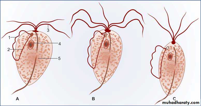
Trichomonas species. (A) T. vaginalis, (B) T. hominis, (C) T. tenax. 1—Undulating membrane, 2—Costa, 3—Flagella, 4—Nucleus, 5—Axostyle
3
Trichomoniasis is a flagellate protozoan that parasitizes the human vagina, prostate gland, and urethra. It is sexually transmitted infection caused by Trichomonas vaginalis. It is one of the most common sexually transmitted diseases in the USA. Women with trichomoniasis may experience various symptoms, including a yellow-green vaginal discharge and vulvar irritation, or they may be asymptomatic. Men with trichomoniasis are frequently asymptomatic.
4
Genital flagellates
Trichomonas vaginalis
Epidemiology
Worldwide distribution, the annual incidence of trichomoniasis is about 170 million cases annually. There are more than 7 million cases of Trichomoniasis each year in the USA.The incidence of trichomoniasis in Europe is similar to that in the USA. In Africa, the prevalence of trichomoniasis may be much higher.
Sexual transmission is the usual mode of infection. Babies may get infected during birth. Fomites such as towels have been implicated in transmission.
5
Most sexually-transmitted infections are more prevalent among adolescents and young adults; however, Trichomoniasis has a similar prevalence among sexually active women of different age groups.
The infection is highly associated with the presence of other sexually T vaginalis transmitted infections, including gonorrhea, chlamydia, and HIV.
6
MORPHOLOGY
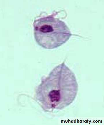
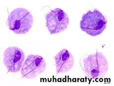
Morphologically T. vaginalis has only the trophozoite stage, resembling other trichomonads.All the trichomonads are morphologically similar, having a pear-shaped body 7- 23 μm long, a single anterior nucleus, three to five forward-directed flagella, and a single posteriorly directed flagellum that forms the outer border of an undulating membrane.
7
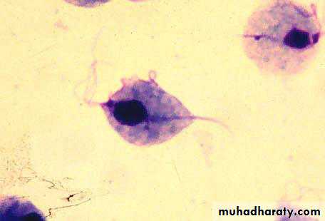
Trophozoit
Pathogenesis
T. vaginalis is around the size of a white blood cell (about 10 μm in diameter), although its size may vary with physical conditions. Its flagellum allows it to move around vaginal and urethral tissues.T. vaginalis attaches to the vaginal epithelium. T. vaginalis produce mediator substances that enable the attachment to epithelial surfaces, which facilitate this binding.
8
After binding, T. vaginalis triggers detachment of cells through proteolytic activity, cytotoxicity and apoptosis.
Infection is often asymptomatic, particularly in the male.
In females, it may produce severe pruritic vaginitis with an offensive, yellowish, often frothy discharge.
Cervical erosion is common, with endometritis.
The incubation period is 4 days to 4 weeks.
9
Patients infected with T. vaginalis produce circulating (IgG) and secreted (IgA) antibodies that recognize adhesions and prevent parasite adhesion; however, protection is only short-term as re-infection rates as high as 30% have been observed.
10
Clinical findings
Women signs:The disease in Women show a frothy yellowish-green vaginal discharge, abnormal vaginal odor, vulvo-vaginal itching and soreness, dyspareunia (pain during sexual intercourse), and dysuria (pain during urination).
Cervicitis due to trichomoniasis is characterized by purulent discharge in the endocervical canal and easily induced endocervical bleeding.
Vaginitis, which is characterized by vaginal discharge, which may be accompanied by vulvar itching, irritation, and odor.
Men signs:
The disease in men range from none to urethritis complicated by prostatitis.Signs of men urethritis include discharge, dysuria, and urethral pruritus. The discharge may be purulent to mucoid in character.
Laboratory Diagnosis
Samples in womenVaginal discharge
Endocervical specimen
Samples in men
Urethral discharge
Prostatic fluid
Early morning first voided urine sediment
Semen
13
Parasitic Diagnosis-Methods of examination
MicroscopyCulture
It can be grown in a variety of solid and liquid media, in tissue culture and in eggs. CPLM (cysteine, peptone, liver, maltose) medium is often used.
Antigen detection ( ELISA )
Molecular diagnosis
DNA probes – more sensitive & highly specific
PCR- highly sensitive & specific
14
Laboratory Diagnosis
Diagnosis by Microscope
Wet mountEasy, useful & economic
80% sensitivity in symtomatic females
T. vaginalis trophozoites seen with characteristic jerky & twitching motility
Acridine orange stain
Rapid & accurate method
Sensitivty same as wet mount
Direct flourescent antibody staining
Rapid & more sensitive
Requires a flourescent microscope
15
Diagnosis by Culture
Most sensitive & Gold standard.Media – Diamond, Lash & kupferberge (contains yeast extract, horse serum & antibiotics), observed for 7 days.
Culture usually positive after 48 hrs.
Done in patients with suspected trichomoniasis but wet mount negative.
16
Treatment
Metronidazole is effective in both males and females.Clotrimazole topical.
Personal hygiene.
17