• 25-11-2018
Medical Parasitology3rd class
HaemoflagellatesLeishmaniasis
• Dr. Abdul Rahman Dahham, PhD
• Department of Microbiology• College of Medicine
• Nineveh University
1
Leishmaniasis is an important protozoal vector-borne diseases that affects both humans and animals, caused by many species of Leishmania. The disease transmitted through the bite of an infected sandfly. The genus Leishmania is named after Sir William Leishman who discovered the flagellate protozoon causing kala-azar, the Indian visceral leishmaniasis.
Leishmaniasis
2
All members of the genus Leishmania are obligate intracellular parasites that pass their life cycle in two hosts, the mammalian host and the insect vector, female sandfly.
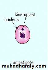
In human and other mammalian hosts, they multiply within macrophages, in which they occur exclusively in the amastigote form, having an ovoid body containing a nucleus and kinetoplast.
In the sandfly, they occur in the promastigote form, with a spindle shaped body and a single flagellum arising from the anterior end.
3
The parasite may survive for decades in asymptomatic infected people, who are of great importance for the transmission since they can spread visceral leishmaniasis indirectly through the sandflies.
The parasites can also be transmitted directly from person to person through the sharing of infected needles.
4
Leishmaniasis is a major public health problem in many parts of the world.
According to the WHO Report around 1.5 million cases of cutaneous leishmaniasis and 500,000 cases of visceral leishmaniasis occur every year, spread over 82 countries.About 350 million people are at risk of leishmaniasis, with 12 million people currently infected.
5
Kingdom: Animalia
Phylum: ProtozoaClass: Zoomastigophora
Order: kinetoplastida
Family: Trypanosomatidae
Genus: Leishmania and Trypanosoma ( blood tissue species )
Species: donovani, tropica, mexicana, braziliensis, major, aethiopica
6
Leishmania species considered pathogenic for humans
Leishmania donovaniLeishmania tropica
Leishmania major
Leishmania aethiopica
Leishmania mexicana
Leishmania brazilliensis
7
Morphology and Life Cycle
The parasite exists in two forms, the amastigote form in humans and other mammals, and the promastigote form in the sandfly and in artificial cultures.8
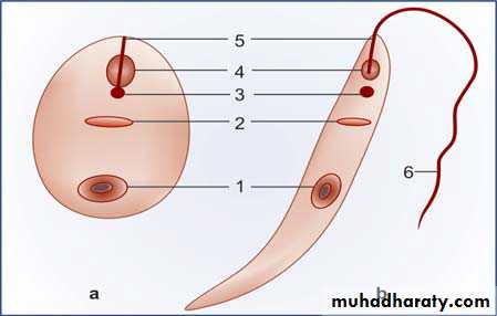
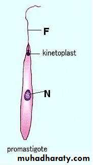
Morphology of Leishmania donovani.
a. Amastigote b. Promastigote. 1. Nucleus 2. Parabasal body 3. Blepharoplast 4. Vacuole 5. Axoneme 6. Flagellum• 1. Amastigote shape:- found in final host, infect the Liver ,Spleen ,bone marrow ,lymph nodes and macrophage. It is Roundish to oval in shape, Consist of a nucleus and kinetoplast. The large single nucleus is typically located off-center.
9
• • 9
The dot like blepharoplast is attached to a small axoneme, this axoneme extends to the edge of the organism. The single parabasal body is located adjacent to the blepharoplast.
10
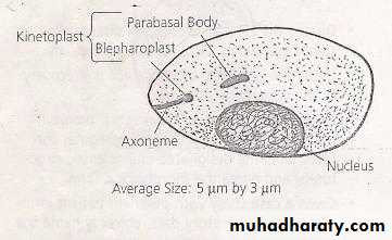
Smears stained with Leishman, Giemsa or Wright stains show a pale blue cytoplasm enclosed by a limiting membrane.
The habitat of the amastigote is the reticulo-endothelial system. They are found mostly within the macrophages in the spleen, liver and bone marrow.
They multiply by binary fission, producing numerous daughter cells that distend the macrophage and rupture it. The liberated daughter cells are in turn phagocytosed by other macrophages and histiocytes.
11
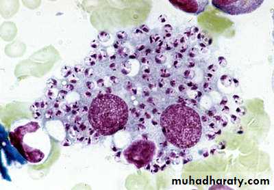
Amastigote from biopsy of macrophages
(Leishman stain)12
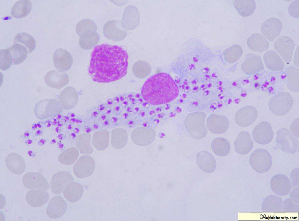
• Ovoid small intracellular parasites in a bone marrow aspirate. The typical rod shaped kinetoplast is seen besides the nucleus.(Giemsa stain).
Leishmania sp.
amastigote stage
13
• When a vector sandfly feeds on an infected person, the amastigotes present in peripheral blood and tissue fluids enter the insect along with its blood meal.
• In the midgut (stomach) of the sandfly, the amastigote elongates and develops into the promastigote form.
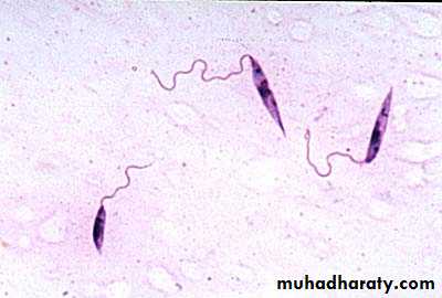
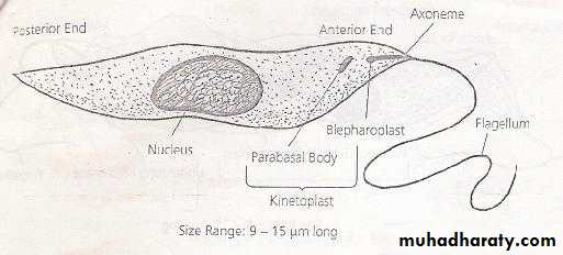
2– Promastigotes shape :
It is Long and slender in appearance.The large single nucleus is located in or near the center.
The kinetoplast is located in the anterior end of the organism.
A single free flagellum extends anteriorly from the axoneme .
15
found in mid gut and Salivary gland for the infective Sand fly or in culture media.
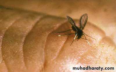
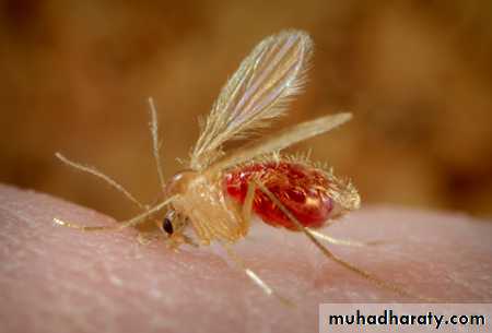
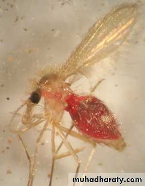
• Infective stage:-Promastigote shape• Final hosts:- human , dogs and rodents.
• Intermediate host:- female Sand fly genus phlebtomus papatsi
16
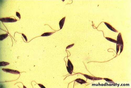
Promastigote culture from
17Promastigote stage
Promastigote stage inside the Sandfly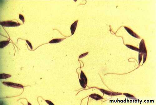
flagella
18The major difference between (Leishmania and Trypanosoma) is that primary diagnostic form found in Leishmania is the amastigote, whereas that of Trypanosoma is the trypomastigote.
The genus Leishmania include flagellate that occur as amastigote form in vertebrate hosts and as promastigote form in invertebrate hosts and cultures.
19
• • 19
Leishmaniasis has three different clinical forms, depending on the parasite species and the cellular immune system of the patient:
20
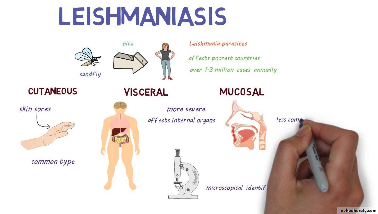
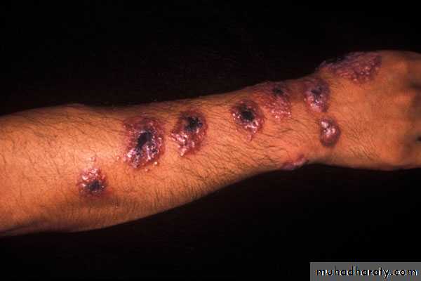
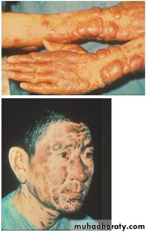
A- Cutaneous form leishmaniasis
Produces skin lesions mainly on the face, arms and legs. Considered as a self-healing form, it can create serious disability and permanent scars.caused by Leishmania tropica
The disease called cutaneous leishmaniasis or oriental sore or Baghdad boil or Delhi boil.21
90% of cutaneous leishmaniasis occurs in Afghanistan, Iran, Saudi Arabia, Syria, Brazil and Peru. 8,7% cases were reported in Iraq in 1992 and 2001.
After recovery or successful treatment, cutaneous leishmaniasis induces immunity to re-infection by the species of Leishmania that caused the disease.
22
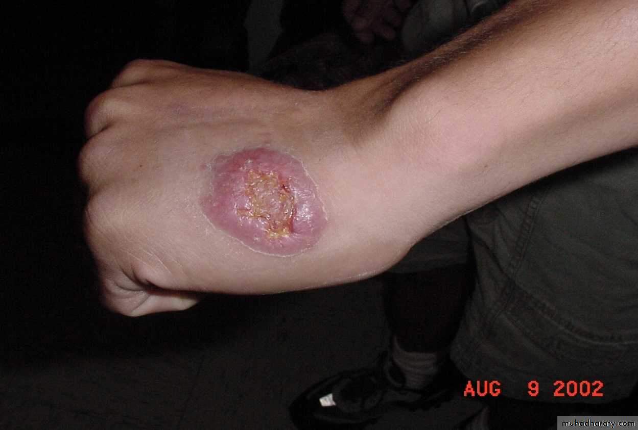
Cutaneous Leishmaniasis
23
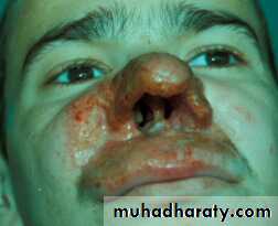
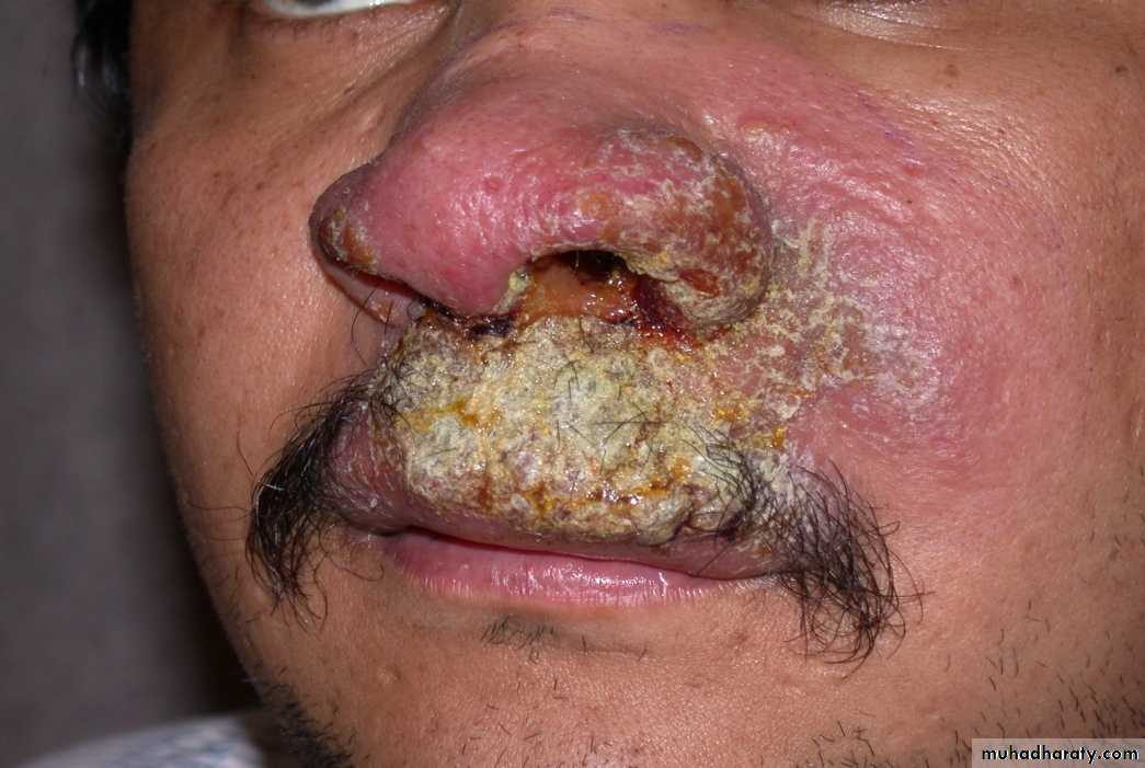
B- Muco-cutaneous leishmaniasis
also called 'espundia' in South America, causes disfiguring lesions to the face; it destroys the mucous membranes of the nose, mouth and throat.The disease called Muco-cutaneous leishmaniasis or American leishmaniasis.
90% of mucocutaneous leishmaniasis occurs in Bolivia, Brazil and Peru.Caused by Leishmania brazilliensis
2425
C- Visceral leishmaniasis
the disease also known as Visceral leishmaniasis or Kala-azar (meaning black sickness), Dum Dum fever, or Black fever.The disesease cased by Leishmania donovani (Involving endothelial tissue, liver, spleen, and bone marrow).
26
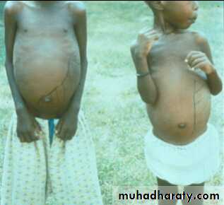
it is characterized by irregular fever, weight loss, swelling of the liver, spleen and anaemia. It is the most severe form of Leishmaniasis, usually fatal if left untreated.
The incubation period can be months or years and, unlike the cutaneous forms of leishmaniasis, it involves the internal organs.
90% of all visceral leishmaniasis occurs in Bangladesh, Brazil, India, and Sudan. 2893 cases were reported in Iraq in 2001.
27
Disease Pathogenesis
The infection is acquired when a person is bitten by an infected sand fly.The parasite is taken up by macrophages and then replicates within the host macrophage. Then the parasites are released from macrophage and are taken up by other macrophages and continue this replication cycle.
This usually results in an ulcerated skin lesion at the site of the original sand fly bite.
28
The infected macrophages will spread from the original site and cause secondary skin lesions or invade mucosal tissue, especially around the mouth and nose, results in a disfiguring disease in which the cartilage of the nose and surrounding tissue is destroyed.
In visceral leishmaniasis the infected macrophages migrate to the bone marrow, liver and spleen and cause a systemic infection known as visceral leishmaniasis.
29
The spleen is the organ most affected. It is grossly enlarged and the capsule is frequently thickened, soft and friable and cuts easily without resistance, due to absence of fibrosis.
The cut section is red or chocolate in colour due to the dilated and engorged vascular spaces. The trabeculae are thin and atrophic.
The liver is enlarged. The Kupffer cells and vascular endothelial cells are heavily parasitised.
The bone marrow is heavily infiltrated with parasitised macrophages which may crowd out the haemopoietic tissues.
30
Leishmaniasis Life-Cycle
Sand fly Stages1
5
4
3
2
6
7
8
Human Stages
Sand fly takes a blood meal
(Injects promastigote
stage into the tissue)
Promastigotes are
Phagocytized by
macrophages
Promastigotes transfer
into amastigotes inside
macrophages

Amastigotes multiply in cells
(Including macrophages) of
Various tissues

Sand fly takes
a blood meal(ingest macrophages
Infected with amastigotes )
Ingestion of
Parasitized cell
Amastigotes transform
Into promastigote
stage in midgut
Divide in midgut and
migrate to proboscis
i
d
i
d


Infective stage
Diagnostic stage31
32
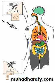
The life cycle of the disease begins when an infected female sand fly bites a person.
When the bite occurs, the sand fly injects the promastigotes (the infective stage) into the person.
Saliva of sand fly containing anti-coagulant is injected into the wound to prevent the blood from clotting, the promastigotes are transferred to the host.
The leishmania Life cycle
• • 32
Once in the host the promastigotes are taken up by the macrophages where they rapidly revert to the amastigote form. causing tissue destruction, and eventually leading to the lysis of the macrophages.
regards the internal organs, ultimately all the organs containing macrophages and phagocytes are infected, especially the spleen, liver and bone marrow, and enters into the circulation system of their host
33
• • 33
At this point, if a sand fly chooses the infected human as the source of a blood meal, it will again ingest the parasite (becomes infected).
The macrophages are ingested by the fly during the blood- meal and the amastigotes are released into the stomach of insect. The amastigotes transform back into the promastigote form in the fly midgut. These promastigotes multiply and migrate into the salivary gland of the fly and transferred to a new victims.
34
Cutaneous leishmaniasis :
Tissue sample (scraping, aspirate or punch biopsy) for smear and cultureVisceral leishmaniasis :
Bone marrow biopsy or splenic aspirate for smear and culture.(N.N.N) V.L.(anemia , leukopenia , glubuline/albumine is high (Hypergammaglobulinia)
Serology ( ELISA ) ( IFAT ).
PCR
Skin test
Inoculate serum of infected person in lab. animals.
• Laboratory Diagnosis of leishmaniasis :
35
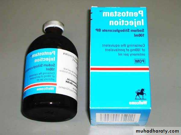
Treatment
– self-healing lesions
Medical:
Pentavalent antimony (Pentostam), Amphotericin B
Antifungal drugs
+/- Antibiotics for secondary bacterial infection.
Surgical:
Cryosurgery
Excision
Curettage
36
Kala azar is treated today essentially as it was in 1940. The major drug is Sodium stibogluconate or Pentastam, a derivative of antimony, which was developed in 1930.
Amphotericin is used with or after an antimony compound for visceral leishmaniasis unresponsive to the antimonial alone.
Pentamidine isotionate has been used in antimony-resistant visceral leishmaniasis.
Recently a new drug was developed, miltefosine. This is a membrane signaling pathway inhibitor. This can be taken orally and is very effective against visceral leishmaniasis.
37