Lab 3
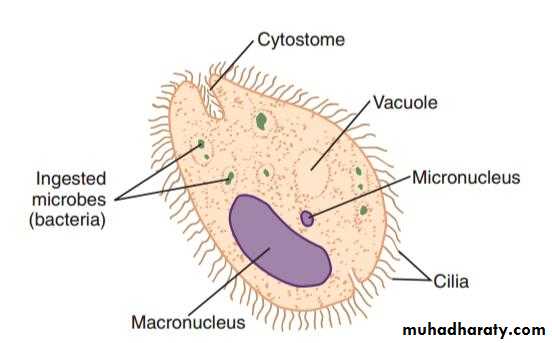
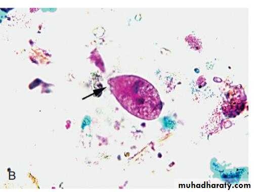
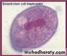
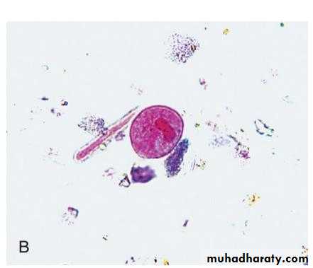
• Balantidium coliTrophozoite morphology
• 50-150 mic• Cilliated parasite
• Oval shape
• Greenish yellow color
• Kidney or bean shape Macronucleus
• Small micronucleus
• Retractile food vacule
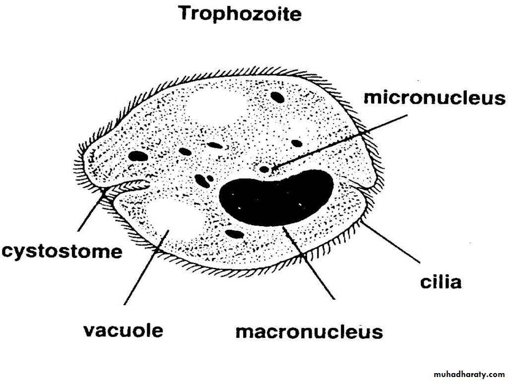
• Trophozoite Balantidium coli
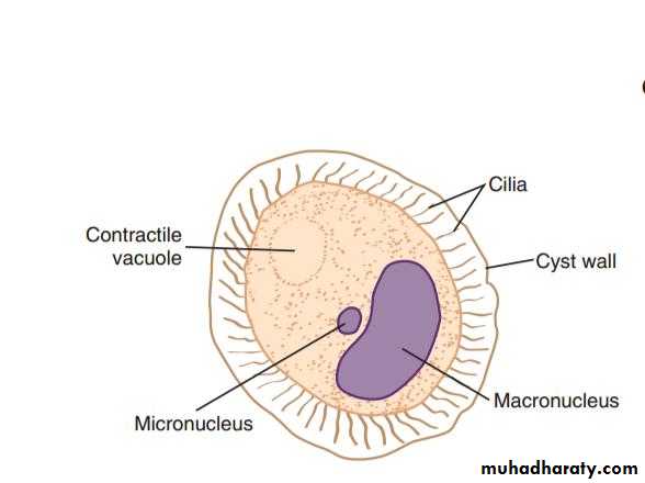
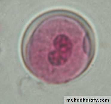
Cyst morphology
45-55 micSpherical shape
Cyst wall is thick consist of 1-2 layers
No phagosome
Macronucleus
Contractile vacules
No cilia
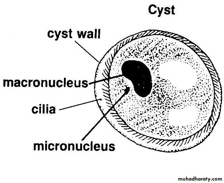
Balantidium coli Cyst
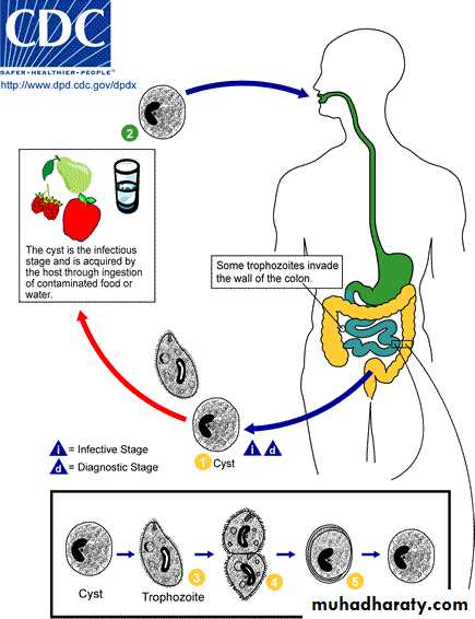
Life cycle
• Cyst exyst in the small intestine releasing trophozoites that migrate to the large intestine. Trophozoites reside in the lumen of large intestine Invade mucosa and sub M.• Feed on mucosal cells, RBC, leukocyte
• where they divide by transverse binary fission.
• Encystation is triggered by dehydration of intestinal content.
Habitate
*Parasite live in L.I specially cecal region.*Cyst formed in large intestine or in outer envirnment.
Diagnosis
Symptoms
Clinical signs could confused by histolytica infection
Finding the typical trophozoites and cysts in the stoolDiagnosis
Finding parasite (cyst or trophozoite) in stool byDirect wet mount method.
Stained smear by iodin.