Lab 2 Amebas
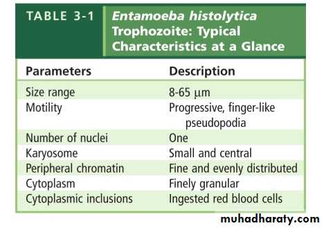
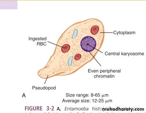
Entameba histolyticaPathogenic
Intestinal
Common diseases :-
• Intestinal amebiasis• Amebic colitis
• Amebic dysentery
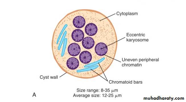
morphology
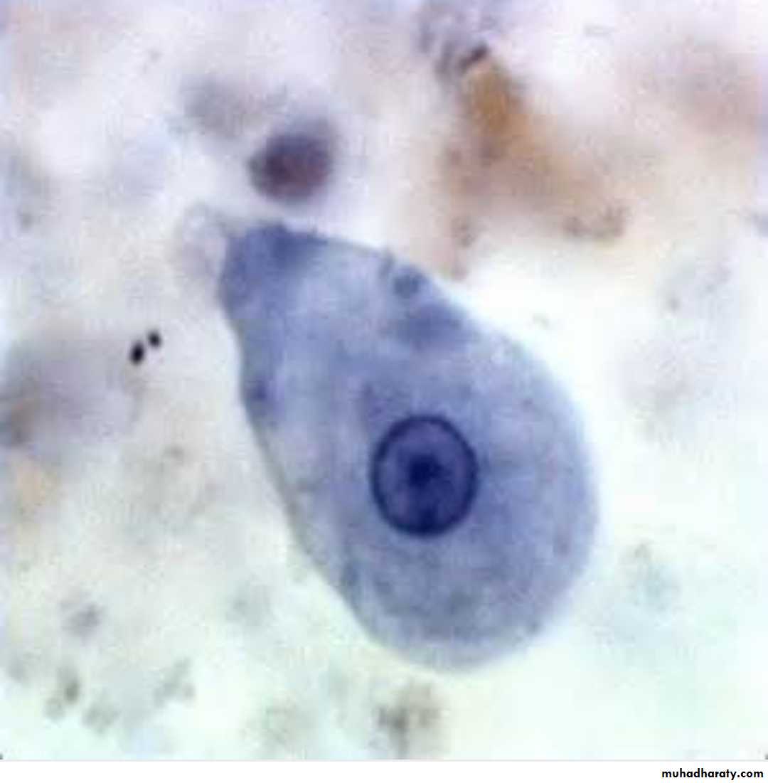
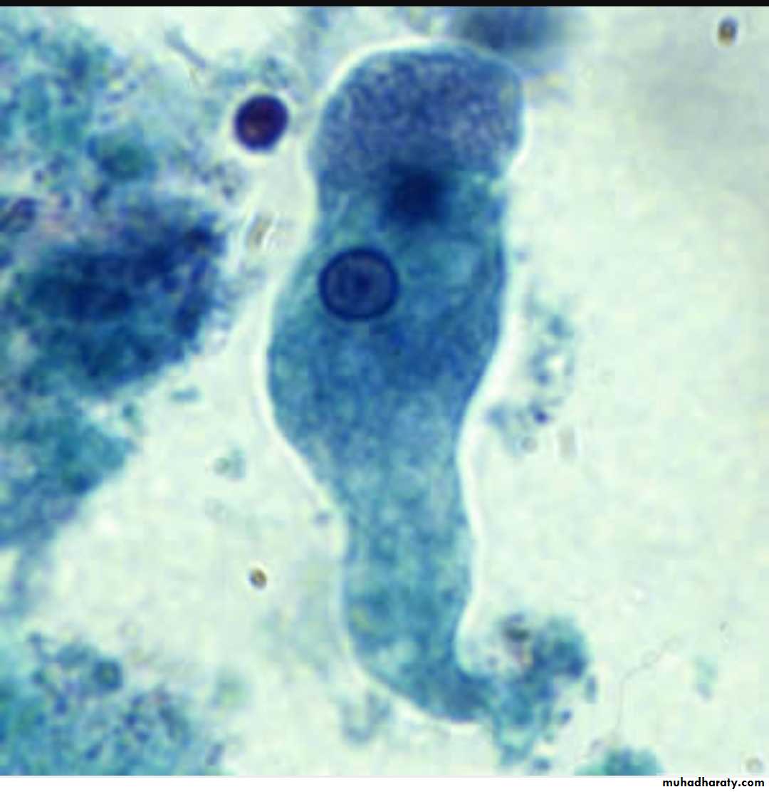
Two stages :-Trophozoite
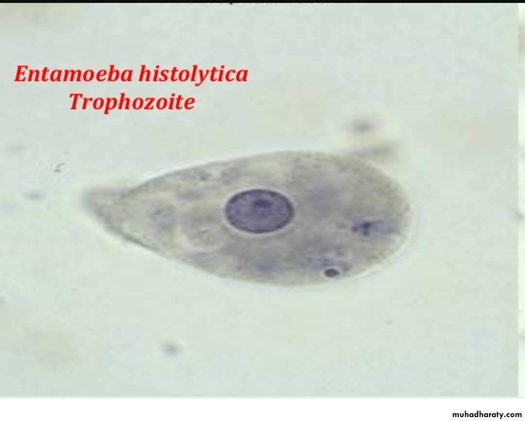
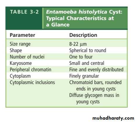
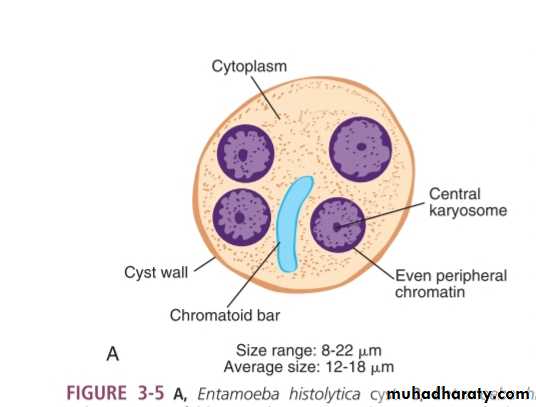
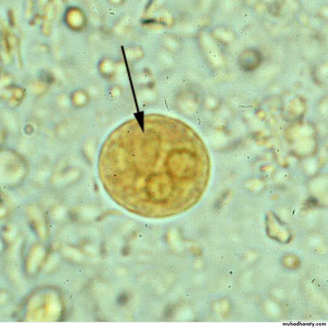
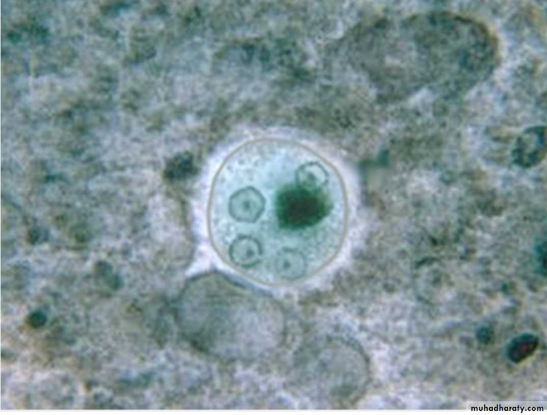
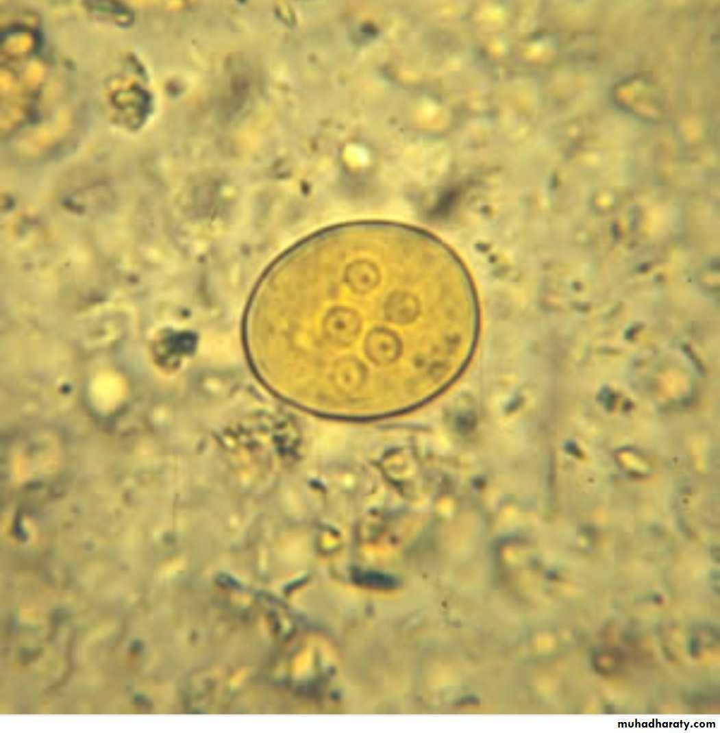
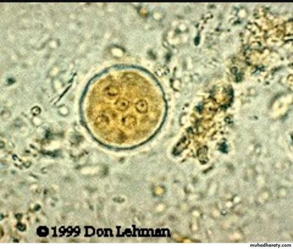
Cyst
Reservoir :- dogs , pigs, monkeys.
Infective stage mature cystIntermediate host :- none
Definitive host :- human
Habitate large intestine (human)
Mode of infection infective cyst in contaminated food and water
Diagnostic stage cyst in formed stool
trophozoite in liquid or loose stoolSource of infection
• Ingestion of infective cyst through hand to mouth.
• Contaminated food or water.
• By vector like flies and cockroaches.
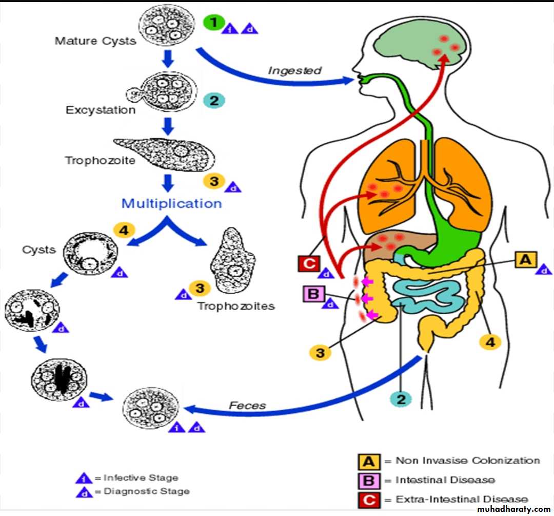
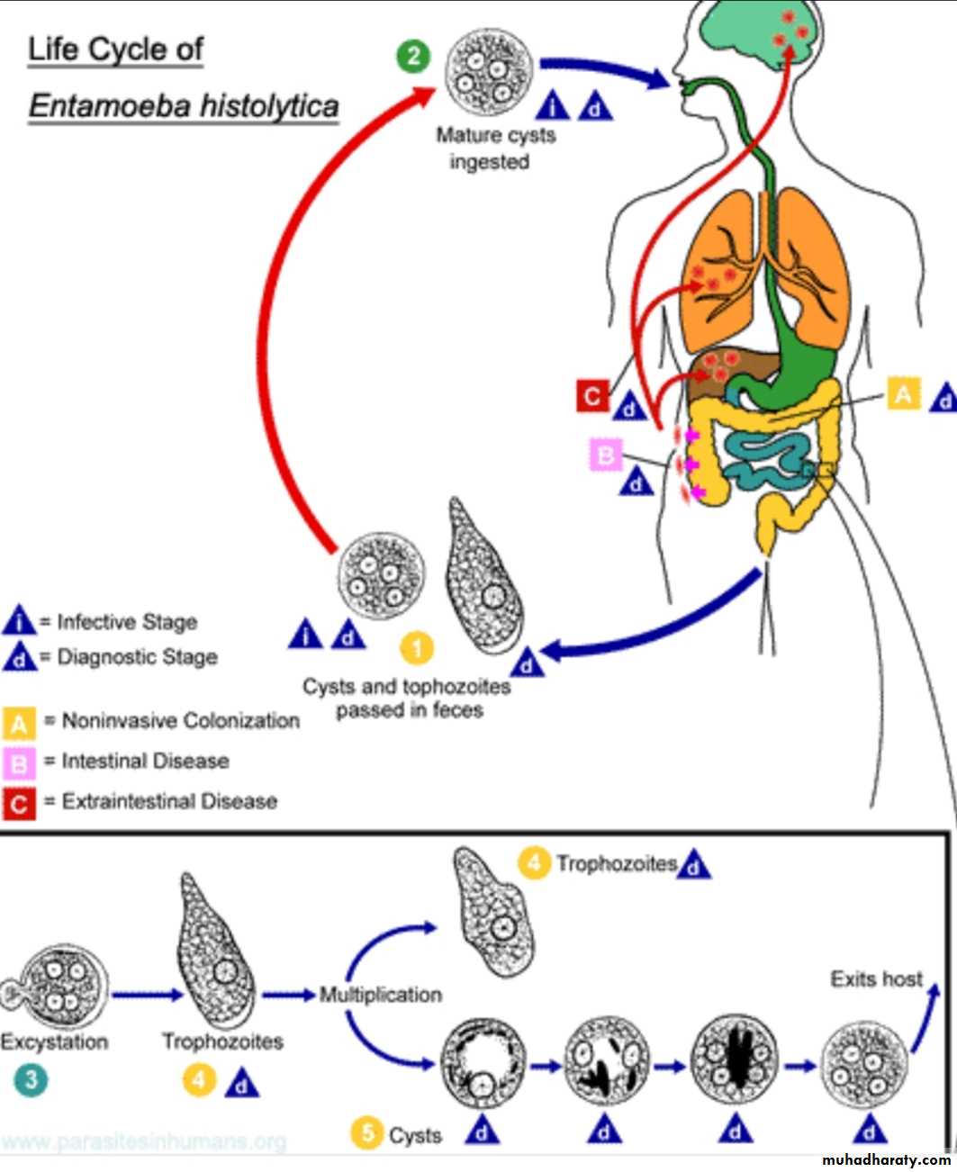
Life cycle
Ingestion of cystExcystation due to nuclear division.
8 motile trophozoite settle in the lumen of the large intestine ( trophozoite migrate to other organs in the body such as liver and may cause liver abscess).
Encystation occurs in the lumen of large intestine and cyst formation complete when four nuclei are present.
Clinical pictures
90 % asymptomatic.10% symptomatic.
• Symptomatic intestinal :-
• Amebic colitis.
• Amebic dysentery (blood, mucus in the stool).
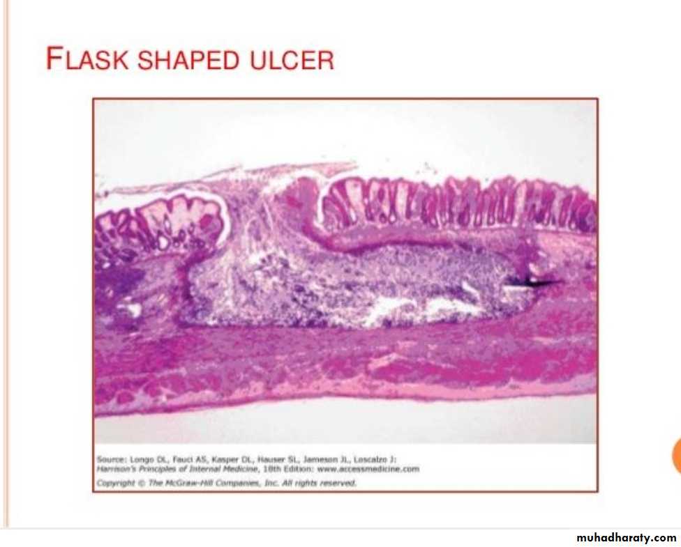
• Flask shape ulcer in the colon, cecum, recto sigmoidal area.
B. Symptomatic extra-intestinal
Trophozoite of Entameba histolytica may migrate to the blood stream, are removed by the liver and lead to formation of liver abscess. Also migrate to other organs like brain, pericardium , lungs and spleen.
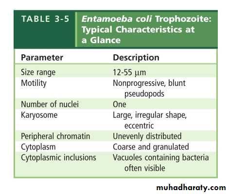
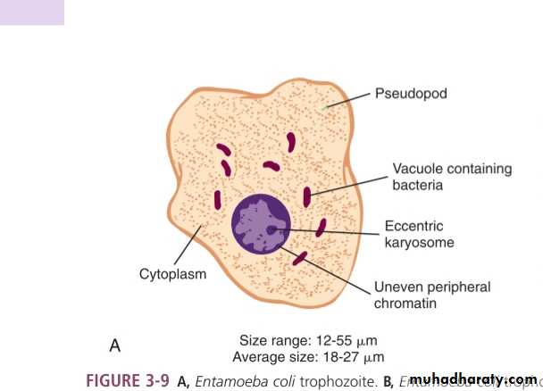
Entameba coli
Non pathogenic
Intestinal
Morphology
Two stages :-Trophozoite
Cyst
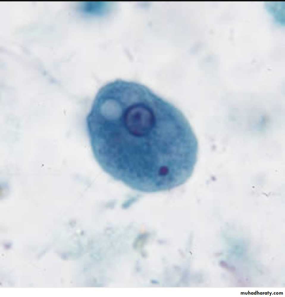
Trophozoite
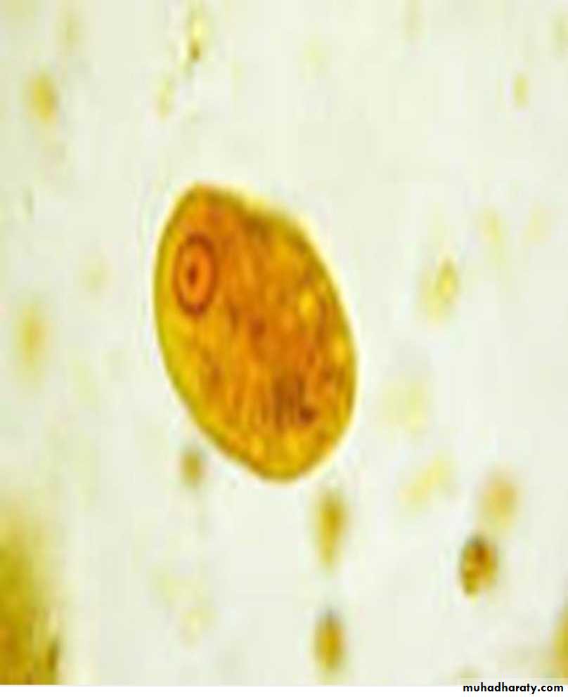
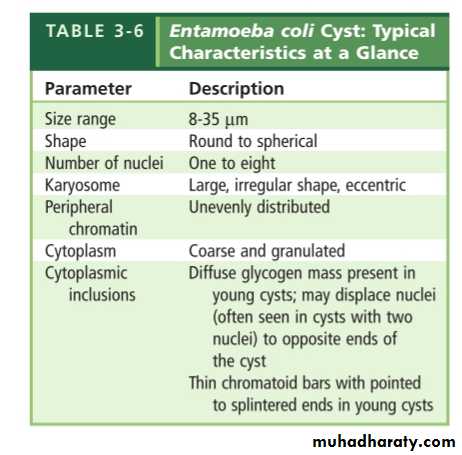
Cyst
Diagnosis
1. Stool examination :-• Direct method for motile trophozoite.
• Iodine stain for cyst .
• The typical amebic stool contain blood , mucus, few WBC , and bacteria.
2. Culture
3. Sigmoidoscopy :- may reveal the characteristic flask shape ulcer.
4. Biopsy
5. Serology :- is very important for the diagnosis of extra-intestinal amebiasis ex PCR , indirect hemagglutination6. Ultrasound , CT scan , MRI can be used to detect hepatic abscess.