Lab - 9
Genus Leishmania• include 4 major species
• Leishmania donovani• Leishmania tropica
• Leishmania Mexicana
• Leishmania braziliensis
• Genus Leishmania
Genus Leishmania
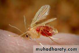
Final host: humanIntermediate host (vector): female of sand fly )phlebotomus)
Infective stage: Promastigote stage
Diagnostic stage: amastigotes stage
Route of infection: skin.
Leishmania donovani
• Disease• L. donovani is the cause of kala-azar (visceral leishmaniasis , black fever).
Leishmania tropica , Leishmania mexicana & Leishmania braziliensis
• Disease
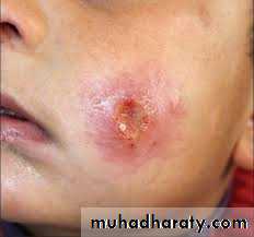
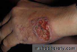
• L. tropica and L. mexicana both cause cutaneous leishmaniasis.
•
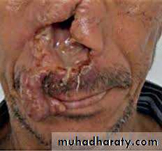
• L. braziliensis causes mucocutaneous leishmaniasis) Espundia(.
Morphological forms of Leishmania
Amastigote (Leishmania) form:• Promastigote (leptomonad) form:
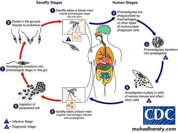
life cycleThe life cycle involves the sandfly as the vector and a variety of mammals such as dogs, foxes, and rodents as reservoirs. Only female flies are vectors because only they take blood meals (a requirement for egg maturation). Shortly after an infected sandfly bites a human, the promastigotes are engulfed by macrophages, where they transform into amastigotes. The infected cells die and release amastigotes that infect other macrophages and reticuloendothelial cells. When the female sandfly (phlebotomus) sucks blood from an infected host, it ingests macrophages containing amastigotes. After dissolution of the macrophages, the freed amastigotes differentiate into promastigotes in the gut. They multiply and then migrate to the pharynx, where they can be transmitted during the next bite. The cycle in the sandfly takes approximately 10 days.
life cycle
Sand fly
Pathogenesis
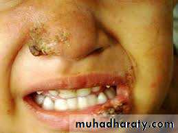
1- L. tropica , L. Mexicana and L. braziliensisThe lesions are confined to the skin in cutaneous leishmaniasis and to the mucous membranes, cartilage, and skin in mucocutaneous leishmaniasis.
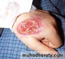
cutaneous leishmaniasis
• Mucocutaneous leishmaniasis) Espundia)
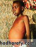
2- L. donovani
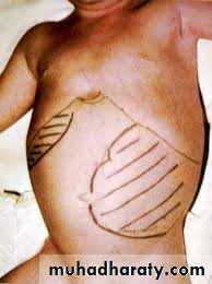
The organs of the reticuloendothelial system (liver, spleen and bone marrow) are the most severely affected.Reduced bone marrow activity with cellular destruction in the spleen.
The striking enlargement of the spleen is due to a combination of proliferating macrophages and sequestered blood cells.
visceral leishmaniasis
Diagnosis of Leishmaniasis
• 1- Microscopic detection of amastigotes (LD bodies: Leishman-Donovan) in Giemsa stained smear of bone marrow, spleen and lymph node or liver.• 2- Cultivation of aspirates in specific culture medium as NNN (Novy-MacNeal-Nicolle) medium → promastigotes seen.
3- Serological method (detection of specific
• anti-leishmanial antibodies):• · IFAT (indirect immunofluorescent antibody test)
• · DAT (Direct agglutination test)
• · ELISA (enzyme linked immunosorbent assay).
• 4- Molecular method (PCR polymerase chain reaction) the most sensitive and specific diagnostic method.
5- Animal inoculation:
6- Leishmanin skin test (Montenegro test):
• 7- In cutaneous leishmaniasis disease Immunological test (serology): has limited role in diagnosis because patient shows no detectable level of circulating antibodies.