Maternal bony pelvis
and fetal head

Objectives of this lecture
1.
Introduction to normal labour and vaginal delivery
passages, passenger, and power.
2.
The student should know the types of female
pelvis.
3.
understand the importance of the dimensions of
the bony pelvis of the pregnant woman in
determining the progress of labour and the mode
of delivery.
4.
What are the methods for assessment of pelvic
dimensions.
5.
Know the dimensions of the fetal skull.
6.
Understand how the attitude of the fetal head
effect these dimensions.

•
Labour
can be defined as the process by
which regular painful contractions bring
about effacement and dilatation of the cervix
and descent of the presenting part, leading
to expulsion of the fetus and the placenta
from the mother.
• A doctor or midwife who manages labour
must be aware of the normal anatomy and
physiology of the mother and fetus, what
distinguishes an abnormal from a normal
labour, and when it is appropriate to
intervene

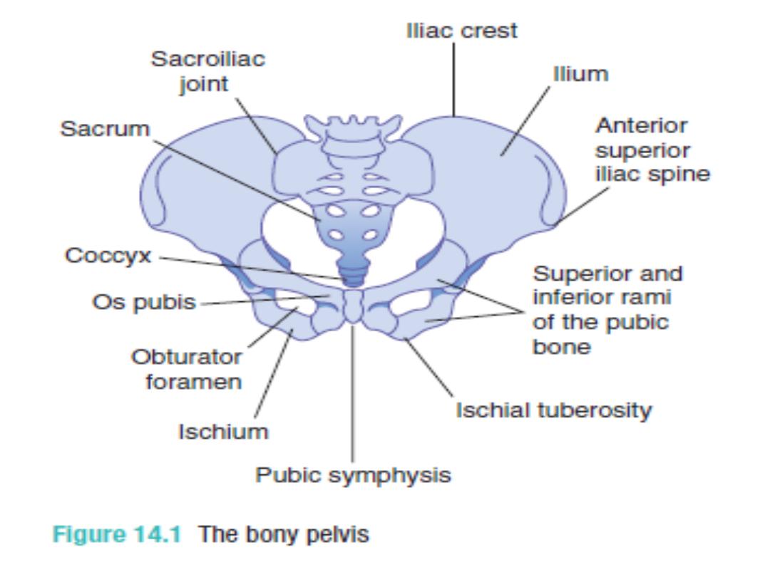
Bony pelvis
• The bony pelvis is made of 4
bones: the
sacrum, coccyx, and
2 innominate bones which are
(composed of the ilium,
ischium, and pubis).
These are
held together by the SIJ, SP, and
the SCJ joints.
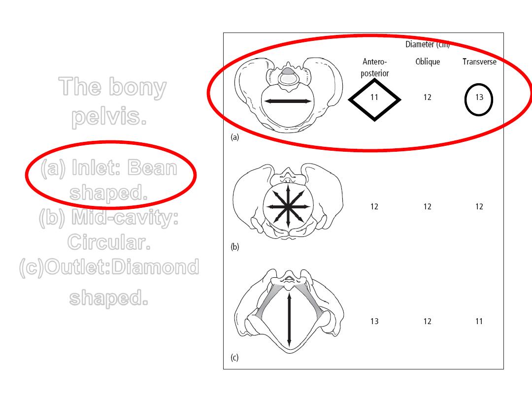
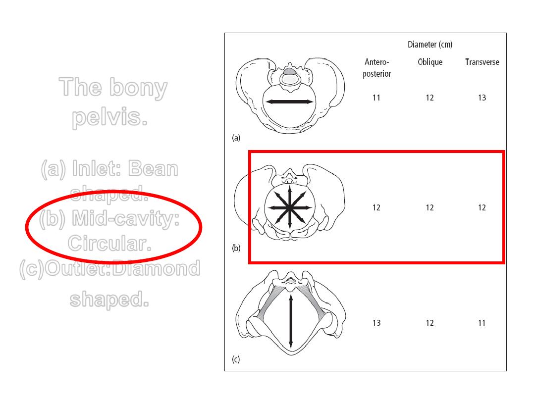
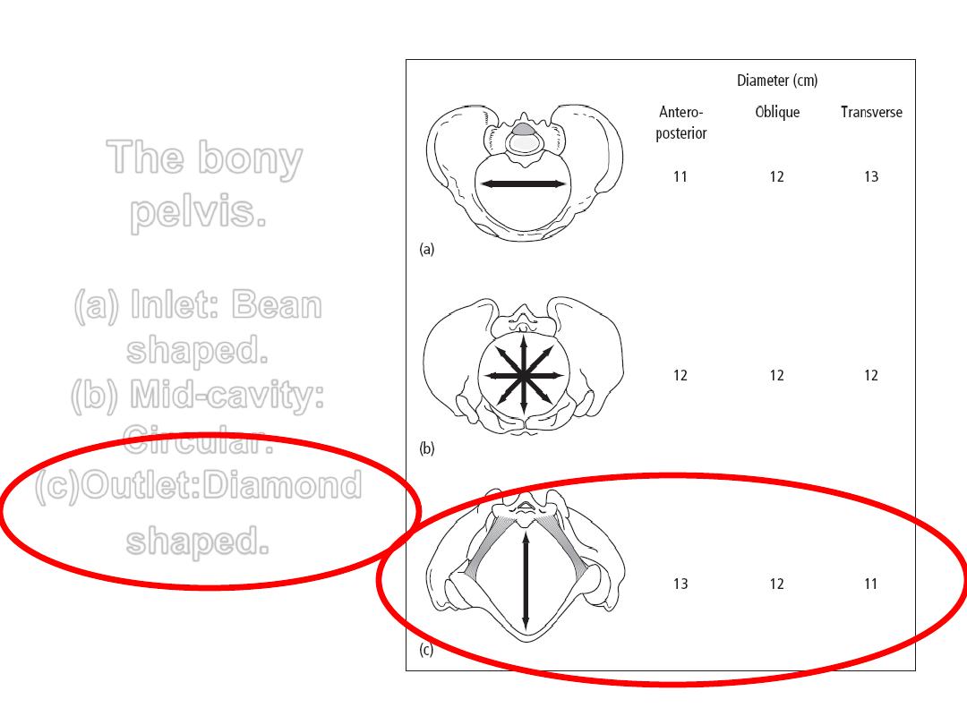
The bony
pelvis.
(a) Inlet: Bean
shaped.
(b) Mid-cavity:
Circular.
(c)Outlet:Diamond
shaped
.
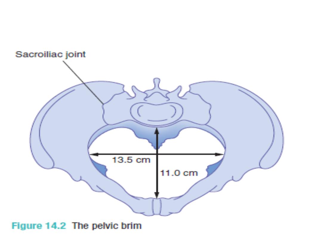
The pelvic brim or inlet
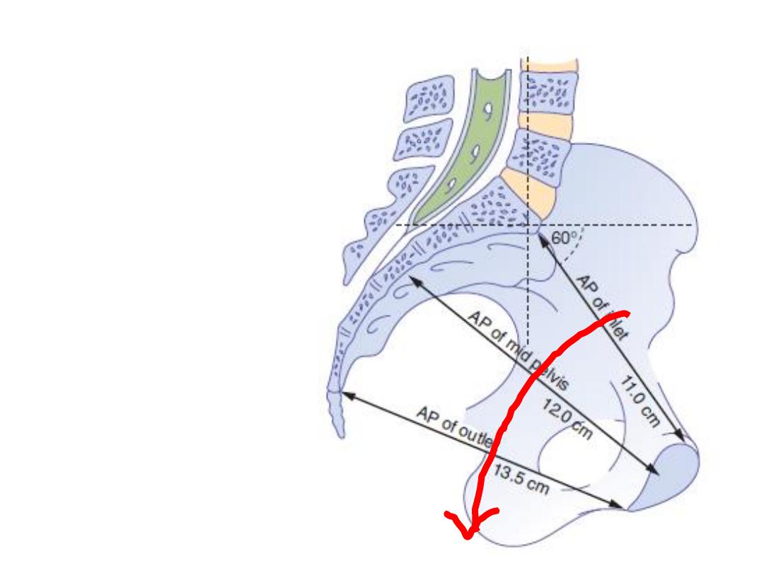
The
pelvic axis
describes
imaginary
curved line,
a path that
the centre
of the fetal
head must
take during
its passage
through the
pelvis

The pelvic mid-cavity
The pelvic mid-cavity can be described as an area
bounded in
front
by the middle of the symphysis
pubis,
on each side
by the pubic bone, the obturator fascia
and the inner aspect of the ischial bone and spines,
and
posteriorly
by the junction of the second and third
sections of the sacrum.
The cavity is almost round, as the transverse and
anterior diameters are similar at 12 cm.
The ischial spines are palpated vaginally and are used as
landmarks to assess the descent of the head on vaginal
examination (station). They are also used as landmarks for
providing an anaesthetic block to the pudendal nerve.
The bony
pelvis.
(a) Inlet: Bean
shaped.
(b) Mid-cavity:
Circular.
(c)Outlet:Diamond
shaped
.
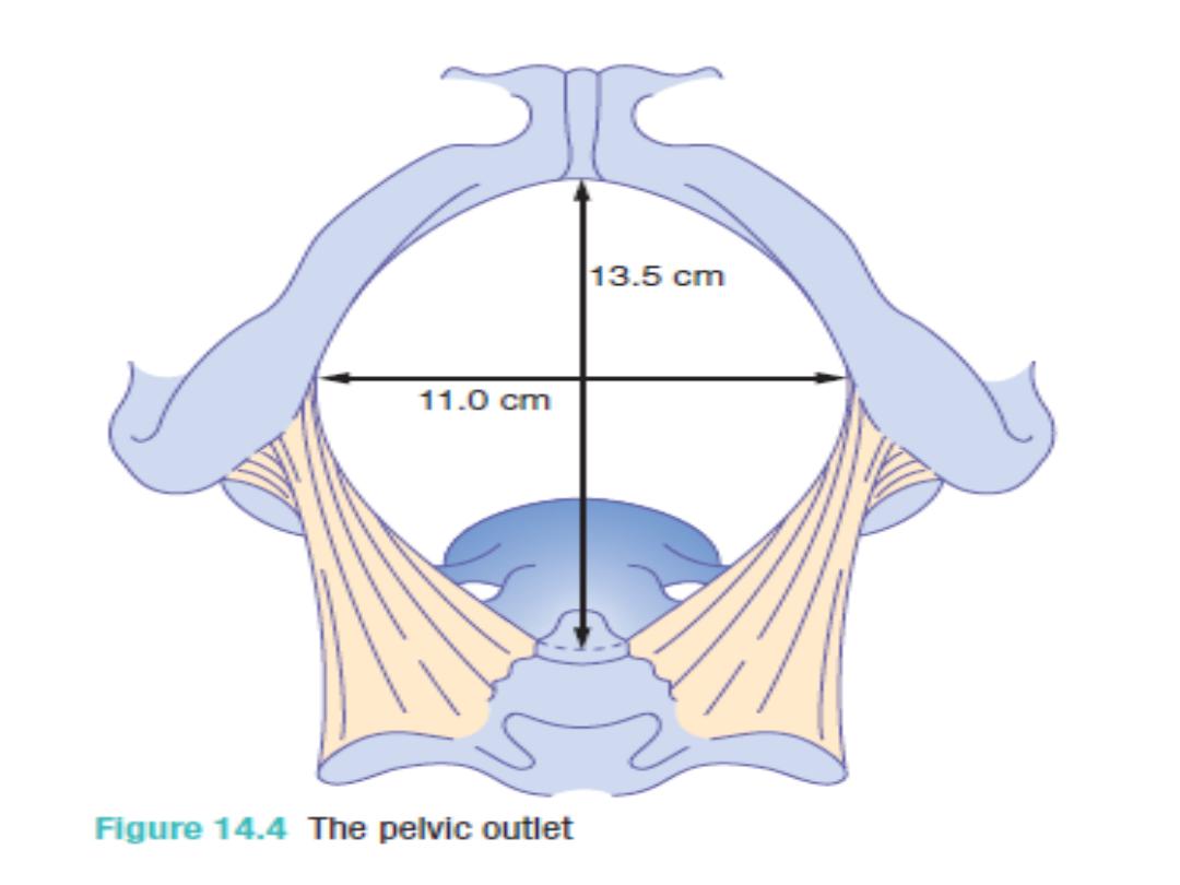
The pelvic outlet
The pelvic outlet is bounded
in
front
by the lower margin of the
symphysis pubis,
on each side
by the descending ramus of
the pubic bone, the ischial tuberosity and
the sacrotuberous ligament,
and
posteriorly
by the last piece of the
sacrum.
The AP diameter of the pelvic outlet is
13.5 cm and the transverse diameter is 11
cm
The bony
pelvis.
(a) Inlet: Bean
shaped.
(b) Mid-cavity:
Circular.
(c)Outlet:Diamond
shaped
.

Pelvic diameters:
These represent the
space available for the
fetal head when it passes
through the pelvis during
labour

1. the obstetric conjugate of the
pelvic inlet: 11 cm
2. the bispinous diameter: 10.5 cm
in the midcavity.
3. the bituberous diameter 11 cm in
the pelvic outlet
4. the curve and length of the
sacrum
5. and finally the subpubic angle

Pelvic shapes (types)
We have 4 types or shapes of the
bony pelvis and these are: the
gynecoid, android,
anthropoid
,
and finally the
platypelloid.
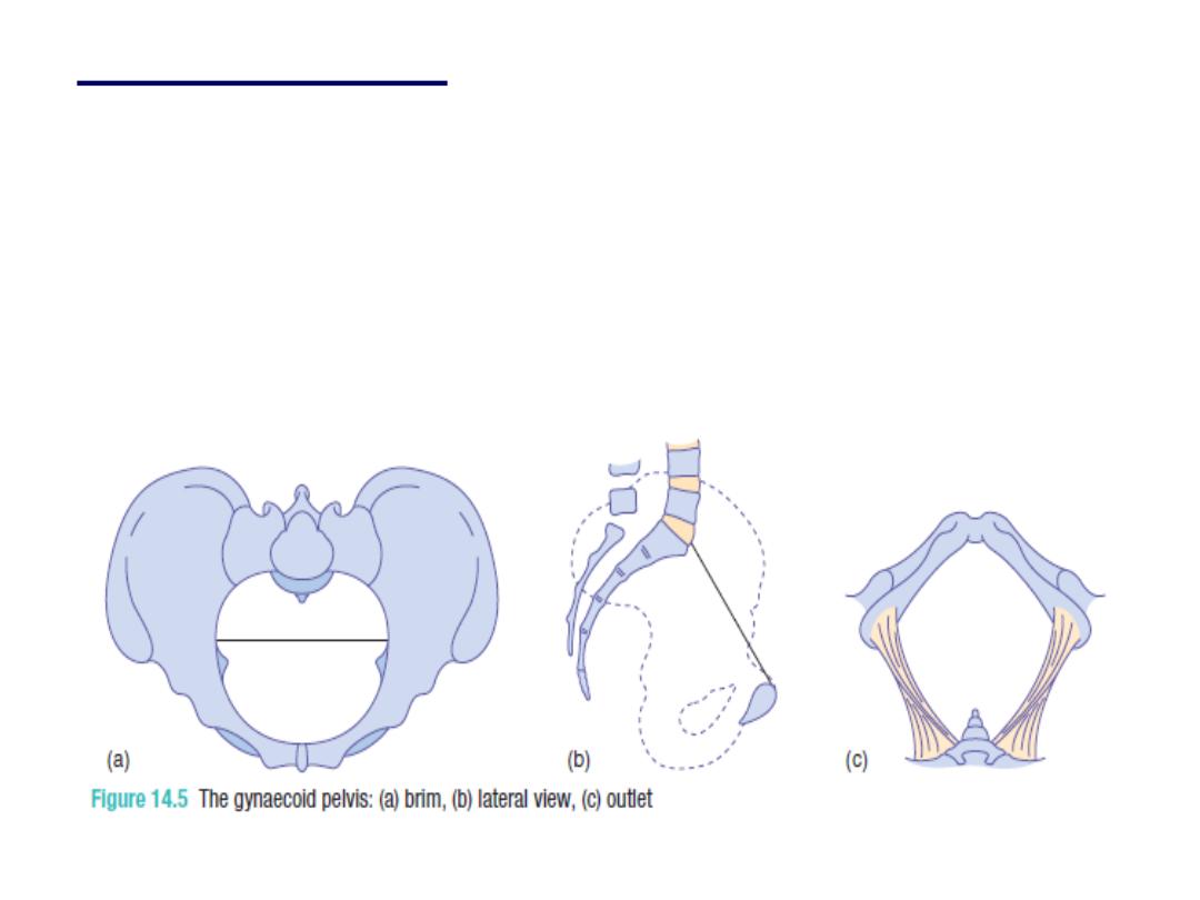
•
1- The gynecoid:
it is the classic female pelvis
and is seen in about 50% of all the women and
characterized by the following:
• Rounded to bean shape inlet, side walls are
straight, ischial spines are of average
prominence, well curved sacrum, wide subpubic
arch , Suitable for vaginal delivery
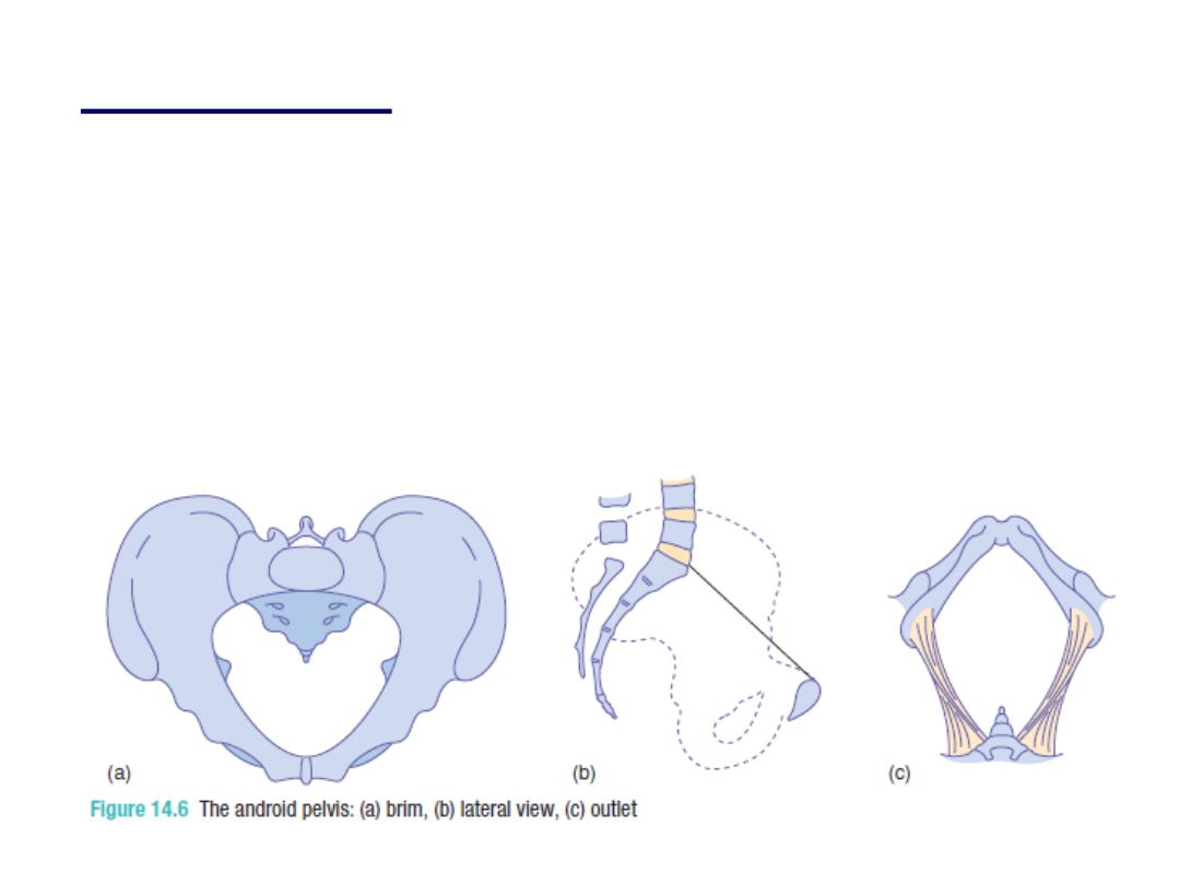
•
android pelvis
:
which is the typical male pelvis
and found in < than 30% of women and
characterized by:
•
Heart shape inlet (triangular), convergent side
wall (funnel shape) with prominent spines,
straight sacrum, and narrow subpubic arch.
•
Associated with deep transverse arrest
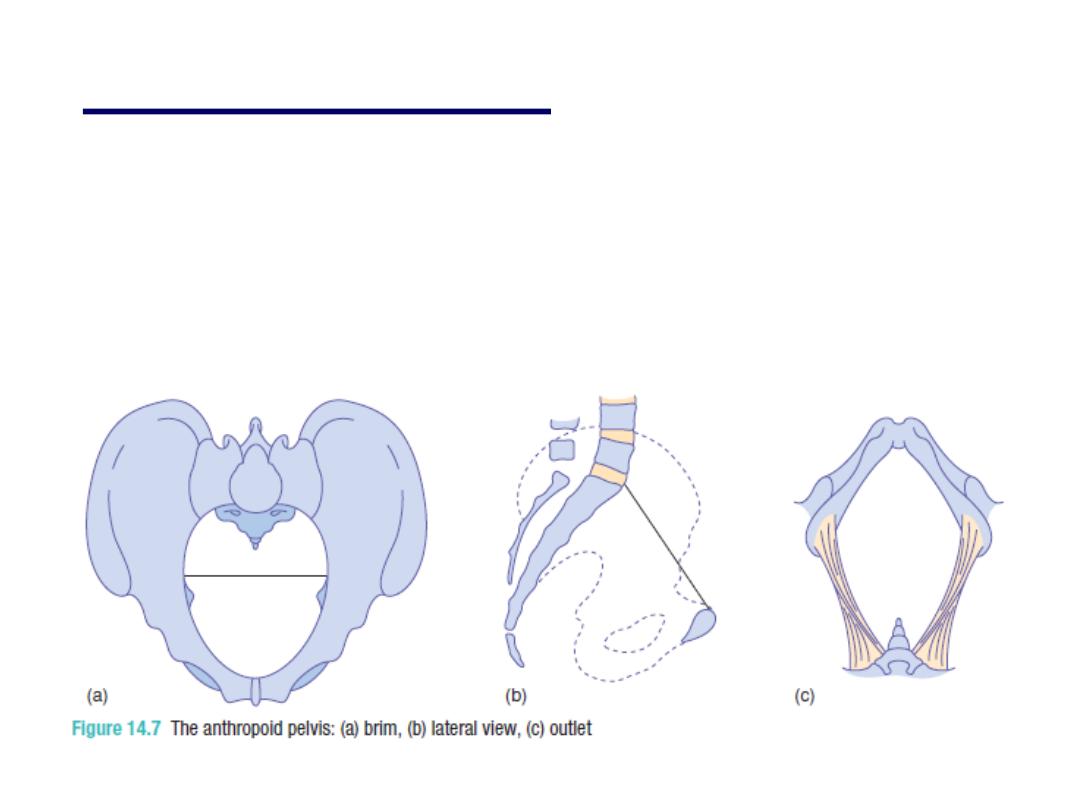
•
anthropoid pelvis:
is found in 20% of
women and
• Associated with occipito- posterior
position during labour
•
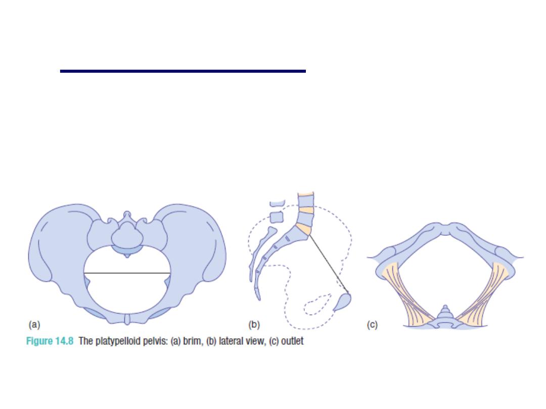
platypelloid pelvis:
which is a
flattened gynecoid pelvis and seen
in 3% of women and is associated
with obstructed labour

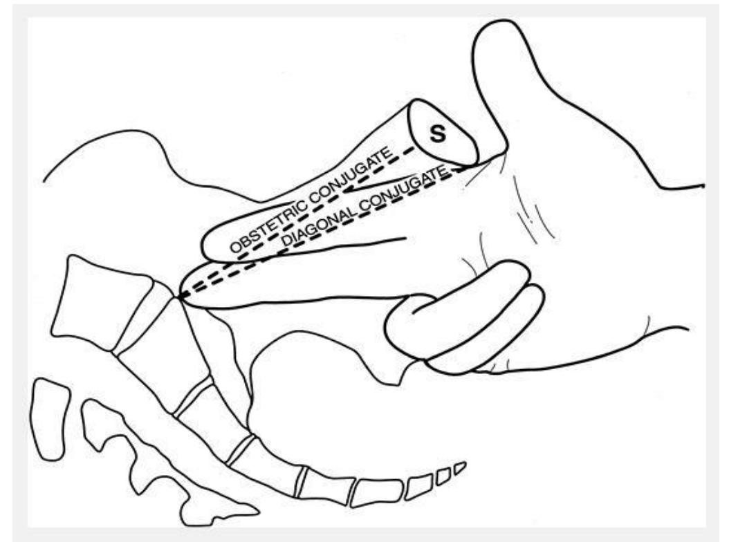
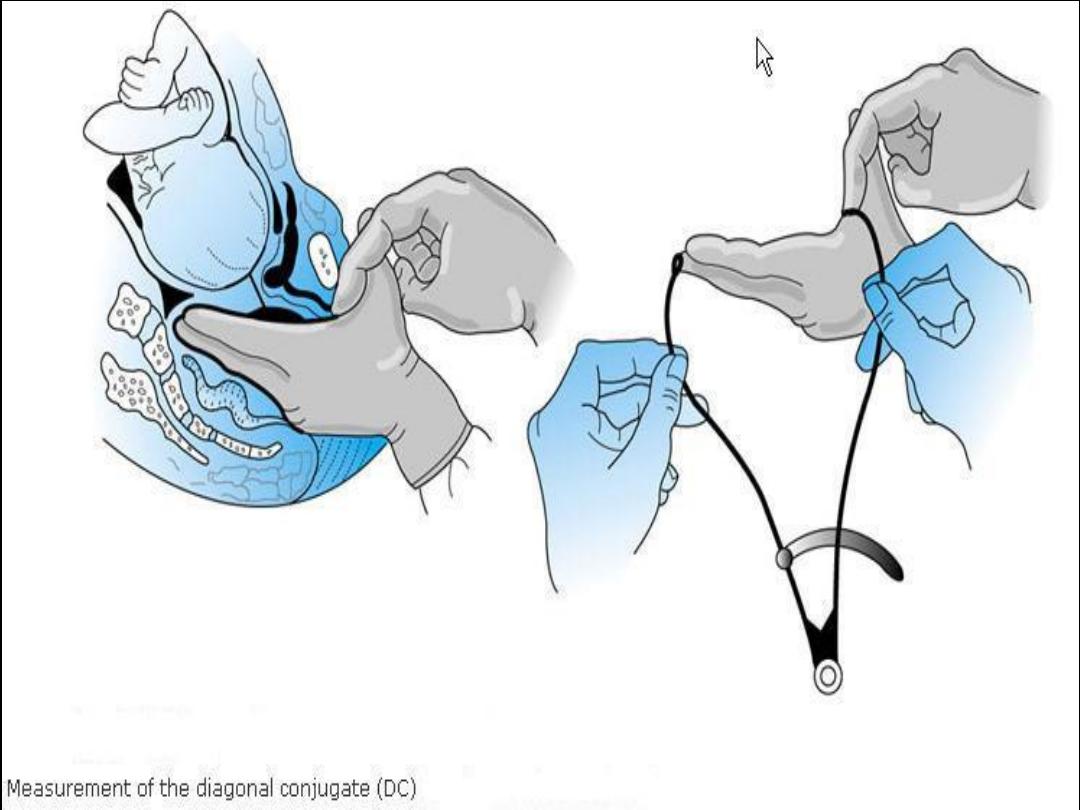
Clinical pelvimetry:
The diameters that can be assessed
clinically are: the obstetric conjugate
of the inlet by clinical assessment of
the diagonal conjugate when the tip
of the middle finger can not meet the
promontory of the sacrum (while the
2 fingers are passed in the vagina
and the index finger meets the pubis)
then we subtract 1.5-2 cm will
corresponds the obstetric conjugate

Then we assess the curvature
of the sacrum by palpating its
anterior surface.
Then the midpelvis is
assessed but it is difficult to
do it clinically unless the
pelvic side walls are
apparently convergent which
indicate narrow pelvic cavity

the bispinous dimension also
can be assessed by palpating
the prominence of the spines,
in addition the width of the
sacrosiatic notch should be
assessed.

And the final step is the
assessment of the outlet by
placing a fist between the
ischial tuberosities, a
dimension of 8.5 cm is
adequate transverse diameter.
And the subpubic arch of less
than 90 degrees usually
associated with narrow
midcavity and outlet

The perineum
The final obstacle to be negotiated by the
fetus during labour is the perineum. The
perineal body is a condensation of fibrous
and muscular tissue lying between the
vagina and the anus. It receives
attachments of the posterior ends of the
bulbo-cavernous muscles, the medial ends
of the superficial and deep transverse
perineal muscles, and the anterior fibres of
the external anal sphincter. It is always
involved in a second-degree perineal tear
and an episiotomy.

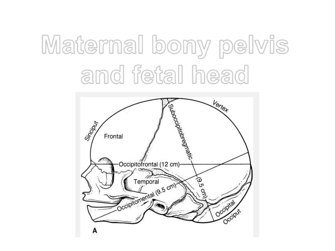
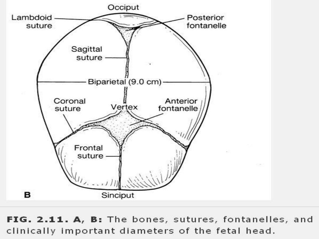
Dimensions of the fetal skull
• The fetal head is the largest and
the least compressible part of the
fetus
• The fetal skull consists of a base
and a vault (cranium) which
consists of the occipital, parietal,
frontal and temporal bones

these are easily compressible
and interconnected by
membranes and these features
allow molding to occur which
means the overlap of these
bones under pressure and
changing their shape to
conform to maternal pelvis
during vaginal delivery

•You have to know many
terms:
*fontanelle [anterior (bregma)
*and posterior (lambda)],
*nasion,
*glabella,
*vertex,
*and the occiput
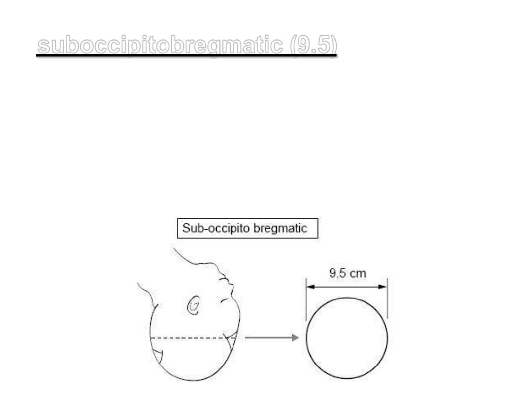
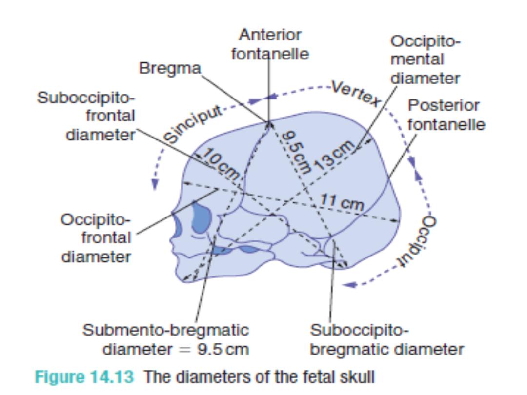
cm this is
)
9.5
suboccipitobregmatic (
the presenting anteroposterior
diameter when the head is well
flexed. It extends from the
undersurface of the occipital bone
to the center of the bregma.
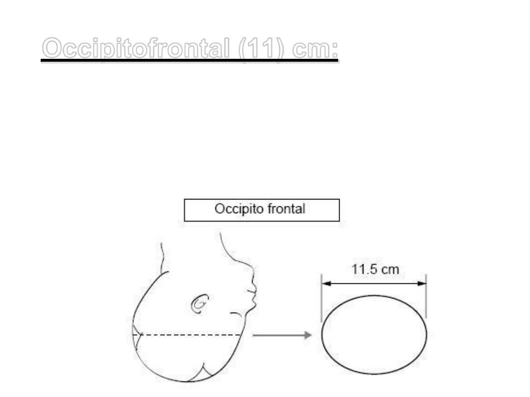
when the
) cm:
11
Occipitofrontal (
head is deflexed. Extend from
the external protuberance of
the occipitalbone to the
glabella.

Mentovertical (13.5) cm when
the head is extended in
brow presentation. It
extends from the vertex to
the chin.
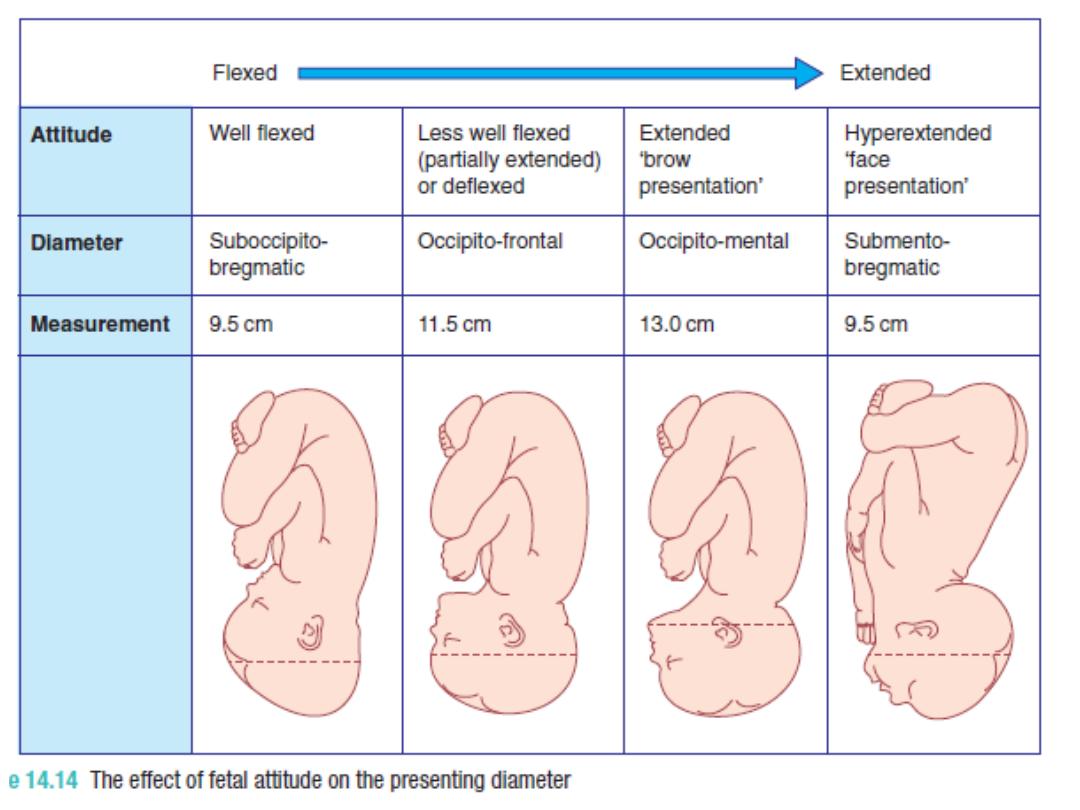
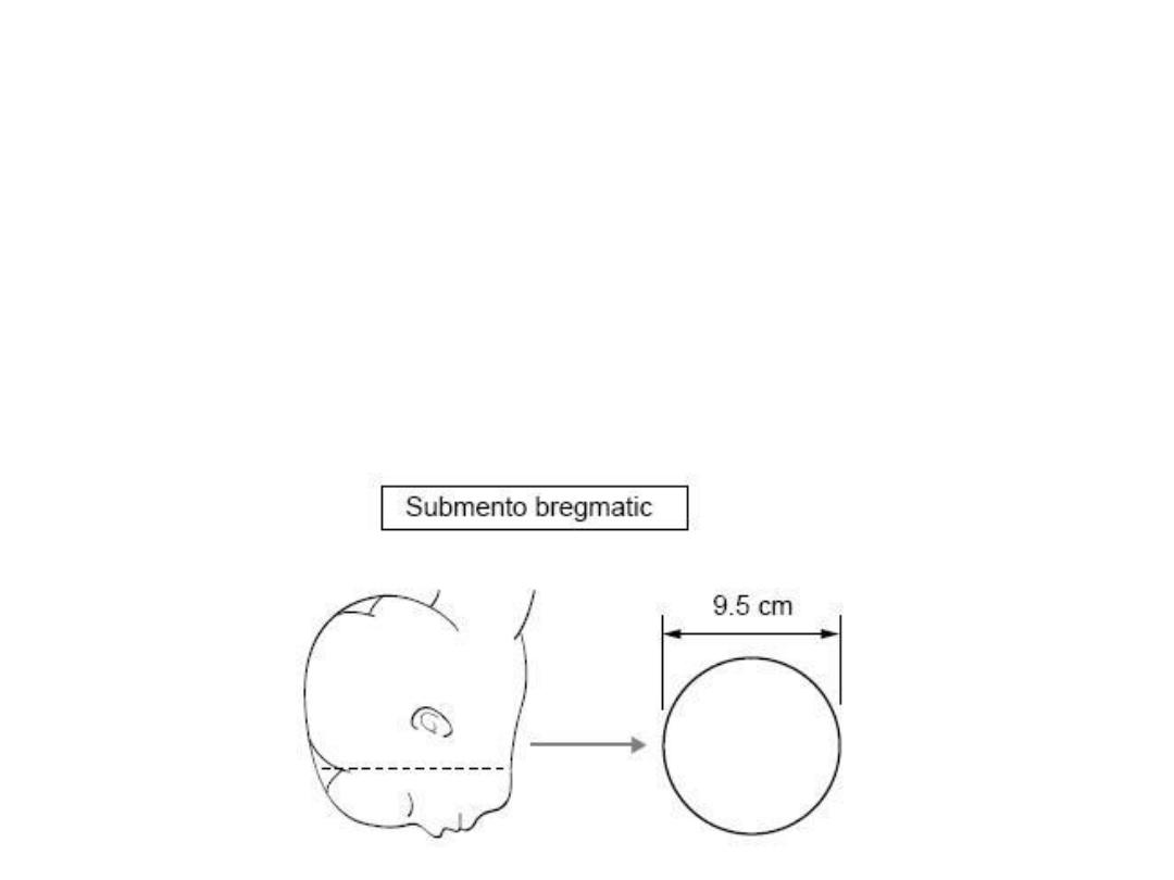
Submentobregmatic (9.5) cm
when hyper extended head
in the face presentation.it
extends from below the
chin to the bregma.
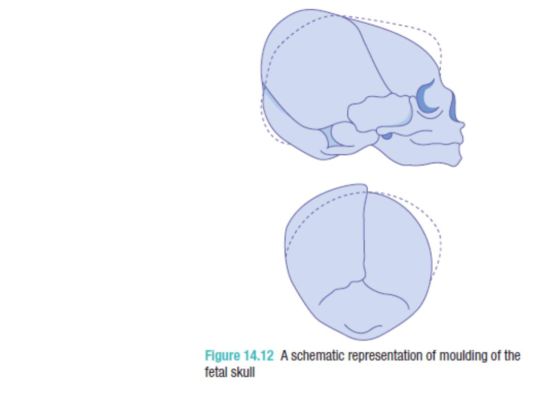
moulding