1
Leiomyoma
Definition :
- Benign tumor of myometrium ( smooth ms & fibrous t. )
- 15 – 20 % ( the commonest pelvic swelling )
- However only few patients will be symptomatic
Etiology :
……… unopposed hyperestrogenemia …………
- Age 30 – 40 years
- Parity NG or low parities
- Race dark races are commoner
Pathology
………. N …….. SSSS …….. CCC ……. M ……….
1. Number solitary or multiple ( may be hundreds )
2. Site
* Corporeal ( 95 % )
- All fibroids that as interstitial fibroids ( 60 % )
- If it grows towards peritoneal cavity subserous fibroid ( 20 % )
it may attach to surrounding structure parasitic fibroids
- If it bulges into uterine cavity submucous fibroid ( 15% )
it may protrude from the cervix fibroid polyp
* Cervical fibroid ( 4 % )
- Starts within the wall , then may
- Bulge into the cervical canal barrel-shaped cervix
- Protrudes through the ectocervix cervical polyp
* Extrauterine ( 1% )
- In broad ligament , round ligament , ovarian ligament .
3. Size From microscopic ( seedling ) to a huge size ( filling perit , cavity )
4. Shape Spherical but as it enlarges it may be compressed
5. Supply Blood vessels enter through
- Capsule .. central areas are > liable to degeneration & necrosis
while – peripheral areas are > liable to calcification
- Pedicle ( in polyps ) .. usually tip of polyp ulcerate infection
6. Capsule Pseudo-capsule of compressed surrounding myometrium
7. Consistency - Firm The more fibrous tissue the more firmer
- Hard if calcification occurs ( womb stone )
- Soft in pregnancy , degeneration .
- Very soft ( brain-like ) if sarcomatous changes occurs
8. Cut section whorly appearance which is paler than surrounding muscle
9. Microscopic exam. Mixture of smooth muscle & fibrous tissue .
Fifth Stage - Gynecology - Dr.Aseel - Lecture 2

2
Secondary ( pathological ) changes
(1) Degenerative
Occur usually near menopause
All will loose their whorly appearance
1. Hyaline ( myxomatous )
- Occurs in large fibroids in the center ( blood supply )
- The tumor homogenous waxy soft material
2. Pseudocystic degeneration
- Area of liquefaction usually following hyaline degeneration
- The tumor becomes soft
3. Fatty change
- More common at periphery ( ppt of lipids from blood vessels )
- The tumor will be yellow , greasy , soft .
4. Calcification
- May follow fatty change hard consistency ( womb-stone )
- X-ray eggshell appearance ( onion – skin )
5. Red degeneration
- Necrobiosis : dead parts – central – + living parts – periheral –
- Etiology it occurs in mid-trimesteric pregnancy ( d.t. vascularity &
hypercoagulability venous obstruction thrombosis )
- C/P severe pain , vomiting , fever ( absorption of Hb )
- TTT : . Analgesics ( anti-PG ) , Antipyretic , Fluids , Bed rest + steroids
. Never surgery (only if failed medical ttt remove only affected fibroids)
6. Atrophy
- Usually occurs after delivery or postmenopausal ( E )
- Postmenopausal growth occurs if calcification , malignancy , HRT .
(2) Vascular
1. Torsion ( axial rotation )
- Pdf (1) pedunculated … (2) moderate-sized (3) subserous fibroids
- Ppt Due to trauma , sudden change in posture , pregnancy .
- Once occurred it maintains itself by the lashing effect of BV pulsations
Acute torsion very painful ( acute abdomen ) up to gangrene
Chronic torsion it may adhere to neighboring organ ( parasitic fibroid )
2. Telangiectasis
( dilatation of blood vessels ) may rupture internal he
3. Lymphangiectasis
(dilatation of lymphatics) may rupture dense adhesions

3
(3) Inflammatory
* Submucous fibroid d.t. trauma & infection ( esp after abortion or labor )
* Fibroid polyp ulceration infection
* subserous fibroid d.t. direct spread from neaby inflamed organ as appendix
(4) Malignant X
* Rare ( 0.5 % ) the association of endometrial cr is > leiomyosarcoma
* Suspected if . rapidly growing , invasive , metastasis
. tumor becomes painful , fixed .
* Macroscopic yellowish , soft , very vascular ( brain like )
* Microscopic > 10 mitotic figures / HPF is diagnostic of leiomosarcoma
Clinical picture
Symptoms
………. ASYMPTOMATIC ( accidentally discovered ) ……
1. Uterine bleeding anemia ( easy fatigability )
Menorrhagia , polumenorrhagia .
- Increased vascularity , size , surface area of endometrium
- Associated hyperplasia & hormonal disturbance .
Metrorrhagia
- Ulcerated submucous fibroid or polyp
- Associated endometrial carcinoma or rarely sarcoma
Polymenorrhea ovarian congestion
If …… Postmenopausal bleeding suspect malignancy
If …… Amenorrhea : Never a feature suspect pregnancy
2. Infertility
* Anatomical On tubes blocking or stretching ( or ectopic )
On uterus prevents implantation ( or habitual abortion )
On cervix may obstruct the canal
* Physiological Associated anovulation , endometriosis
3. Vaginal discharge . Leucorrhea ( pelvic congestion )
. Offensive or mucopurulent ( infected ulcerated polyp )
4. Swelling ( abdominal or vaginal )
5. Pain
. Congestive dysmenorrheal ( pelvic congestion )
. Spasmodic dysmenorrhea ( esp. if submucous )
. Acute abdomen in …….. Torsion …. Inflammation …. Red degeneration
6. Pressure symptoms ……….
7. Pregnancy complications ……….

4
Signs
* General anemia ( pallor )
Rarely - Polycythemia ( erythropoietin from secretory fibroid )
- Pseudo-Meig's syndrome in large subserous fibroids
* Abdominal
* If large pelviabdominal mass which is usually
. Firm ( soft if degeneration , pregnancy , malignancy )
. Mobile ( moves with the movement of cx )
. Not tender ( painful if torsion , inflammation , red deg )
* Auscultation uterine soufflé may be heard .
* Vaginally
* Bimanual examination :
- Symmetrically or asymmetrically ( knobby ) enlarged uterus
- Cervical fibroid small knob ( uterus ) on top a large swelling
* Speculum fibroid polyp protruding from service .
Complications of fibroid
- Pathological ( 2
ry
) changes of fibroid
- Pregnancy complications
- Pressure effects
- Prolapse , chronic inversion
- Infertility ( 1/3 cases ) & anemia .
Differential diagnosis
- D.D. of mass in . Pelvis …. Pelviabdominal mass } symmetrical &
. Mass in DP } asymmetrical
. Broad ligament mass } enlargement
. Mass protruding from cx } of uterus
- Acute abdomen
- Causes of anemia
Investigations
* Preoperative
- Hb % …..
- IVP …… detect pressure on ureter ( cervical or br. lig. fibroid )
* For diagnosis U/S ( exact site , size , number ) +
- Submuncous fibroid HSG , Hysteroscope ,. Sonohysterography
- Subserous fibroid Laparoscopy
* D & C to exclude coexisting malignancy ( if C/O postmenopausal bleeding )

5
Treatment of fibroids
(1) Expectant ttt
- No symptoms , small size e.g.
- Near menopause wait for menopausal atrophy
(2) Medial ttt
1. Young patient or near menopause
2. With mild symptoms & signs
- Iron & tonics for anemia
- Hormonal therapy size ( 25 – 50 % after 3 – 6 months ) e.g.
Anti –'E' ( Tamoxifen ) … Danazol … LH – RH analogues
Progestins … recently not proved to size of fibroid
3. In pregnancy with red degeneration .
(3) Surgical ttt
* Polypectomy and D & C ( for submucous fibroid polyp )
- Using ring forceps or volsellum to catch the polyp )
- Twist several times
- Then D & C for the hyperplastic endometrium
- Can be done endoscopy ( Hysteroscopy …. Or …. Laparoscopy )
* Myomectomy
- Young patient
. Symptomatic ( bleeding ) with signs ( > 12 wks )
. Infertility ( HSG is done to document tubal patency )
- Myomectomy is a very Bloody operation . Therefore :
- Preoperative :
1. Better done postmenstrual
2. Elevate the Hb % prepare blood
3. Some give progestins , Danazol or GnRH for 3 – 6 months
- Intra-operative :
1. Temporary occlusion of :
. Uterine Bonney m. clamp or rubber catheter or assistant hand
. Ovarian occlusion of infundibulo-pelvic lig. By ring forceps .
2. May inject vasopressin around the tumor to cause V.C.
3. Incisions
. Try to have all operation from a single incision
. Better to be anterior in the midline .
. close the bed of one tumor meticulously before opening the next

6
Hysterectomy
…. Commoner than myomectomy ( easier , less blood loss ) ….
* Old patient > 40 years , completed her family .
- Associated endometrial carcinoma
- Suspected malignant change
* Recurrence after myomectomy .
* Uncontrollable haemorrhage in myomectomy
* Multple fibroids if myomectomy would leave a useless mutilated organ
Fibroid with pregnancy
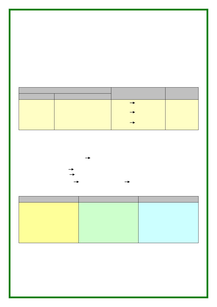
(1) Effect on pregnancy
Pregnancy
Labor
Puerp.
Early
Late
1. Abortion
2. Ectopic
( tubal stretch )
3. Incarcerated
RVF gravid
Uterus
1. Pressure manifestations ..
2. Mal-Presentation
3. Non-engagement of
Presenting part
4. PTL
5. Pain (acute abd ) …,…,…
1
st
stage inertia
2
nd
stage obstruction
3
rd
stage retained pl.
( implanted on fibroid )
- S3
- Inversion
of
uterus
(2) Effect on fibroid
* Due to increased hormones
. size , vascularity
. Softening & degeneration difficulty to identify by palpation
* More liable to complications ( esp 2
nd
trimester )
. Red Degeneration acute abdomen
. Subserous fibroid torsion / intraperitoneal he d.t. ruptured telangicetasia
. SubMucous Fibroid extrusion or trauma ulceration & infection .
(3) Management
( conservative mainly )
Pregnancy
Labor
Prerperium
* No Myoomectomy
* If Red Degeneration
Rest, analgesics, antipyretics
* Surgery Only if
- Failed medical ttt or red
degeneration ( rare )
- If torsion or he from
subserous fibroid
* No obstruction
Allow vaginal delivery
* Bstruction
CS. Bo myomectomy
except of pedunclated
subserous
* If old + multiple fibroids
Cesarean hysterectomy
* Myomectomy
done after
3 – 6 m
( the myoma will be
less vascular )

7
Extras
Q. What are the complications of myomectomy …
Haemorrhage … how to avoid ?
Adhesions … avoid by ventrosuspension ( round lig ) & proper haemostasis
Recurrence … follow up / progesterone
Weak scar …. Rupture in next preg .
Q. What has Bonney done ..
1. Bonney myomectomy Clamp
2. Bonney myomectomy Screw
3. Bonney Hood operation ( trans-mural ) through the fundus
4. Bonney elevation test ( SUI )
Q. How to remove posterior myoma …
1. Trans-cavitary
2. Bonney-hood operation
Q. Indications for surgery in fibroids …
Symptomatic … severe hge . / Infertility
Cervical fibroid … Pressure sympt .
Large size … > 12 – 14 wks .
Pedunculated subserous ( for fear torsion )
Q. What are new lines for therapy …
Myolysis
. Thermal or Cryo-cautery ( via laparoscopy )
. Disadv : extensive adhesions , rupture uterus in next preg .
Bilateral uterine astery embolization
. Via fluoroscopic directed angiography injection of gelfoam
50 % in size
. Disadv : bladder necrosis & fistula
Q. Special types of fibroid …
Cervical ( ant or post to cervical canal )
Broad ligamentary
In both : ureteric injury is a high risk ( distorted anatomy )
Q. Rare fibroids ..
Intravenous leiomyomtosis : Fibroids are found in pelvic veins , due to
Direct spread or arise from SM of vessels rapid metastasis ( lung )
Leiomyoma peritonealis disseminata : Fibroids are found on peritoneum &
omentum ( benign condition )
