Dr.KHALID WISSAM
Lec 1Diseases of Respiratory System
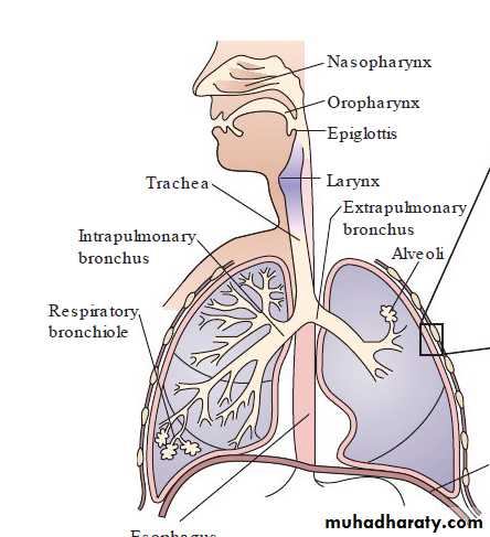
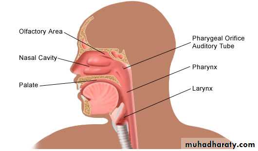
Respiratory System
Diseases of the nose :
Rhinitis : Inflammation of the nasal mucosa usually accompanied by sinusitis ,it may be :1-Acute rhinitis : a- Specific as in diphtheria .
b- Nonspecific as in catarrhal ( common cold ) & allergic rhinitis
2- Chronic rhinitis : a- Chronic specific ( granuloma) as T.B , syphilis & leprosy .
b- chronic non specific as : atrophic rhinitis , hypertrophic rhinitis & allergic rhinitis .
Chronic hypertrophic rhinitis :
The mucosa is thickened, polypoidal & covered by mucopurulent exudates & may form nasal polyp .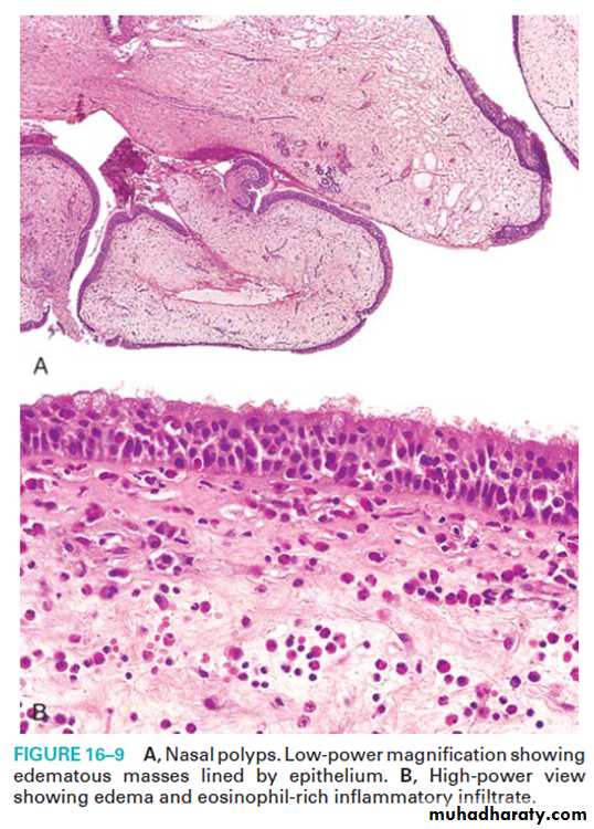
Epistaxis
Epistaxis : Bleeding from the nose.Causes
(1) Local causes :
(a)trauma.
(d) nasal polyps.
(2) Systemic causes :
(a) Hypertension.
(b)leukemia's.
(c) Hemorrhagic blood diseases as purpura
(d)Acute infections e.g. typhoid fever and measles .
(e) Deficiency of vitamin C & K .
Tumors of the Nose, Sinuses,and Nasopharynx
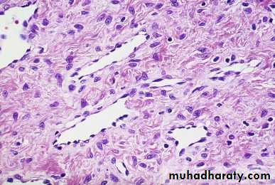
Nasopharyngeal Angiofibroma is a benign, highly vascular tumor that occurs almost exclusively in adolescent males who are often fair-skinned and red headed.Sinonasal (Schneiderian) Papilloma. is a benign neoplasm arising from the respiratory mucosa lining the nasal cavity and paranasal sinuses.
Asociated with HPV DNA, often types 6 and 11.
pathological features: papillomatous proliferation of squamous epithelium.
Sinonasal (Schneiderian) Papilloma
Nasopharyngeal AngiofibromaOlfactory Neuroblastoma
arise from the neuroectodermal olfactory cells present within the mucosa, particularly in the superior aspect of the nasal cavity.There is a bimodal age distribution with peaks at 15 and 50 years of age.
The patients typically present with nasal obstruction and/or epistaxis.
Histologically, olfactory neuroblastomas are composed of nests and lobules of well-circumscribed cells that are separated by a fibrovascular stroma.
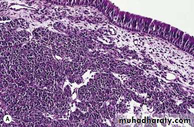
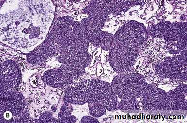
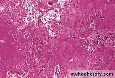
Nasopharyngeal carcinoma:
Rare neoplasm, has strong epidemiologic link to EBV (Epstein–Barr virus) common in Chinese.It has 3-histological variant :
* keratinizing squamous cell carcinoma .
* non keratinizing squamous cell carcinoma .
* undifferentiated carcinoma which compose of large malignant cell in addition there is marked infiltration with lymphocytes
It is a radiosensitive tumor and the 5-year survival rate is 50% even for advanced cancer.
Nasopharyngeal carcinoma:
Laryngitis
Laryngitis either acute or chronic.causes:
allergic,viral, bacterial, or chemical.
but it is more commonly part of a generalized upper respiratory tract infection.
environmental toxins such as tobacco smoke.
excess use of voice , The vocal cords are diffusely thickened & may show polypoidal mass called laryngeal polyp ( singer’s nodule).
in association with gastroesophageal reflux due to the irritating effect of gastric contents.
systemic infections, such as tuberculosis and diphtheria.
Croup : laryngoepiglottitis, caused by respiratory syncytial virus, Haemophilus influenzae, or β-hemolytic streptococci may induce sudden swelling of the epiglottis and vocal cords with laryngeal obstruction. In particular, in infants and young children with their small airways, so it is considered a medical emergency in infancy or childhood.
Laryngeal tumors:
Benign tumors :It includes (vocal cord polyp and laryngeal papilloma).
Vocal cord polyps are smooth protrusion less than 0.5cm in diameter usually located on the true vocal cord.
M: composed of core of edematous fibrous tissue cover by stratified squamous epithelial , contain chronic inflammatory cell infiltrate . These lesions occur usually on heavy smoker or singers (singers nodule) so it could be due to chronic irritation .
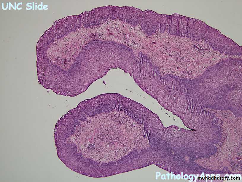
Squamous cell papilloma:
Usually arise from true vocal cord as soft pink rarely >1cm in diameter.
M: multiple finger like projection of fibro vascular cords cover by stratified squamous epithelial.
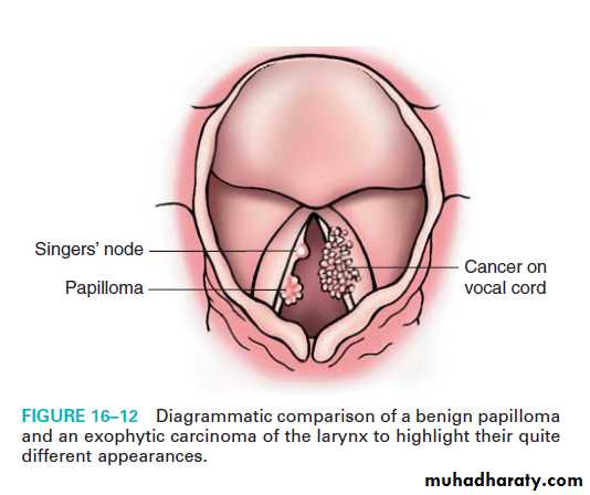
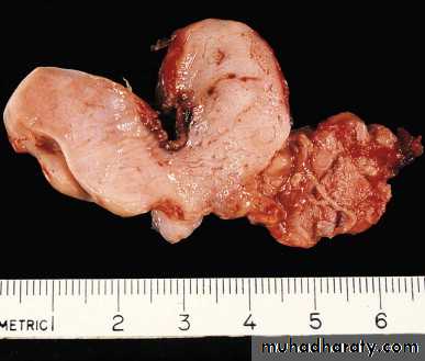
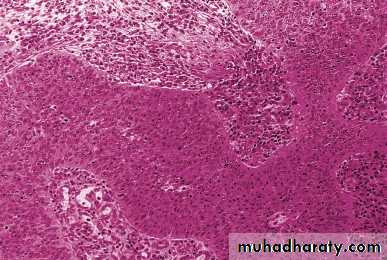
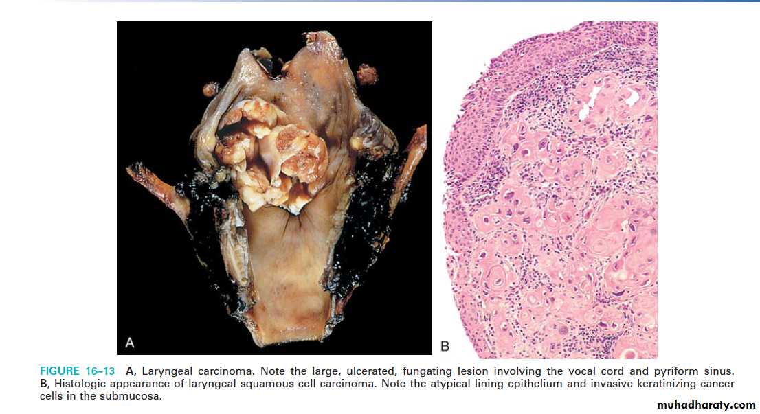
Carcinoma at the larynx:
* Usually occur after the age of 40, affect male > female.* causes : Environmental influence are very important in its causation nearly all cases occur in smoker while alcohol and asbestos exposure are also play a role.
* 95% are of squamous cell carcinoma ,rarely it is adenocarcinoma.
Sqamous cell carcinoma :
In the majority of cases it arise from the vocal cord (glottic) ,next it may arise from above the vocal cord (supraglottic), next it may arise from below the vocal cord (subglottic tumor) .
Carcinoma of larynx usually present as persistent hoarseness of voice .
90% of glottic tumor has excellent prognosis because it affect the motility of the vocal cord so the patient present early also because this region has no lymphatic supply so spread outside the vocal cord is rare.
In contrast to supraglottic which is rich with lymphatic supply also the supraglottic tumor usually remain silent and the patient present late so it has poor prognosis .
Carcinoma at the larynx:
Pneumonia :
Pneumonia :Inflammation of the lung :Predisposing conditions :
local defense mechanisms are impaired , including:
• Loss or suppression of the cough reflex as a result of coma, anesthesia, neuromuscular disorders, drugs.
• Injury to the mucociliary apparatus due to cigarette smoke, inhalation of hot or corrosive gases, viral diseases, or genetic defects of ciliary function (e.g., the immotile cilia syndrome)
• Accumulation of secretions in conditions such as cystic fibrosis and bronchial obstruction.
• Interference with the phagocytic or bactericidal action of alveolar macrophages by alcohol, tobacco smoke, anoxia, or oxygen intoxication
• Pulmonary congestion and edema
the systemic resistance of the host is lowered by chronic diseases, immunologic deficiency, treatment with immunosuppressive agents, and leukopenia.
Pneumonia

Pneumonia :
The histological spectrum of pneumonia syndrome :1- fibrinopurulent alveolar exudates seen in acute bacterial pneumonias.
2- mononuclear interstitial infiltrates in viral pneumonia .
3-granulomas and cavitations in chronic pneumonias.
Acute bacterial pneumonias can manifest as one of two anatomic and radiographic patterns, referred to as bronchopneumonia and lobar pneumonia
lobar pneumonia :part or all of a lobe are homogeneously filled with an exudates.

Bronchopneumonia :
a patchy distribution of inflammation that generally involves more than one lobe.Lobar pneumonia
streptococcus pneumonia is responsible for more than 90% of lobar pneumonias.Morphology
It pass into 4 stages but with the use of antibiotic it can arrest at any stage depend on onset of treatment .
1-Stage of congestion : last 1-2 days
Gross:lobes are heavy & red
Mic :
Congestion of alveolar capillary .
Alveolar space contain inflammatory exudates , red blood cells , bacteria & few neutrophilis .
Lobar pneumonia
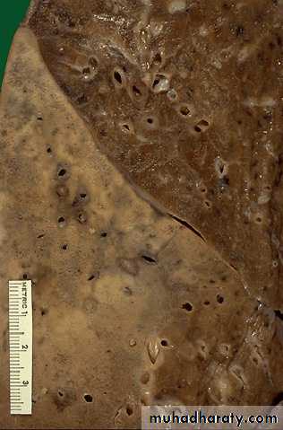
2-Stage of red hepatization ;Gross : lobe is heavy , red , firm similar to the liver .
Mic : capillary congestion , alveolar space contain red blood cell , neutrophils & fibrin .
Pleural cavity usually show fibrinous or fibrinopurulant exudates .
3- stage of grey hepatization:
Gross : lobe is grey , firm heavy
Mic : congestion disappear , alveolar space fill with neutrophils .
4- Stage of resolution :
The exudates is liquefied by enzymes then it either coughed or absorbed &re- aeration of alveoli take place .
Complication :
Complete healing is usual with proper antibiotics & complication is rare in both types of pneumonia:
(1) organization of the intra alveolar exudates may convert areas of the lung into solid fibrous tissue .
(2) pleural effusion & empyema;
(3)Lung abscess is a rare complication in lobar pneumonia .
(4) bacteremia causing meningitis.
bronchopneumonia :
foci of inflammatory consolidation are distributed in patches throughout one or several lobes, most frequently bilateral and basal.Histologically :
* the reaction consist of focal suppurative exudates that fills the bronchi, bronchioles, and adjacent alveolar spaces.* The intervening areas are normal .
* Pleural involvement is less common than in lobar pneumonia .
Complications :
Complete resolution is uncommon ,Same complication of lobar pneumonia may occur
Bronchiectasis may occur .