
FIFTH STAGE
INTERNAL MEDICINE
DR.FADHIL – LECTURE 9
1
BEHCET'S SYNDROME + BONE DISORDERS
BEHCET'S SYNDROME
Behcet's syndrome : vasculitis of unknown cause affecting characteristically venules.
There is a striking geographical distribution, it being most common in Turkey,
Iran and Japan.
The prevalence per 100000 is 10-15 in Japan and 80-300 in Turkey. There is a
link to the HLA-B51 allele (a split antigen of B5), with a relative risk of 5-10.
CLINICAL FEATURES
there is a wide range of clinical features. The disease is characterized by
unpredictable exacerbations. There is no definite investigation & the diagnosis
is made depending on clinical criteria:
*recurrent oral ulcerations – minor aphthous , major aphthous or herpitiform
ulceration at least 3 times in a 12 month period.
PLUS 2 OF THE FALLOWING :
1. recurrent genitalulceration(60-80%)
2. eye lesions : anterior uveitis , posterior uveitis , cells in vitreous on slit-lamp
examination, retinal vasculitis. The involvement is usually bilateral
3. skin lesions: erythema nodosum, pseudofolliculitis, papulopustular lesions, acne
form nodules, migratory thrombophlebitis& vasculitis
4. positive pathergy test: involves intradermal skin pricking with a needle, a
pustule develops within 48 hours.
Other manifestations include a self-limiting peripheral mono-or oligoarthritis affecting
knee, ankle, wrist and elbows; gastrointestinal symptoms of diarrhea, abdominal pain
and anorexia; pulmonary and renal lesions; brainstem syndrome; organic confusional
state and meningo-encephalitis.

2
All the common manifestations are self-limiting
except ocular attacks. Repeated attacks of uveitis
can cause blindness.
The pathology is highly specific to Behcet's disease.
Recurrent thrombosis can occur. Renal involvement
is rare.
TREATMENT
Steroid(topical), immunosuppressants& cyclosporine are used for chronic
uveitis& the rare neurological complications, colchicine for skin disease&
arthralgia, thalidomide 100-300 mg/day for 28 days for oral & genital ulceration
, but this drug is highly teratogenic(phocomelia)& neurotoxic.
Systemic manifestations require systemic steroids& immunosuppressive drugs.
OSTEOMALACIA (ADULT RICKETS)
Inadequate and delayed mineralization of osteoid in mature compact and
spongy bone
Major deficit is in Vitamin D , which is required for Ca++ uptake in intestines
Decreased Ca++ stimulates PTH, which does increase Ca++, but also increases
phosphate excretion by kidney
When phosphate levels too low, mineralization cannot occur
4 categories of osteomalacia &rickets can be identified based on the underlying
cause:
1- deficiency of v.D or defects in v.D metabolism.
2- hypophosphatemia.
3- drug induced inhibition of bone mineralization.
4- defects in pyrophosphate metabolism.
ETIOLOGY
More prevalent in extreme preemies, elderly, those following strict
macrobiotic vegetarian diets and persons on anticonvulsant Rx

3
Pancreatic insufficiency
Hepatobiliary diseases
Lack of bile salts decreases absorption of Vit D
Malabsorption syndromes
Hyperthyroidism
Rare in US due to fortification of foods
Common in GB and Middle Eastern Countries
CLINICAL PRESENTATION
Generalized body aches /LBP as well as hip pain
Lower extremity pain & deformity
Physical examination
Scoliosis / kyphosis of spine
Deformities of weight bearing bones
Muscle weakness leading to classic waddling gait
Generalized Malaise
Patients with chronic renal failure cannot synthesize the active metabolite of
v.D(1,25(OH)2D3) due to renal damage& this causes secondary hyperparathyroidism
& in some cases osteomalacia.
DIAGNOSIS
Serum Ca++ –↓ or Normal
Serum inorganic Phosphate ↑> 5.5
Vitamin D ↓
BUN & creatinine ↑
Alkaline Phosphatase & PTH ↑
Bone bx to determine aluminum levels

4
X-Rays
Demineralization
Pseudofractures
Bowing of long bones
Radiological examination is of limited value unless in advanced cases where focal
radiolucent area ; pseudofracture or looser's zones are seen in ribs, pelvis& long
bones. Radiological osteopenia &crush vertebral fracture may cause confusion with
osteoporosis.
Diagnosis of osteomalacia is confirmed with bone biopsy which shows pathognomonic
increased thickness of osteoid seams.
CLINICAL MANAGEMENT
Correcting serum Ca++ & phosphorous
Chelating bone aluminum if needed
Suppressing hyperthyroidism
Supplement with Vitamin D
Administer Ca++ carbonate to ↓ hyperphosphatemia
Renal dialysis/transplant for renal osteodystrophy
Correction of associated intestinal disorders
The response will be rapid clinically& radiologically. After 3-4 months, the
treatment can be stopped or the dose of v.D is reduced to the maintenance .
Patients with chronic renal failure require 1-alpha (OH)D or 1,25(OH)2D to
bypass the metabolic defect in 1- alpha hydroxylation of 25(OH)D.
Screening of serum Ca& alkaline phosphatase is needed during treatment to
avoid hyperCa. Alkaline phosphatase returns to normal after treatment.

5
PAGET’S DISEASE (OSTEITIS DEFORMANS)
Excess of bone destruction & unorganized bone formation and repair. The 2
nd
most common bone disorder in the U.S.
The etiology is unknown
Usually affects the axial skeleton, vertebrae and skull, although the pelvis, tibia,
femur are the other common sites of disease.
Most persons are asymptomatic & diagnosis is incidental.
Vascularity is increased in affected portions of the skeleton. Lesions may occur
in one or more bones, does not spread from bone to bone.
Deformities & bony enlargement often occur. Bowing of the limbs & spinal
curvature in persons with advanced disease.
Bone pain- is the most common symptom. Is usually worse with ambulation or
activity but may also occur at rest. Involved bones may feel spongy & warm
because of increased vascularity.
Skull pain is usually accompanied with headache, warmth, tenderness &
enlargement of the head.
Pathologic fractures- because of the increased vascularity of the involved bone-
bleeding is a potential danger.
Alkaline phosphatase levels- markedly elevated as the result of osteoblast
activity.
Serum calcium are normal except with generalized disease or immobilization.
Gout and hyperuricemia may develop as a result of increased bone activity,
which causes an increase in nucleic acid catabolism.
Radiograph reveals radiolucent areas in the bone, typical of increased bone
resorption. Deformities & fractures may also be present.
Goals of the treatment- to relieve pain & prevent fracture & deformities.
Pharmacologic agents are used to suppress osteoclastic activity.
Bisphosphonates & calcitonin are effective agents to decrease bone pain & bone
warmth & also relieve neural decompression, joint pain & lytic lesions.
Use of analgesics & NSAIDs. Assistive devices, including cane, walker.
6
Deformities may be corrected by surgical intervention (osteotomy). ORIF may be
necessary for fractures.
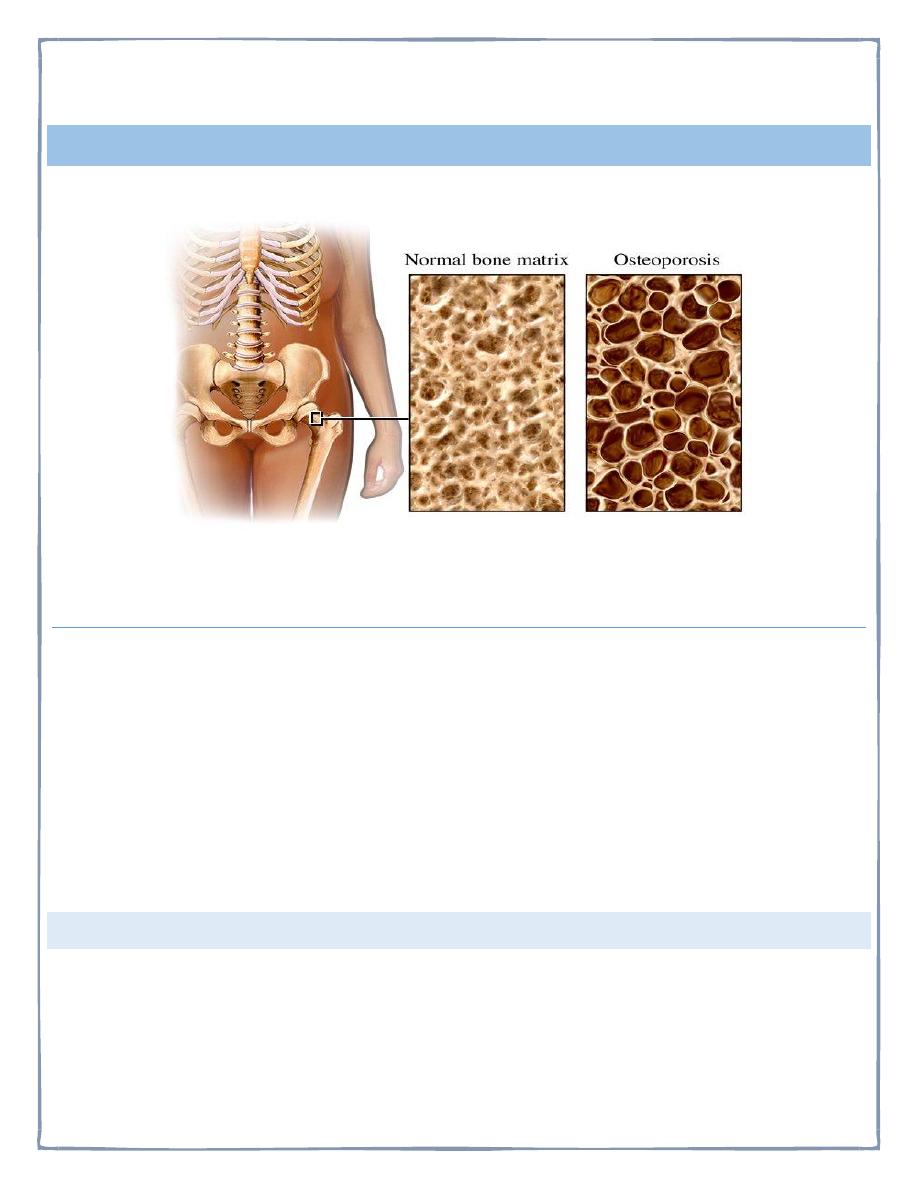
OSTEOPOROSIS
Characterized by low bone mass and structural deterioration
Normal homeostatic bone remodeling is altered – the rate of bone resorption is greater
than the rate of bone formation.
Eight times more common in women than men for several reaso s:
1. Lower calcium intake than men
2. Less bone mass because of smaller frame
3. Bone resorption begins earlier and accelerates after menopause
4. Pregnancy and breastfeeding deplete woman’s skeletal reserve of calcium
5. Longevity increases likelihood of osteoporosis; women live longer than
men
RISK FACTORS
Excess alcohol intake
Cigarette smoking
Anorexia

7
Oophorectomy
Sedentary lifestyle
Insufficient calcium intake
Low testosterone levels (hypogonadism in men)
ETIOLOGY AND PATHOPHYSIOLOGY
• Peak bone mass is achieved before age 20
• Bone loss after midlife is inevitable but rate of loss is variable
• Bone resorption exceeds bone deposition
• Bones become weakened and prone to fracture, loss of height, and kyphosis.
• Diseases associated with osteoporosis
• Intestinal malabsorption
• Kidney disease
• Rheumatoid arthritis
• Hyperthyroidism
• Chronic alcoholism
• Cirrhosis of the liver
• Hypergonadism
• Diabetes mellitus
Clinical Manifestations: Known as silent disease
DIAGNOSIS
Bone Mineral Density (BMD)
Dual-energy x-ray absorptiometry (DEXA)
History and physical examination
Quantitative ultrasound

8
TREATMENT AND NURSING CARE
• Diet Therapy
• Weight bearing Exercises
• Decrease Risk Factors
• Quit smoking and decrease consumption of alcohol
DRUG TREATMENT OF OSTEOPOROSIS
• Estrogen Replacement Therapy
• Calcium & Vitamin D supplements
• Calcitonin
• Biphosphonates (Fosamax, Didronel, Actonel, Boniva, Aredia, Bonefos, Skelid)
• Selective Estrogen receptor modulator – Evista
• Teriparatide (Forteo)
Portion of parathyroid hormone
First drug to stimulate new bone formation
• Hormone Replacement Therapy – Estrogen
Controversy over use. Should discuss with health care provider
• Calcium
There are a variety of calcium supplements available
• Calcium carbonate should be taken with ___food____ _ to aid in
absorption since it needs high gastric acidity to be absorbed
properly.
*parathyroid hormone: the main action is bone formation. The hormone should
be given in an intermittent dosing to alleviate the risk of bone resorption in cases
of sustained hyperparathyroidism. The available drug is teriparatide which is
given as single S.C. daily dose.
Thank you