Reproductive systems
Dr. Zainab .W. A. AlhayaliCollege of medicine (Ninevah)
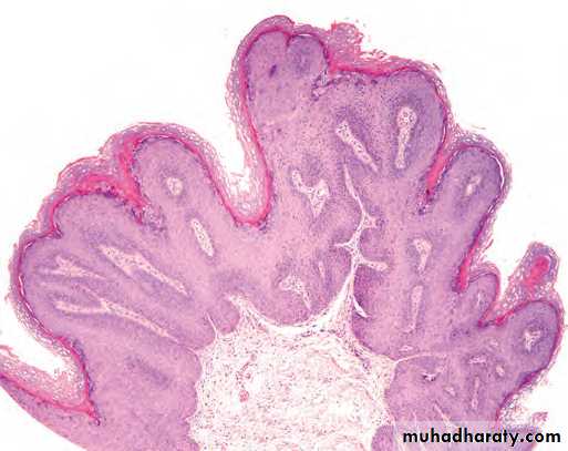
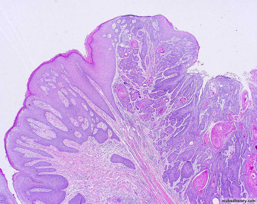
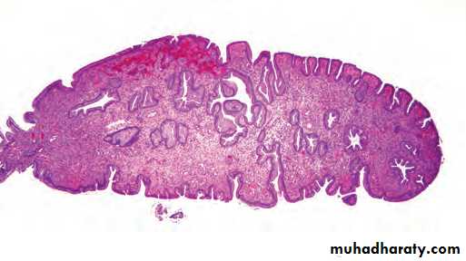
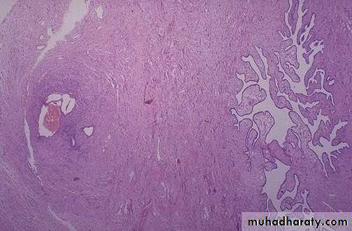
Condyloma acuminatum. Low-power view showing exophytic, papillary architecture.
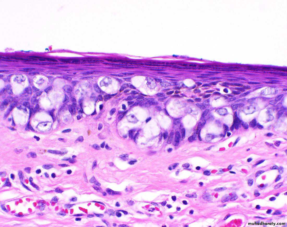
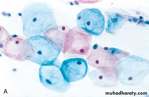
High-power view reveals HPV cytopathic effect (koilocytic atypia) characterized by atypical, enlarged, hyperchromatic nuclei with perinuclear halos (arrow).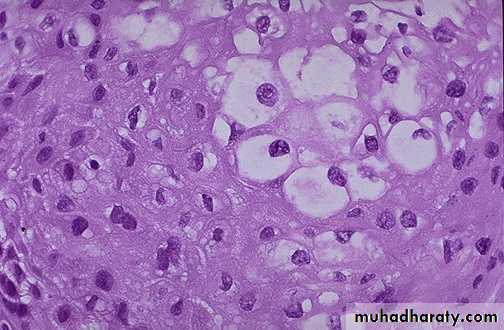
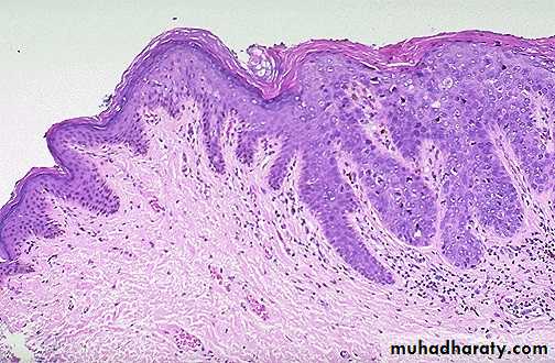
VIN: Dysplasia of the vulvar epithelium, seen here at the right with overlying hyperkeratosis , with more normal keratinizing squamous epithelium at the left.
Histologically is identical to its counterparts on the skin with varying degree of anaplasia and depth of invasion at the right , with normal keratinizing squamous epithelium at the left.
Paget’s Disease of the Vulva
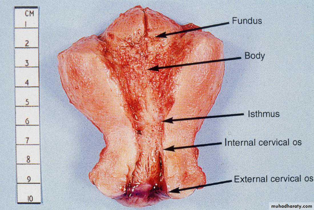
Pathology of the Uterus
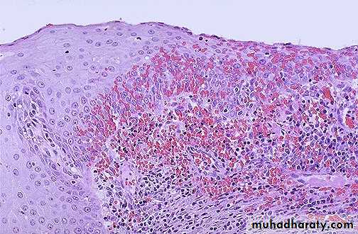
chronic cervicitis:
Small round dark lymphocytes are seen in the submucosa, and there is also hemorrhage.Endocervical polyp composed of a dense fibrous stroma covered with endocervical columnar epithelium.
Pap smear is by far the most effective cancer screening and prevention technique in use
Cervical cancerArose in squamocolumnar junction
Squamous cell carcinoma of the cervix
There are scattered neutrophils in glands and stroma, indicative of acute endometritis
Tuberculous Endometritis
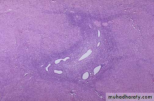
In the upper fundus of the uterus is a nodule protruding into the endometrial cavity that proved to be endometrial polyp .
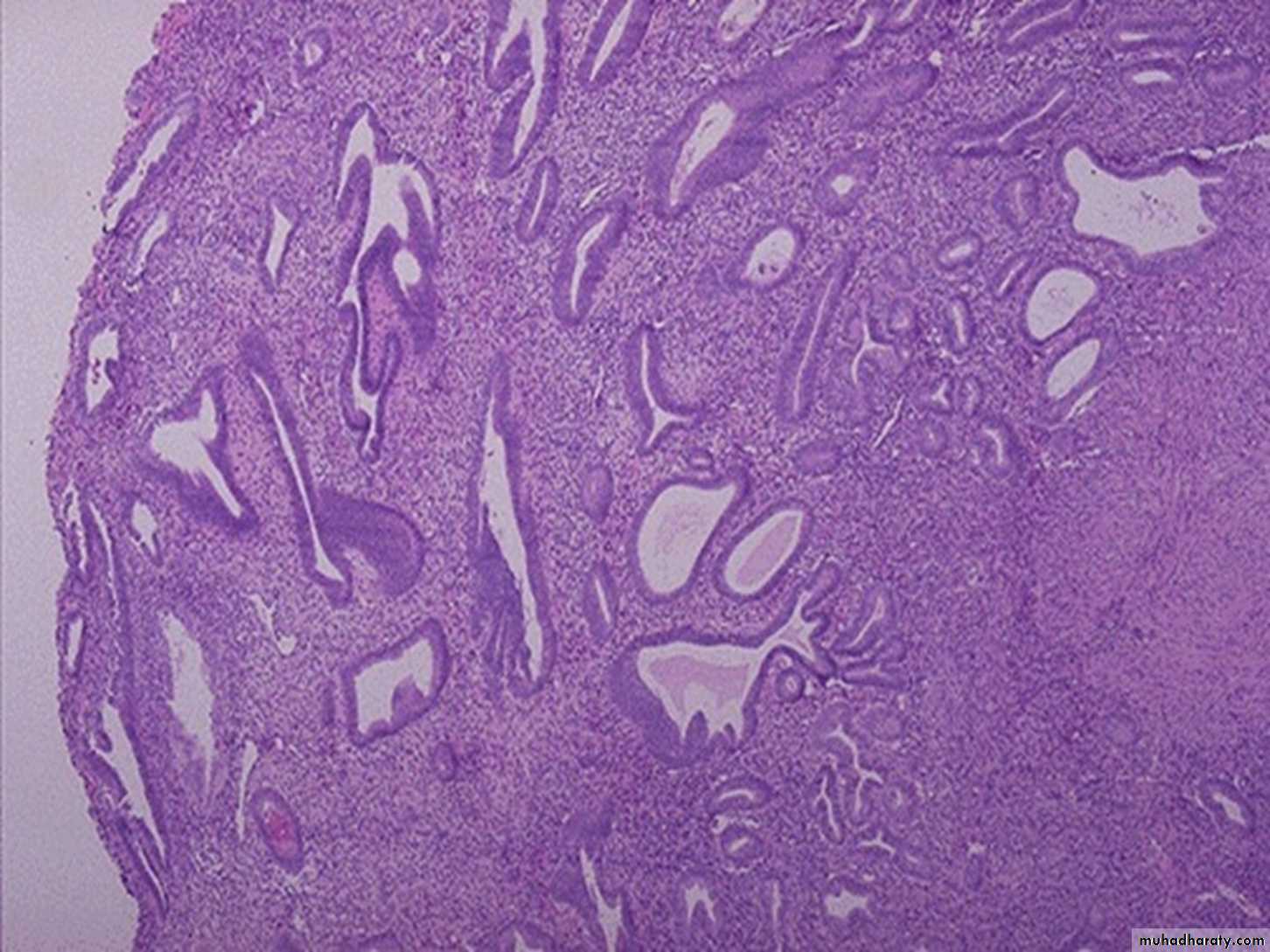
Simple hyperplasia without atypia
Complex hyperplasia with atypia
This is endometrial adenocarcinoma which can be seen invading into the smooth muscle bundles of the myometrial wall of the uterus. The glandular and cribriform endometrioid pattern fits with type I endometrial carcinoma. This neoplasm is superficially invasive into the myometrium at the right.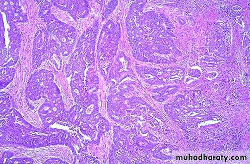
Endometriosis : small cluster of endometrial glands and stroma with hemorrhage are seen at the left near the surface of the fallopian tube. The lumen of the tube is at the right.
Adenomyosis occurs when endometrial glands and stroma are found in the myometrium, not just in the endometrium where they belong. This condition leads to uterine enlargement and irregular bleeding
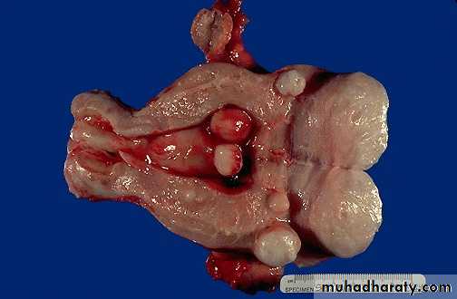
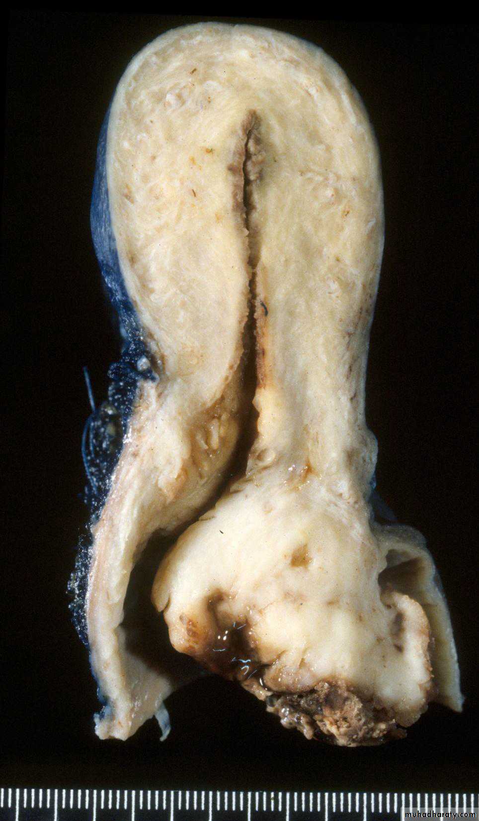
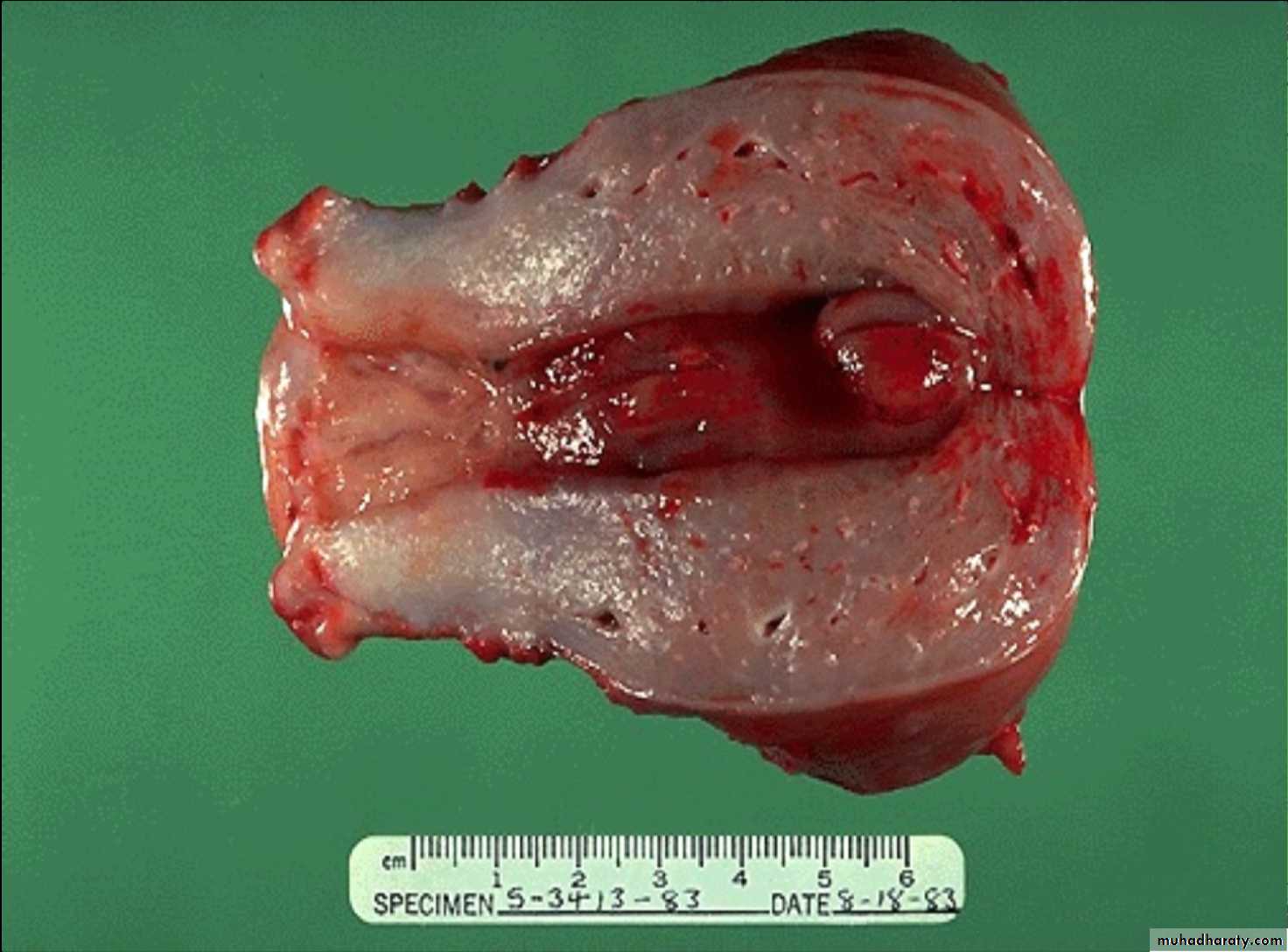
Smooth muscle tumors of the uterus are often multiple. Seen here are submucosal, intramural, and subserosal leiomyomata of the uterus.
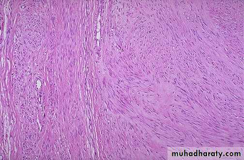
Here is the microscopic appearance of a benign leiomyoma. Normal myometrium is at the left, and the neoplasm is well-differentiated so that the leiomyoma at the right hardly appears different. Bundles of smooth muscle are interlacing in the tumor mass.
leiomyosarcoma : spindle cells have much more pleomorphism and hyperchromatism than the benign leiomyoma. An irregular mitosis and multinucleated giant cell is seen in the center.
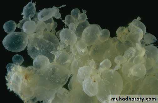
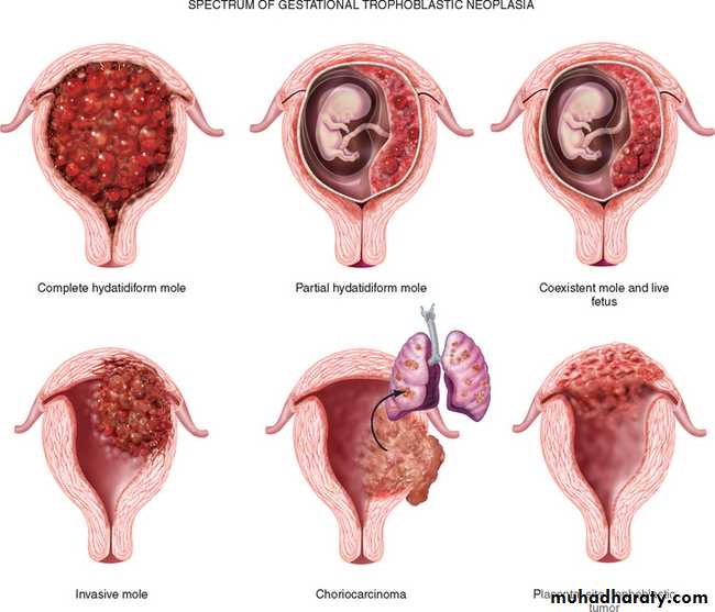
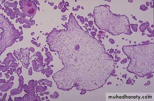
Complete mole : The stroma of the villi is markedly edematous often with cistern formation .
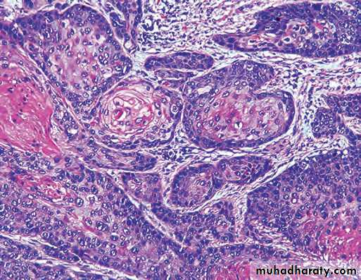
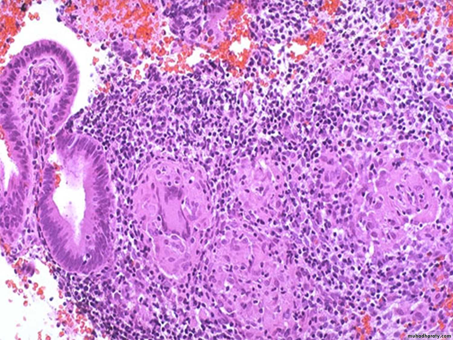
A constant feature is the presence o f a variable degree of atypical villous trophoblastic hyperplasia
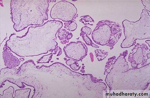
In partial moles, some villi appear normal, whereas others are swollen, avascular, and grape-like (though not as large as a complete mole). There is minimal trophoblastic proliferation. In fact, most placentas in cases of triploidy do not have grossly recognizable grape-like vill
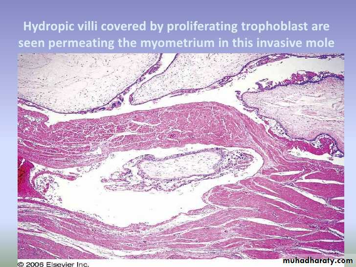
Invasive mole : hydropic villi within the myomatrium
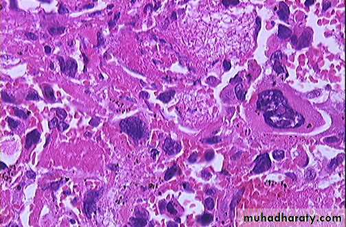
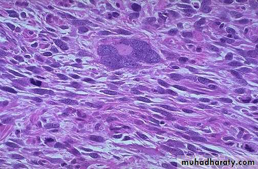
choriocarcinoma
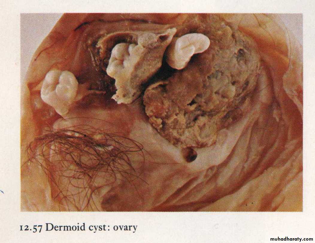
Mature teratoma : Dermoid cyst containing tooth, cartilage & hair