Reproductive system
Dr. Zainab .W. A. AlhayaliCollege of medicine (Ninevah)
Female Genital System
Objectives
• What is the most common malignant tumor of the vulva?• What is cervical dysplasia?
• Extra mammary Paget’s Disease of the vulva
• What are the epidemiologic risk factors for cervical cancer?
• Role of human papillomavirus (HPV) in the patho genesis of cervical neoplasia.
• What causes endometrial hyperplasia?
• Pathogenesis of hydatiform mole
• pathogenesis of endometriosis
• World Health Organization (WHO) of ovarian tumors?
• What are Krukenberg tumors?
Female Genital System
Part I:
Vulva, Vagina, and Cervix
1. Diseases of the Vulva
– Neoplastic2. Diseases of the Vagina
– Infectious
– Neoplastic
3. Diseases of the Cervix
– Infectious
– Non-neoplastic lesions
– Neoplasms
• Carcinoma
• Dysplasia
VULVAL TUMOURS
Benign tumors :Condyloma Acuminatum ( Anogenital warts )
Benign papillary lesions of squamous epithelium which can be transmitted to male sex partner.Solitary but more frequently are multiple forming soft warty masses.
The common locations are the anus, perineum, vaginal wall, and vulva.
They are induced by human papilloma virus (HPV), particularly types 6 and 11.
Histologically The papillary projections consist of fibrovascular stoma lined by stratified squamous epithelium with perinuclear vacuolisation called koilocytosis, indicative of HPV infection.

Condyloma acuminatum. Low-power view showing exophytic, papillary architecture.
High-power view reveals HPV cytopathic effect (koilocytic atypia) characterized by atypical, enlarged, hyperchromatic nuclei with perinuclear halos (arrow).
Premalignant and Malignant Neoplasms:
• Vulvar Intraepithelial Neoplasia (VIN)• Squamous cell carcinoma
• Extra mammary Paget’s Disease of the vulva
Vulvar Intraepithelial Neoplasia (VIN)
A typical cytological changes (dysplasia) in the layers of the vulval squamous epithelium .Graded as VIN-1, 2, or 3 (mild, moderate, severe dysplasia /carcinoma in situ or Bowen disease).
– Oncogenic HPV 16, 18 play a role in the pathogenesis .
– 50% asymptomatic• White patch (leukoplakia), sometimes red or even hyperpigmented
• May remain non-invasive for many years.
• Treatment: surgery, laser, chemicals.
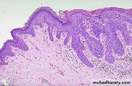
VIN: Dysplasia of the vulvar epithelium, seen here at the right with overlying hyperkeratosis , with more normal keratinizing squamous epithelium at the left.
Vulval Squamous cell carcinoma
• 90% of vulvar cancers• Risk factors
– Human papilloma virus– HIV
– VIN
– Lichen sclerosus.
– History of cervical cancer
Histologically is identical to its counterparts on the skin with varying degree of anaplasia and depth of invasion .
HPV-positive tumours are more often poorly-differentiated carcinoma.
Histologically is identical to its counterparts on the skin with varying degree of anaplasia and depth of invasion at the right , with normal keratinizing squamous epithelium at the left.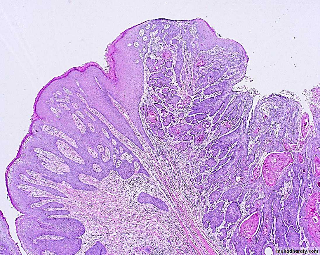
“Extramammary” Paget’s disease
Unique form of intraepithelial adenocarcinoma histologically indistinguishable from Paget’s disease of the nipple, this has a very different biology.–– Grossly : pruritic, red, crusted, sharply demarcated map-like areas often mistaken for inflammation clinically
–– Histologically, all tumor cells limited to epidermis.
it reveals single anaplastic tumor cells with in the epidermis. These cells are characterized by having clear spaces (“halos”) between them and the adjacent epithelial cells.
–– Prognosis excellent unless invasive cancer found.
Paget’s Disease of the Vulva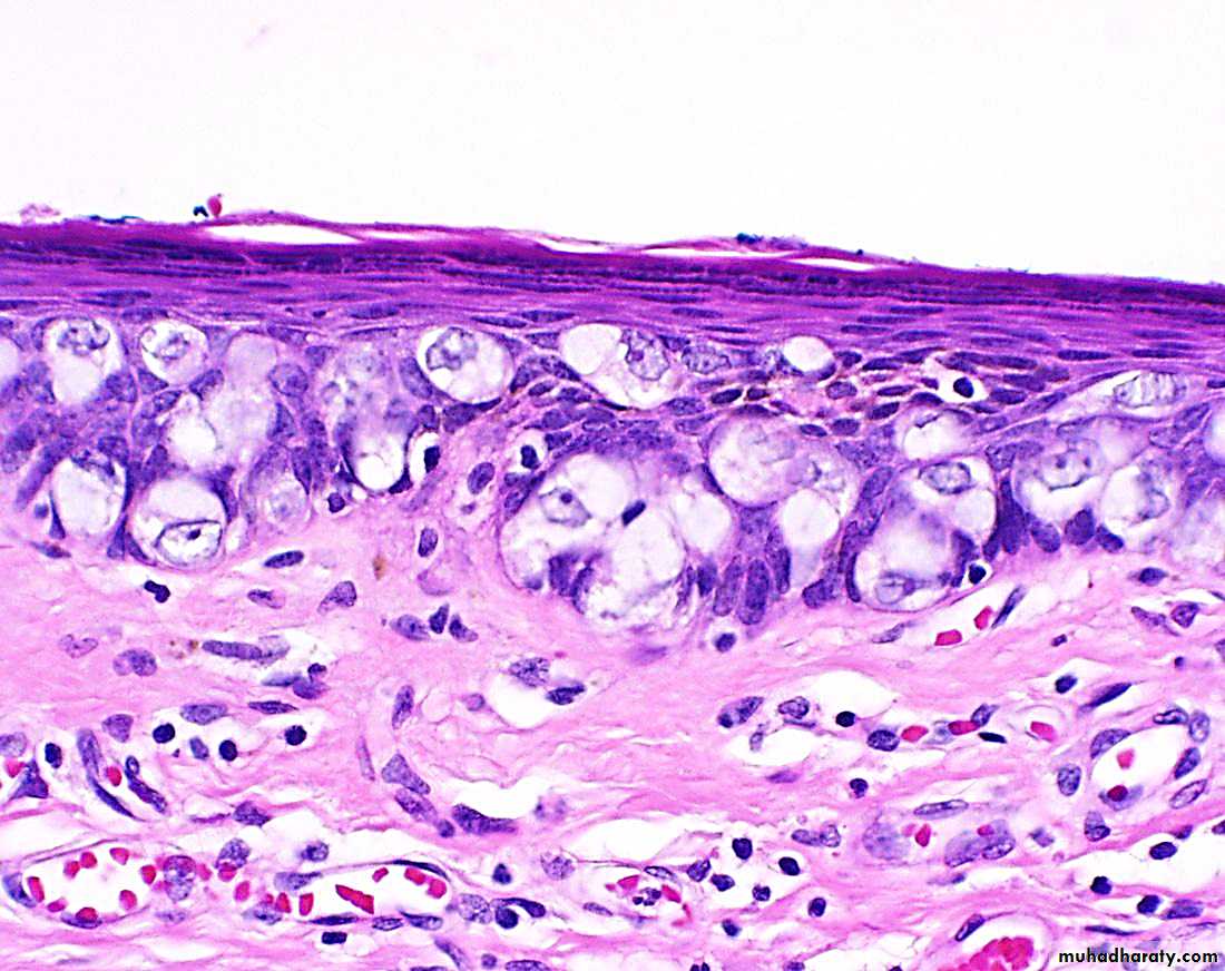
Diseases of the Vagina
• Relatively rare site of significant disease– Congenital anomalies
– Infections
– Cancers
Gartner’s Duct Cysts
They are relatively common lesions found along the lateral walls of the vagina & derived from wolffian duct rests.
They are 2cm fluid filled cysts that occur submucosally
Vaginal Intraepithelial Neoplasia and Squamous Cell Carcinoma
Primary carcinoma of the vagina are very uncommon1% female genital tract with 95% are sq. cell carcinoma.
Secondary spread from adjacent sites are more common.
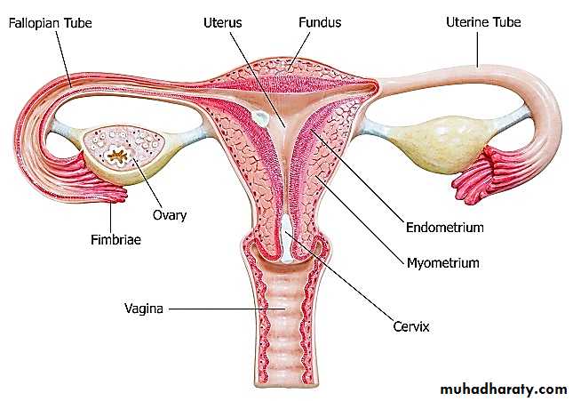
Pathology of the Uterus
Pathology of the Uterus• Cervix
• Endometrium
• Myometrium
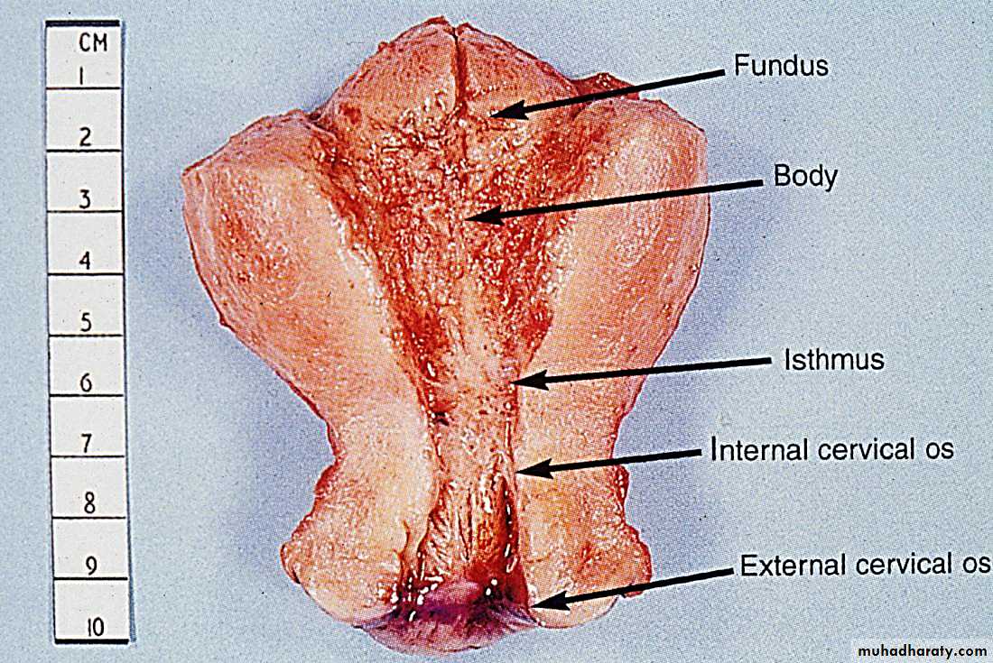
Cervix
• Infections– cervicitis
– Any vaginitis will also involve cervix
• Non-neoplastic lesions
– Squamous metaplasia– Nabothian Cysts
– Cervical polyp
• Neoplasms
– Dysplasia– Cancer
Cervicitis
Definition : inflammation of the cervical mucosa.Many cervical infections are of a non-specific and probably mixed bacterial nature.
Specific infections with Neisseria gonorrhea , Herpes virus ,Chlamydia , Mycobacteria ,Trichomonas vaginalis & Treponema pallidum
chronic cervicitis:
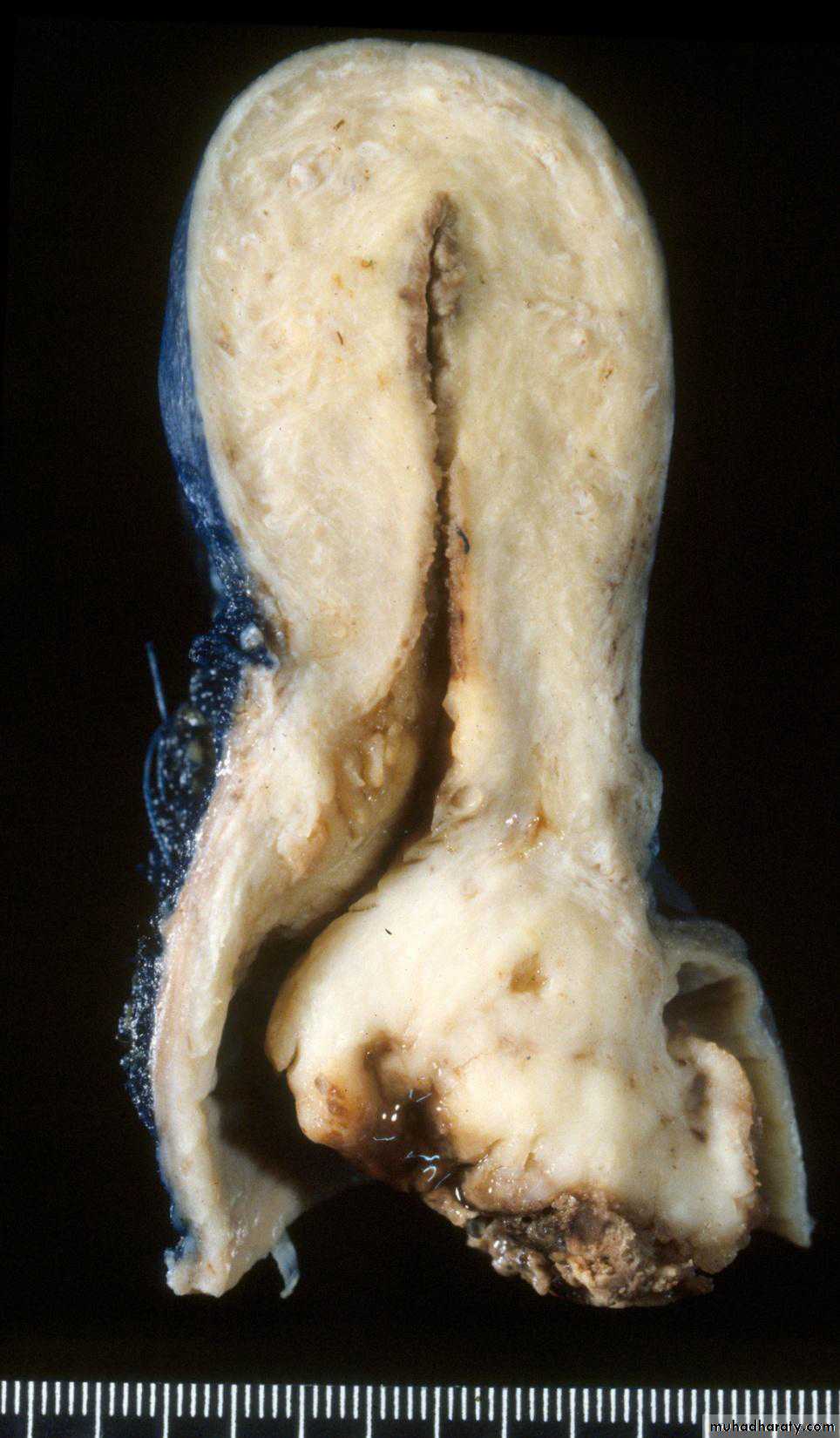
Small round dark lymphocytes are seen in the submucosa, and there is also hemorrhage.CERVICAL POLYPS
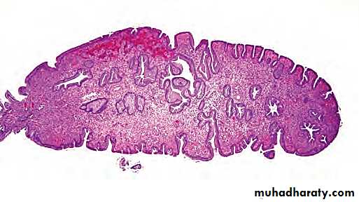
localized benign proliferations of endocervical mucosa though they may protrude through the external os.Found in 2-5% of adult women and produce irregular vaginal spotting.
Grossly: small (up to 5 cm in size), bright red, fragile growth which is frequently pedunculated but may be sessile.
Microscopically :most cervical polyps are endocervical polyps and are covered with endocervical epithelium which may show squamous metaplasia.
Endocervical polyp composed of a dense fibrous stroma covered with endocervical columnar epithelium.
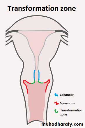
Squamous Metaplasia
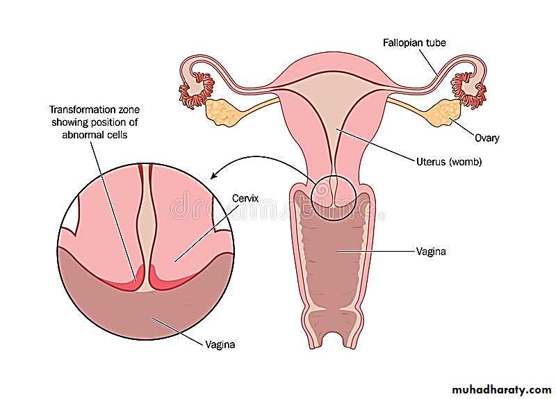
• Process by which endocervical mucinous columnar epithelium changes to squamous epithelium.• This area of sq. metaplasia is often known as the “Transformation Zone” and it is of considerable importance because it is the site of cervical dysplasias and cancers.
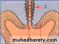
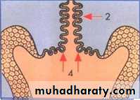

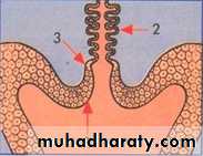
Before puberty
At pubertyAfter puberty and pregnancy
At menopause
Immature
squamous
Cells
Squamocolumnar
JunctionColumnar
glandularcells
Mature squamous cells
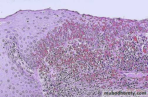
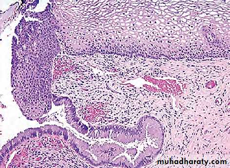
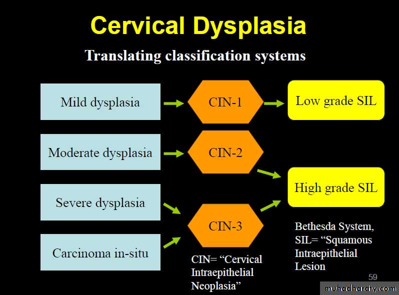
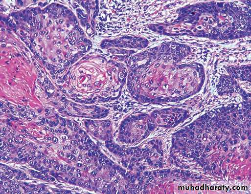
CERVICAL INTRAEPITHELIAL NEOPLASIA (CIN).
DYSPLASIA This term has been commonly used for atypical cytological changes in the layers of squamous epithelium, the changes being progressive.Depending upon the thickness of squamous epithelium involved by atypical cells, dysplasia is conventionally graded as mild, moderate and severe.
Carcinoma in situ is the full-thickness involvement by atypical cells, or in other words carcinoma confined to layers above the basement membrane.
CIN An alternative classification is to group various grades of dysplasia and carcinoma in situ together into cervical intraepithelial neoplasia (CIN) which is similarly graded from grade I to III.
Cervical Dysplasia
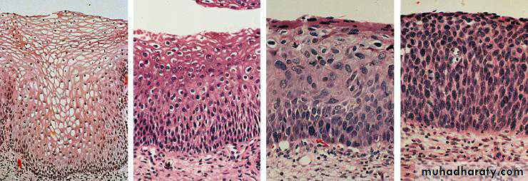
Normal CIN I CIN II CIN III
SIL Currently, the three grades of CIN are readjusted into two grades of squamous intraepithelial lesions (SIL)
—Low-grade SIL (L-SIL)
—High-grade SIL (H-SIL)
Many cases of CIN either remain stationary or regress with probably no more than 1/3 of cases of CIN III advancing to invasive stage .
All cases of CIN have to be regarded as potentially invasive ,fortunately these epithelial lesions , though asymptomatic are readily detected by cytological examination of the cervical smear preparations (Pap smear)
Treatment & eradication by techniques such as cryocautory & laser therapy are relatively simple.
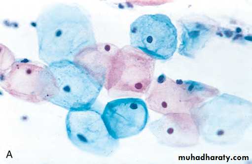
Pap smear is by far the most effective cancer screening and prevention technique in use
Cervical cancer
Risk factors
1) Early age of sexual activity.2) Family history of cervical cancer
• HPV infection with high-risk types of oncogenic virus.
• Weakened immune system
• Socio-economic status
• Oral contraceptives
• Potential role of high risk male sexual partner having history of penile condyloma and those lack of circumcision
Grossly : Invasive cervical carcinoma may present 3 types of patterns: fungating, ulcerating and infiltrating.
Microscopically: The following patterns are seen:
1. Squamous cell carcinoma This type comprises vast majority of invasive cervical carcinomas (about 70%).
2. Adenocarcinoma Adenocarcinomas comprise about 20-25% of cases.
3. Others The remaining 5% cases are a variety of other patterns such as adenosquamous carcinoma .
Cervical cancerArose in squamocolumnar junction
Squamous cell carcinoma of the cervix
HPV DNA sequences are often integrated into the genome of dysplastic or malignant cervical epithelial cells. What is the molecular mechanism associated with this process?HPV viral proteins E6 and E7 bind and inactivate the gene products of p53 and Rb, both tumor suppressor genes, thus allowing the cells to accumulate DNA damage.