
Endocrine System 3
Third year class
By Dr.Riyadh A. Ali
Department of Pathology
TUCOM

Parathyroid glands
Normal Parathyroid
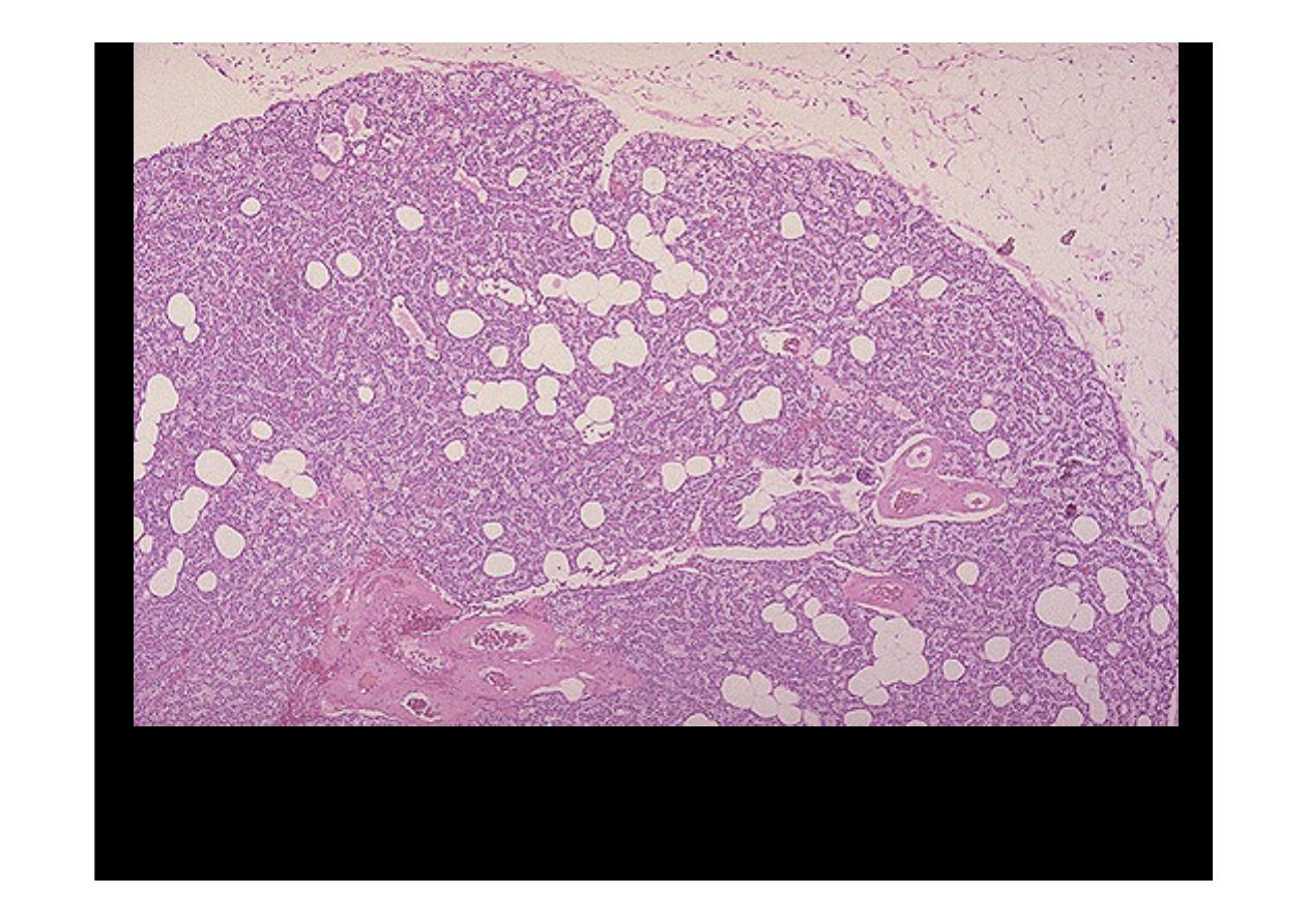
Here is a
normal parathyroid gland
for comparison. Adipose tissue
cells are mixed with the parathyroid tissue.
Parathyroid hyperplasia
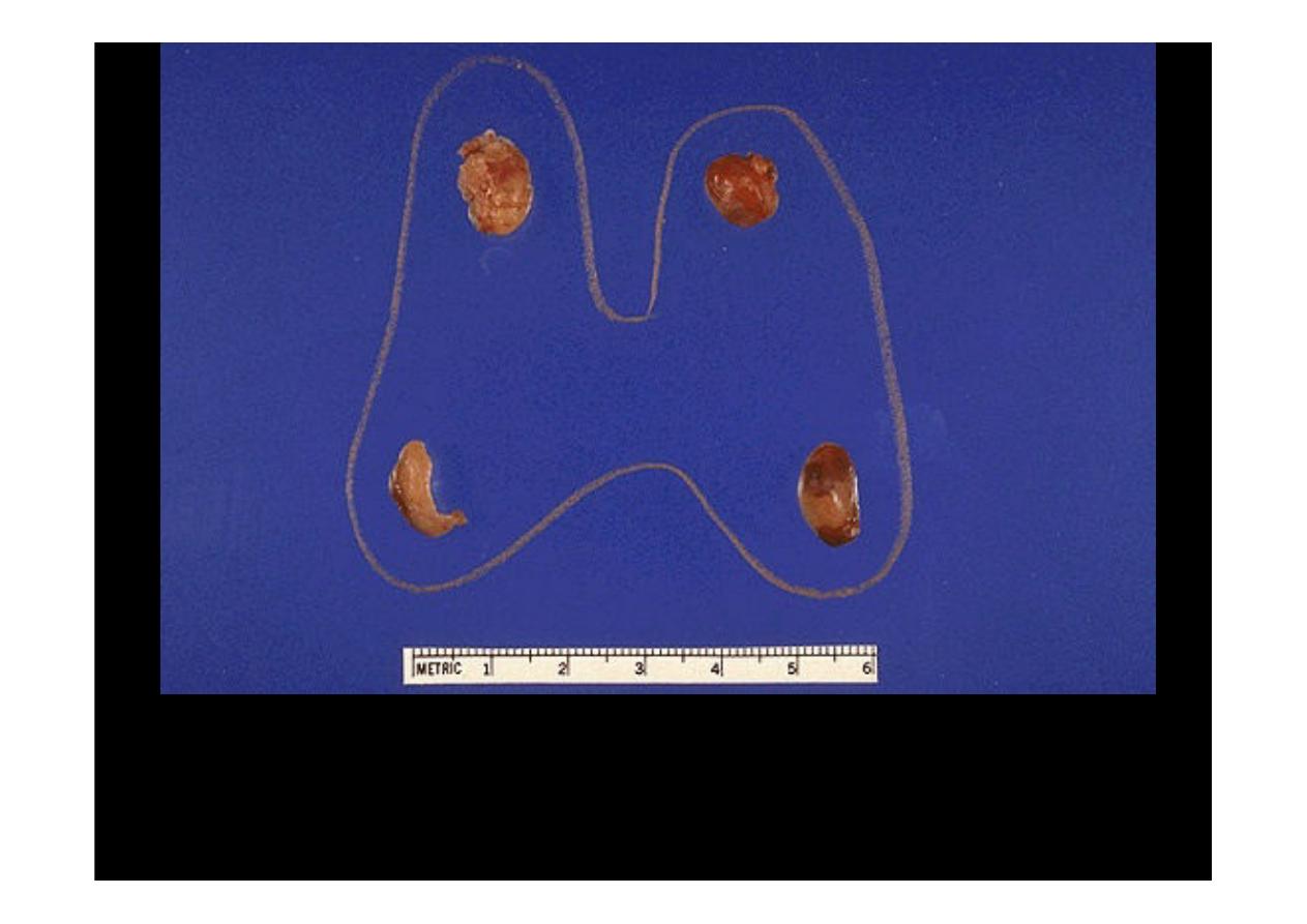
Parathyroid hyperplasia
is shown here. Three and one-half glands have
been removed (only half the gland at the lower left is present). Parathyroid
hyperplasia is the second most common form of primary hyperparathyroidism,
with parathyroid carcinoma the least common form.
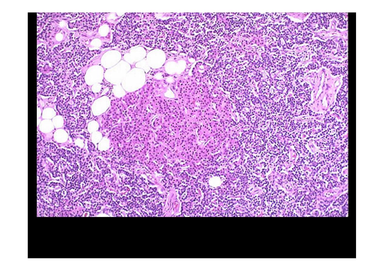
In
parathyroid hyperplasia
, there is little or no adipose tissue, but any or all
cell types normally found in parathyroid are present. Note the pink oxyphil cells
here. the parathyroids secrete more parathormone

Parathyroid
Adenoma
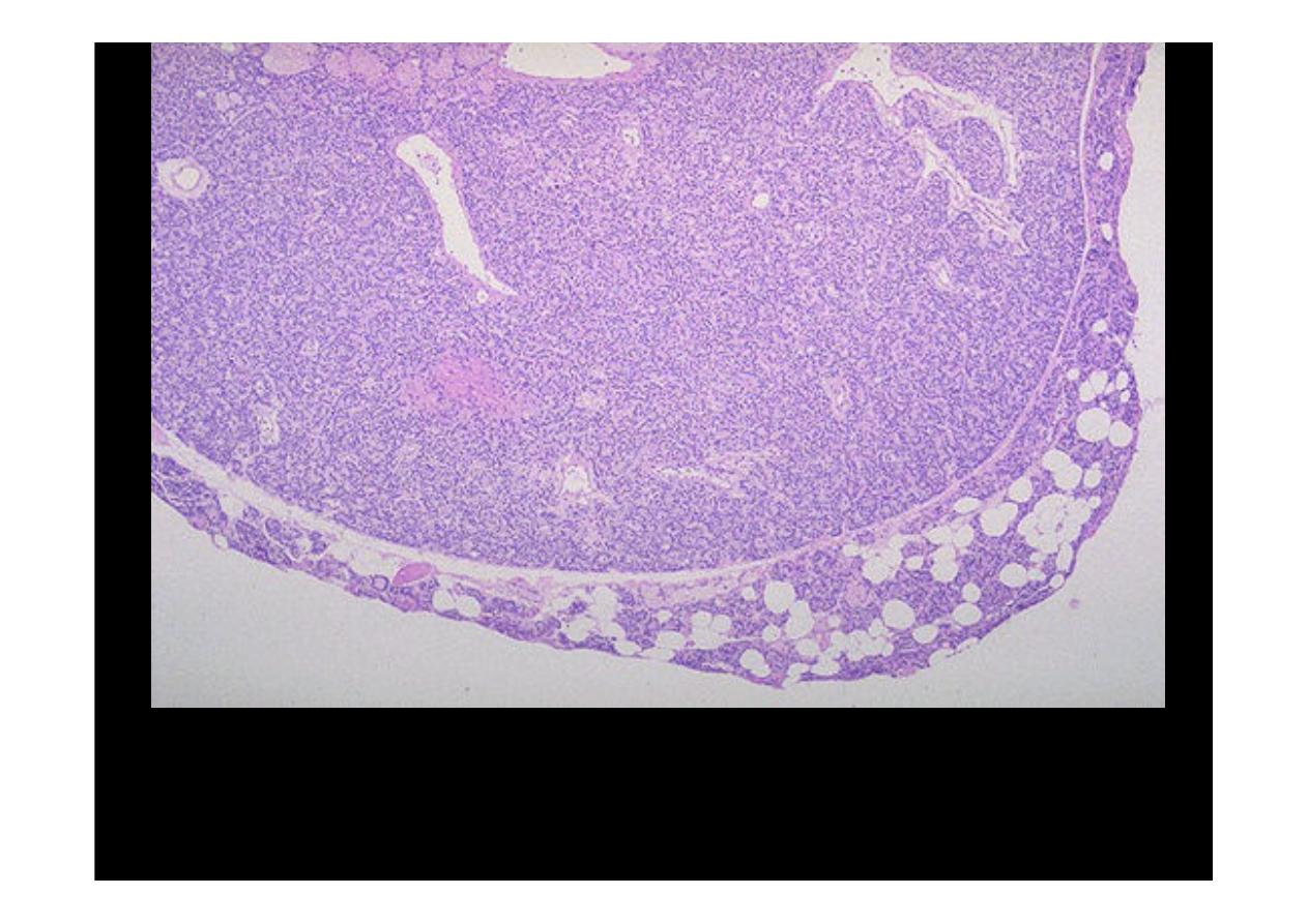
Here is a
parathyroid adenoma
, the most common cause for primary
hyperparathyroidism. A rim of normal parathyroid tissue admixed with
adipose tissue cells is seen compressed to the right and lower edge of the
adenoma.
Adjacent to this
parathyroid adenoma
is a rim of normal parathyroid
tissue (with a pink oxyphil cell nodule) at the upper right, and a small
benign parathyroid cyst (an incidental finding) is at the upper left
.
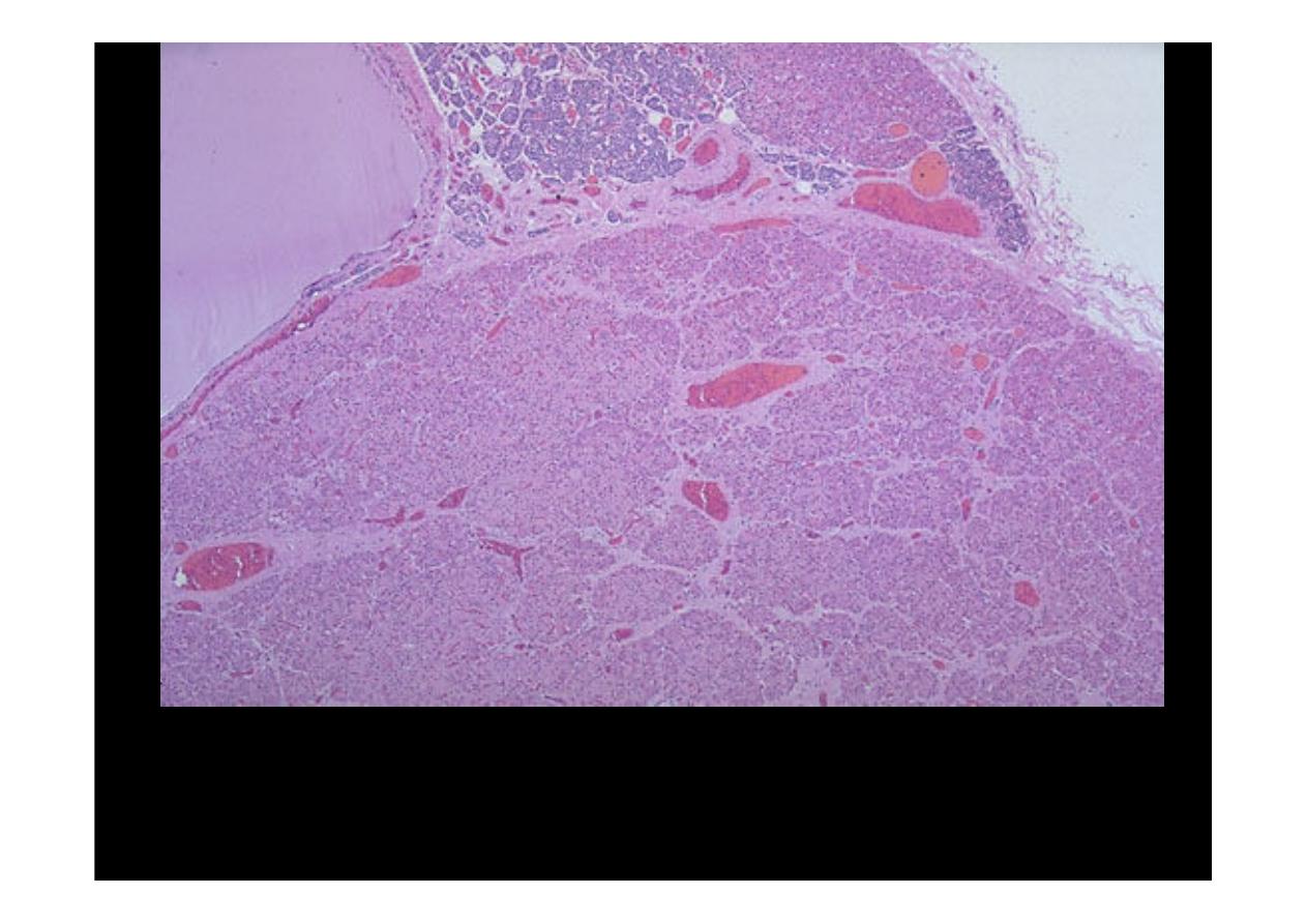
Parathyroid
carcinoma
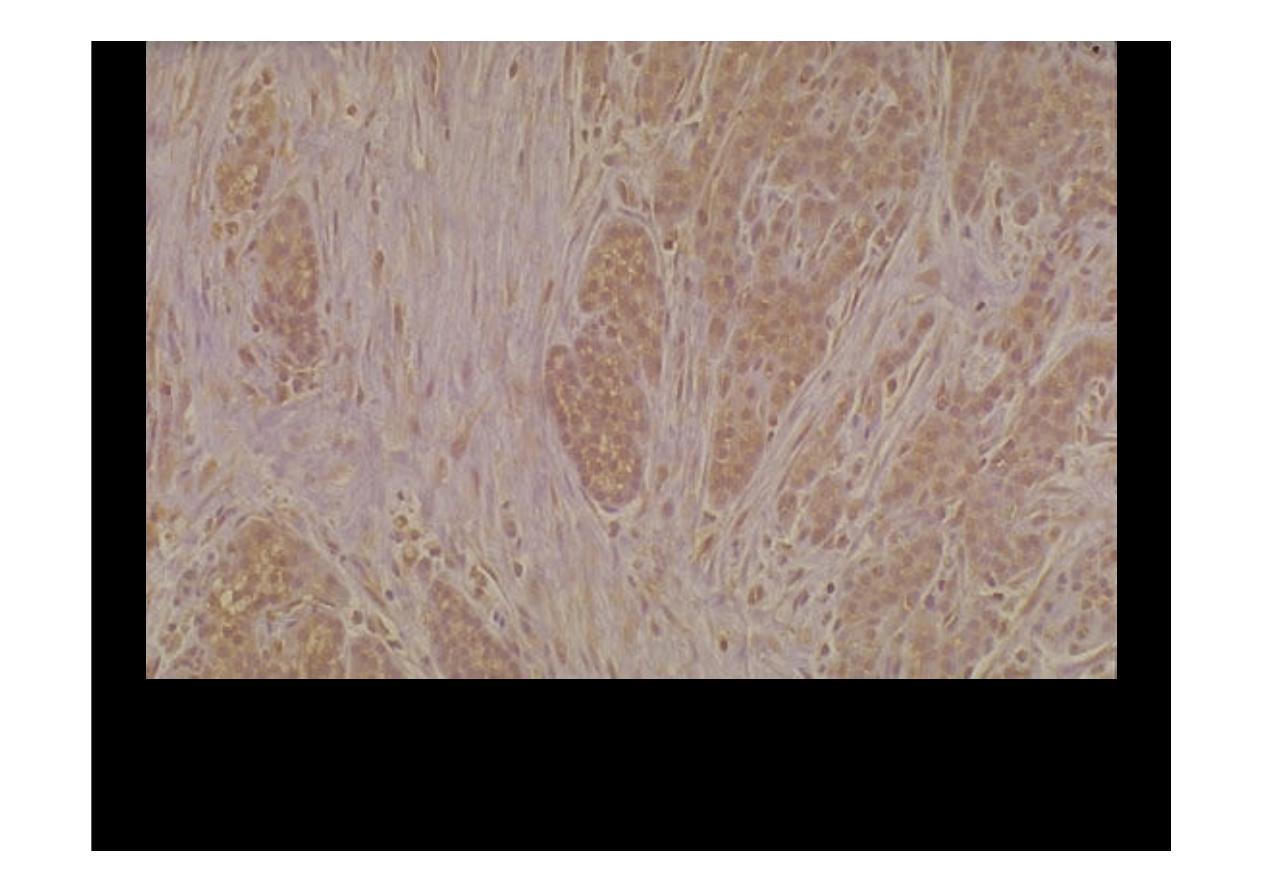
This is a
parathyroid carcinoma
with nests of neoplastic cells that are not
very pleomorphic and that are demonstrating positive immunoperoxidase
staining with antibody to parathormone. Note the bands of fibrous tissue
between the nests.