PATHOLOGY OF DENTINE CARIES
The earliest stage of dentine caries starts deep to a carious enamel lesion before any clinical evidence of cavitation.Diffusion of acid from the enamel lesion into the dentine causes demineralisation of the mineral component but leaves the collagenous dentine matrix intact.
However, once bacteria have penetrated the enamel, they spread along the amelodentinal junction to attack the dentine over a wide area.
The lesion is therefore conical with a broad base at the enamel junction and its apex toward the pulp.
Infection of dentine is facilitated by the dentine tubules, which form a pathway open to bacteria once they have been slightly widened by acid attack. After demineralization the dentine matrix is progressively destroyed by proteolytic enzymes secreted by bacteria.
Infection of the dentinal tubules.
This electron photomicrograph shows bacteria in the lumen of the tubules.
Between the tubules is the collagenous matrix of the dentine
Caries of dentine. Infected tubules and fusiform
masses of bacteria have expanded into the softened tissue.
Adjacent tubules in the demineralised dentine have been bent
and pushed aside by these masses.
Advanced dentine caries. The dentine is disintegrating (left). To the right is a large focus of destruction and tubules packed with bacteria.
Key events in the development of dentinecaries:
Non-bacterial, pre-cavitation, acid softening of matrix.Widening of tubules by demineralization
Migration of pioneer bacteria along tubulesDevelopment of a mixed proteolytic bacterial flora in the dentine
Distortion of tubules by expanding masses of bacteriaBreakdown of intervening matrix forming liquefaction foci
Progressive disintegration of remaining matrix tissueProtective reactions of dentine and pulpunder caries
Translucent dentine:A form of reactionary dentine, reduces the diameter of the dentinal tubules, preventing bacterial penetration and generating a more heavily mineralised dentine by ‘tubular sclerosis’.Regular reactionary dentine: Forms at the pulp–dentine interface and retains the tubular structure of dentine. Forms in response to mild stimuli.
Irregular reparative dentine:Forms in response to moderate or severe insult.
Dead tracts:Formed when odontoblasts die and their tubules become sealed off.
Translucent dentine in dentine caries:
The dentinal tubules are seen in cross-section. Those in the centre of the picture have become obliterated by calcification; only the original outline of the tubules remains visible, and the zone appears translucent to transmitted light.
On either side are patent(open) tubules filled with stain.
Translucent dentineThere is early occlusal caries in the fissure, and below it peritubular dentine has sealed off the
pathway to the pulp to produce a zone of translucent dentine.
When dye is put into the pulp chamber, it cannot pass along the tubules in the translucent dentine as it does elsewhere. The translucent zone is thus rendered less permeable to bacteria and acid.
Regular reactionary dentine
below the occlusal caries,bacteria extend more than half the distance from the amelodentinal junction to the pulp, and the underlying pulp horn has been obliterated by reactionary dentine.The reactionary dentine bulges into the pulp.
Note: the lack of inflammation in the pulp,
organised tubular structure and odontoblast layer.
Regular reactionary dentine.
Regular, tubular, secondary dentine has formed under a carious cavity.A line marks the junction of the primary and secondary dentine where the tubules change direction.
Bacteria spreading down the tubules of the primary tissue have extended along the junction and into the tubules of the reactionary dentine.
Root surface caries lesions may start along the cervical region when gingiva starts toretract. No probing is needed.
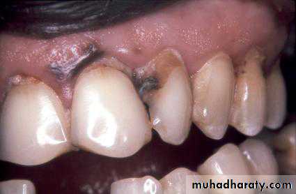
Inactive root surface caries lesions which are entirely dark and hard on probing.These are scar tissue resulting from previousactive caries lesions! Imagine if a dentist had drilled and filled such lesions
A sharp probe has been jammed into the white spot lesion on the buccal aspect of this extracted molar. (a) The lesion before probing. (b) The probe and the resulting damage. On the occlusal surface the enamel lesion has formed on the walls of the fissure. Heavy use of a sharp probe would be similarly destructive on this surface.
Iatrogenic:induced by a physician’s words or therapy(used especially of a complication resulting from ttt).
A sharp probe is useful in diagnosis. It can be used very gently to draw across the lesion and detect the slight roughness of the active white spot lesion. Remember, it is a delicate instrument, not a bayonet!
The gentle use of the probe explains why these methods are described as visualtactile criteria.
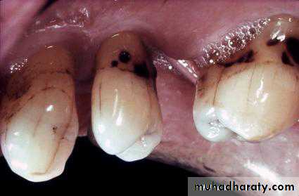
These lesions are on the root surface close to the gingival margin. They are darkly coloured and leathery in texture. These are slowly progressing lesions and some are cavitated. This woman is in her 70s. She has a dry mouth and rheumatoidarthritis (secondary Sjögren’s syndrome). It is not easy for her to clean, but with helpwith cleaning, fluoride toothpaste, and fluoride varnish application, these lesions can be arrested.
Ditched amalgam restorations
The enamel around the amalgam restorations on the palatal aspect of the upper lateral incisors is discoloured. Is this discolouration due to caries or corrosion of the amalgam? A decision was made to replace these restorations and removal of the amalgam revealed discoloured, hard, dentine. This was corrosion and the replacement was necessary.