
GIT 2
Third Year Class
By Dr.Riyadh A. Ali
Department of Pathology
TUCOM
Titles
• Normal Stomach
• Gastritis
• Gastric Ulcer

Normal Stomach
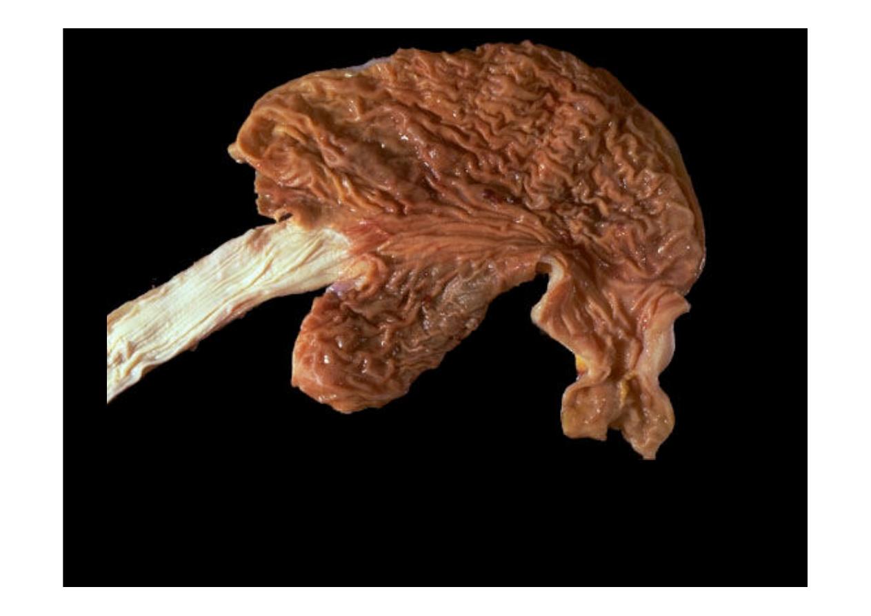
This is the
normal appearance of the stomach
, which has been opened along the
greater curvature. The esophagus is at the left and the pylorus emptying into the first
portion of duodenum is at the lower right.
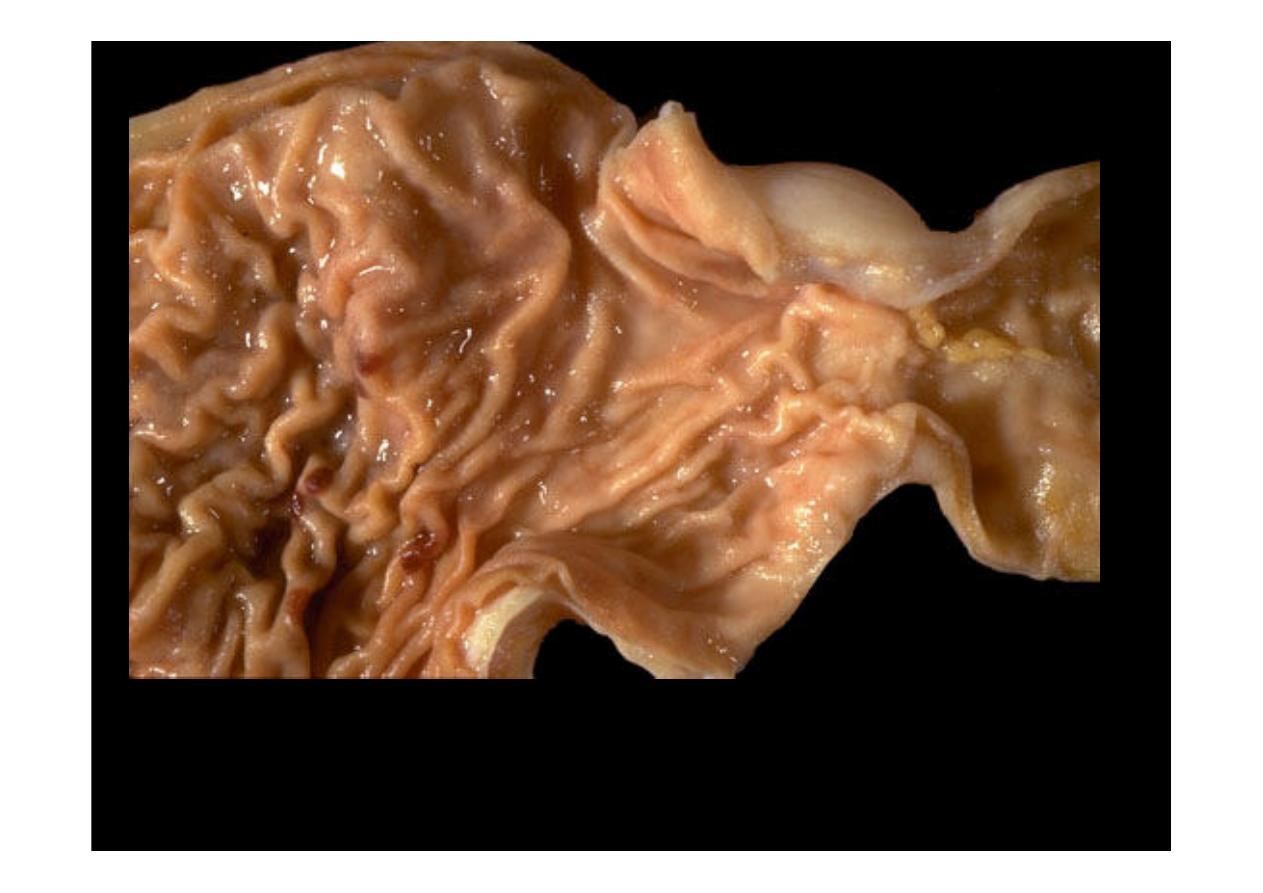
This is the
normal appearance of the gastric antrum
extending to the pylorus
at the right of center. The first portion of the duodenum (duodenal bulb) is at
the far right.
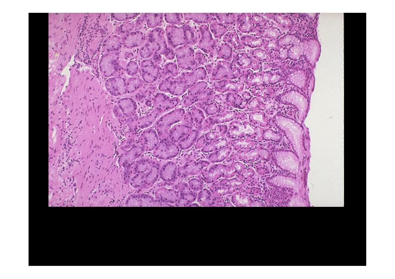
This is the
normal appearance of the gastric fundal mucosa
, with short
pits lined by pale columnar mucus cells leading into long glands which contain
bright pink parietal cells that secrete hydrochloric acid.

Gastritis
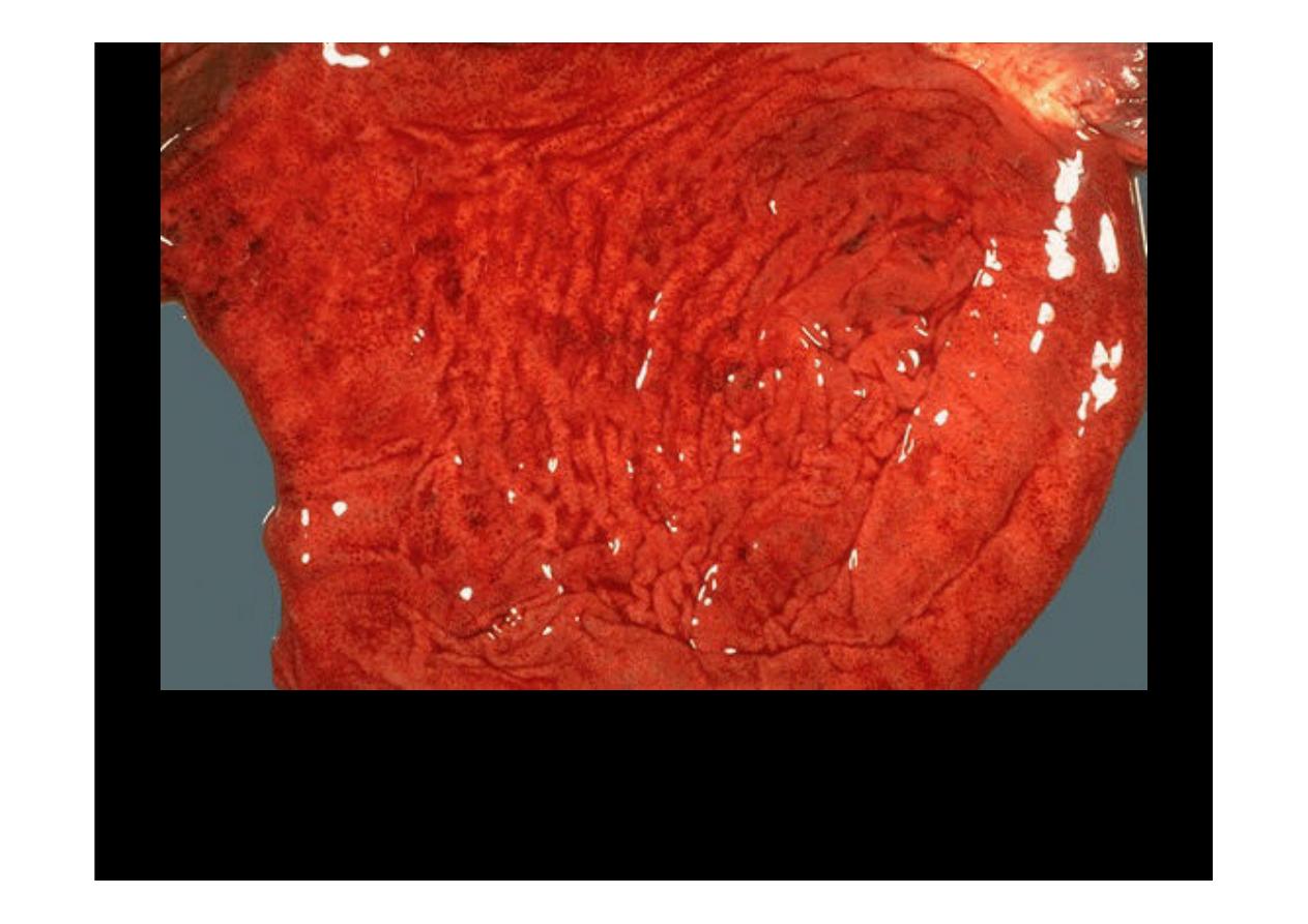
This is a more typical
acute gastritis
with a diffusely hyperemic gastric mucosa.
There are many causes for acute gastritis: alcoholism, drugs, infections, etc.
Here are some larger areas of gastric hemorrhage that could best be termed
"
erosions
" because the superficial mucosa is eroded away.
At high power, gastric mucosa demonstrates infiltration by neutrophils.
This is
acute gastritis.
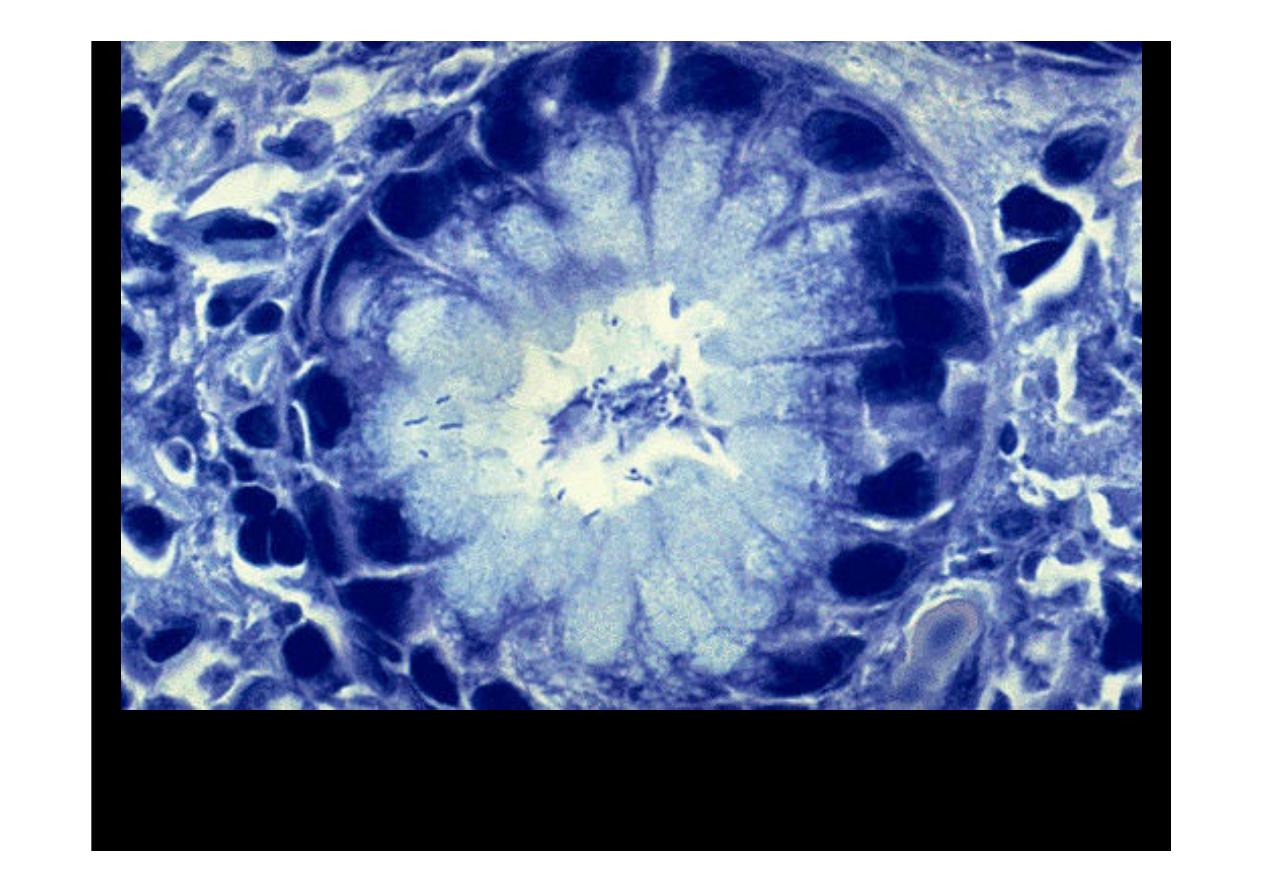
Gastritis
is often accompanied by infection with Helicobacter pylori. This small curved
to spiral rod-shaped bacterium is found in the surface epithelial mucus of most patients
with active gastritis. The rods are seen here with a methylene blue stain.

Gastric Ulcer
Gastric Ulcer
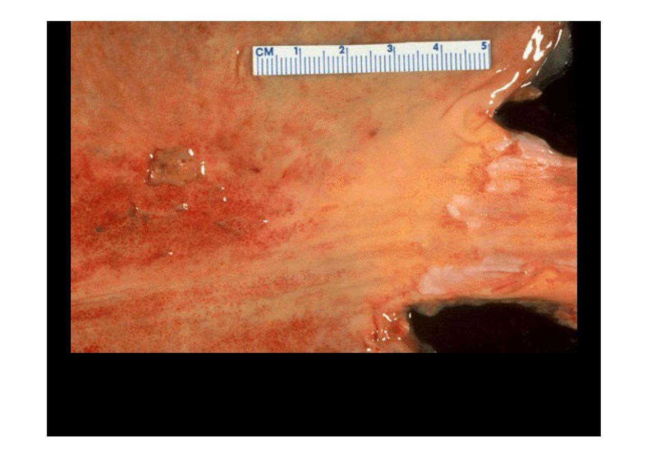
A 1 cm
acute gastric ulcer
is shown here in the upper fundus. The ulcer is shallow and
sharply demarcated, with surrounding hyperemia. It is probably benign. However, all gastric
ulcers should be biopsied to rule out a malignancy.
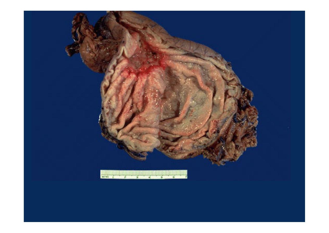
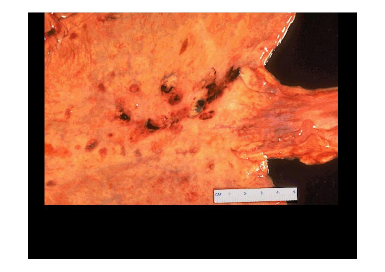
Here is a much larger 3 x 4 cm gastric ulcer that led to the resection of the stomach
shown here. This ulcer is much deeper with more irregular margins. Complications of
gastric ulcers (either benign or malignant) include pain, bleeding, perforation, and
obstruction.
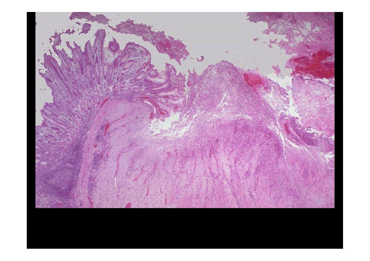
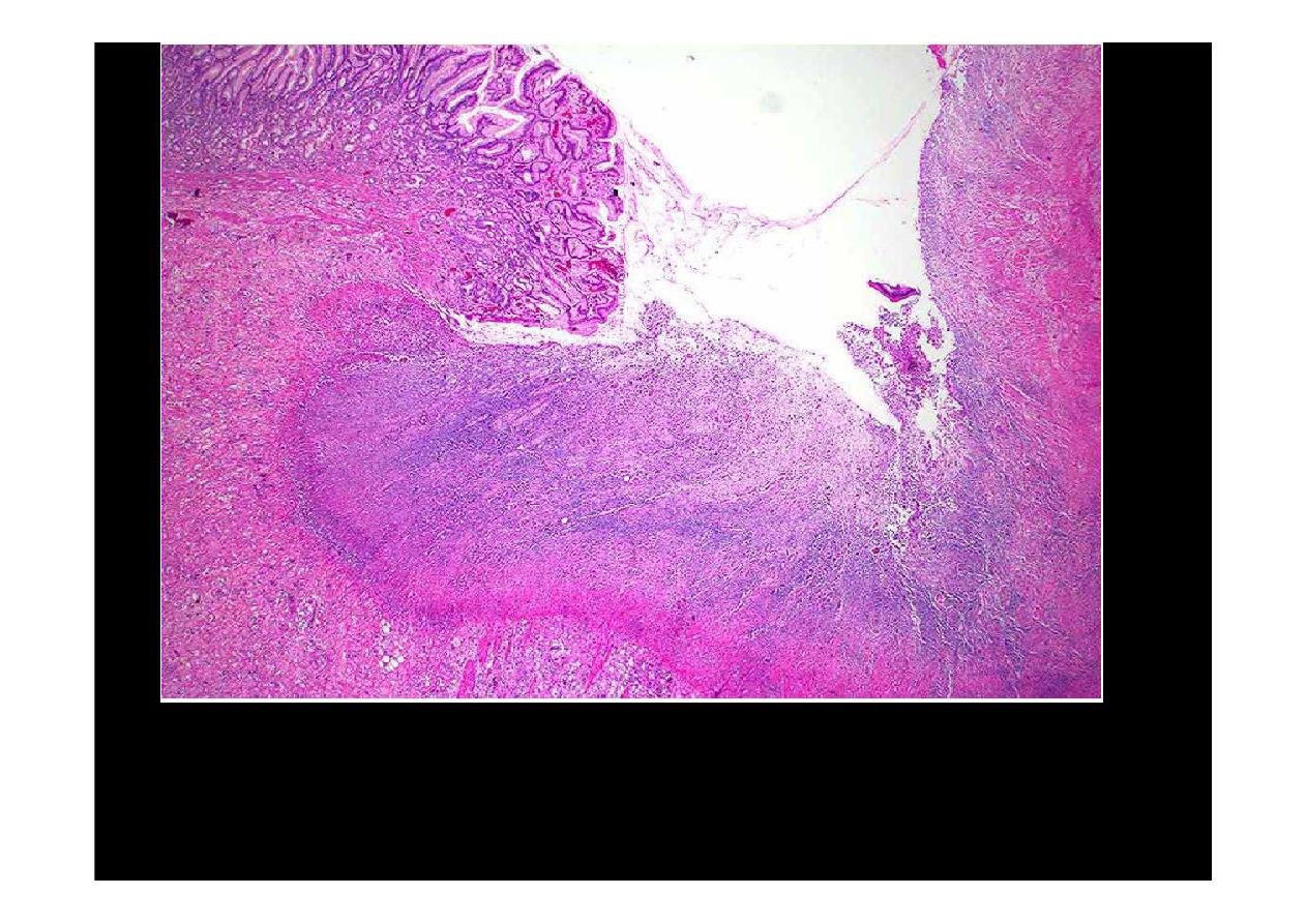
Microscopically, the ulcer here is sharply demarcated, with normal gastric mucosa on the
left falling away into a deep ulcer whose base contains inflamed, necrotic debris. An
arterial branch at the ulcer base is eroded and bleeding.
(Gastric ulcer)
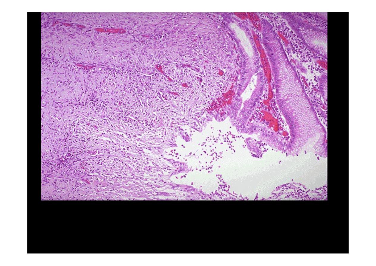
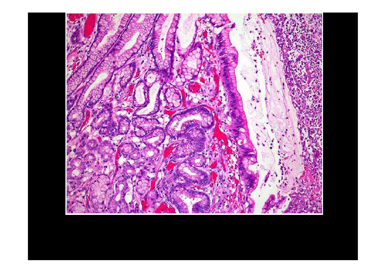
The mucosa at the upper right merges into the
ulcer
at the left which is eroding through
the mucosa. Ulcers will penetrate over time if they do not heal. Penetration leads to pain.
If the ulcer penetrates through the muscularis and through adventitia, then the ulcer is said
to "perforate" and leads to an acute abdomen. An abdominal radiograph may demonstrate
free air with a perforation.
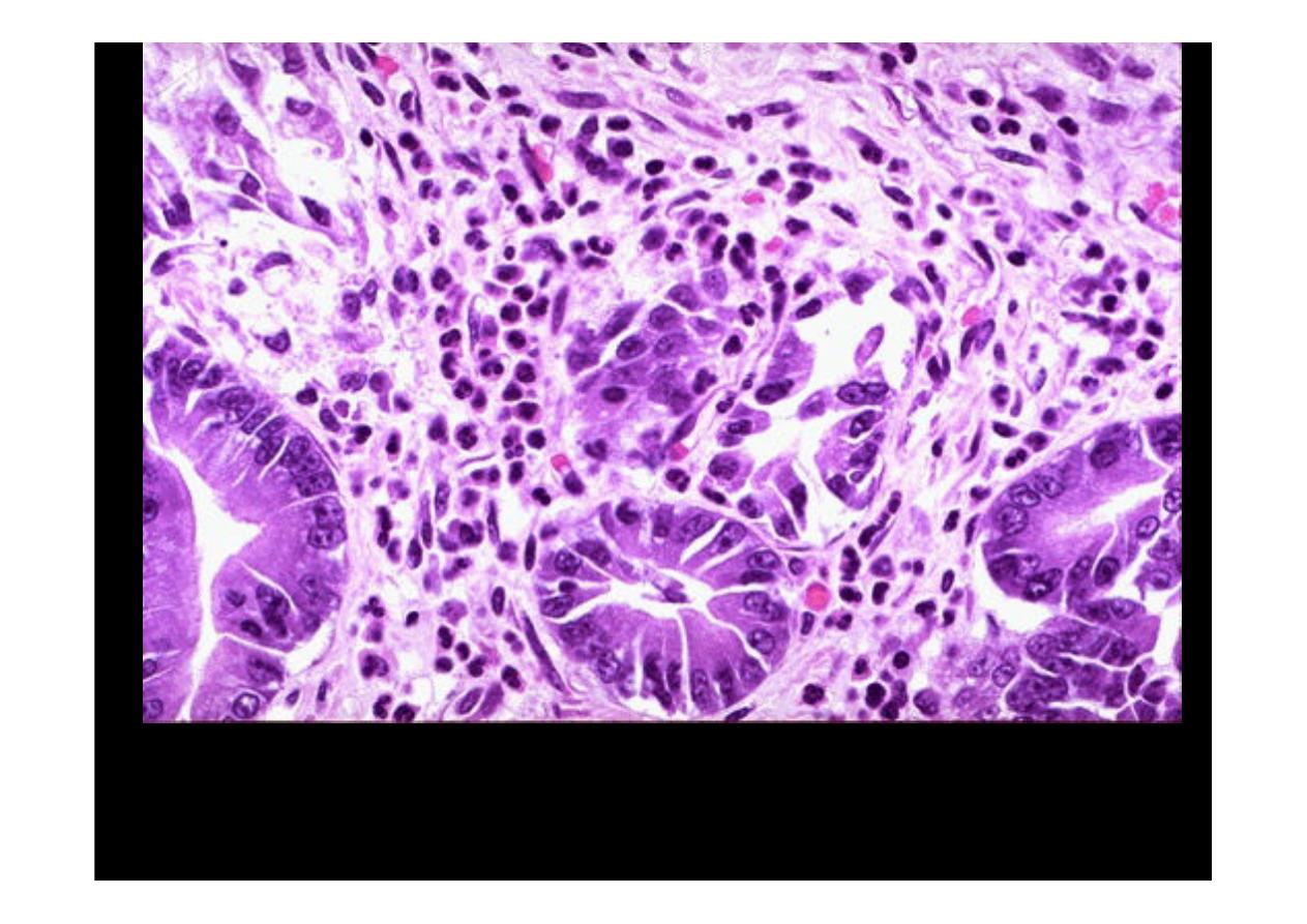
Acute erosions will show loss or necrosis of superficial epithelium with associated
hemorrhage, will show organization of surrounding tissue (fibroblast proliferation,
collagen deposition) and a collection of inflammatory cells.
Gastric Ulcer
Necrotic debris and fibrin are usually present on the ulcer surface. The epithelium adjacent
to the ulcer will show reactive epithelial changes, with enlarged vesicular nuclei and
prominent nuclei.
Gastric Ulcer