•

IMAGE RECEPTOR
• In dental radiography after the beam pass through the teeth and adjacent structure it reach the x-ray film the dental x-ray film servers as a recording medium or image receptor
• Types of dental x-ray film
• Intraoral film
• Extra oral film
Intra oral x-ray film
It’s a double emulsion type of the film therefore its require less radiation exposure to produce an image
Introduction
1
• Size 0:pedo-type
• Size 1:adult anterior type• Size 2: adult posterior type
• Size 3:bitewing type (0,1,2&3)
• Size 4: occlusal type
Intra oral film size
2
Dental x-ray films vary in speed or sensitivity to the radiation that required to produce a radiograph of standard density.
The film speed is determined by the following:
Size of silver halide crystals.
Thickness of the emulsion.
Intra oral film speed have been standardized on the alphabetical basis; A,B,C,D,E&F(A the lowest and F the fastest)
Intra Oral Film Speed
3
• Film Base: Is a flexible pieces of polyester plastic that measure 0.2 mm thickness and constructed to withstand heat, moisture and chemical exposure. The film base is transparent and exhibit a slight blue tint that is used to emphasize contrast and enhance image quality. The primary purpose of the base is to provide mechanical support to the delicate emulsion.
• Adhesive Layer: Is a thin layer material on both sides of film base and serve to attach the emulsion to the base
• Film Emulsion: Is a homogenous mixture of gelatin and silver halide crystal, is coating and attached on both side of film base
Film composition
4
• Gelatin: Is used to support and evenly disperse of silver halide crystals over the film base, during processing the gelatin serve to absorb the processing solution and allow the chemical to react with the silver halide crystal.
• Halide Crystals: Is a chemical compound that is sensitive to radiation and light, the halide in dentistry usually made of silver plus a halogen either bromine or iodine. Silver bromide form 80-90% and silver iodine form 10% of entire silver halide crystal, halides crystals absorb and store energy of radiation.
• Protective Layer: Is a thin transparent coating placed over the emulsion. Its protect emulsion surface from mechanical and processing damage.
Film composition
5
Film composition
6• Its either one or two film package.
• In one corner of the intra oral film a small raised bump known as the identification dot is used to determine film orientation. After the processing the raised dot used to distinguish between the left and right sides of the patient.Film Package
7
• Outer Package Wrapping: Is a soft vinyl or paper wrapper that seals the film, protective black paper and lead foil sheet. Its serves to protect the film from light and saliva.
• The outer wrapper of the film have two sides
• Tube Side; Is solid white and has raise dump in one corner. Its stippled to aid in preventing the film package from slipping when it is held against the oral mucosa by the patient fingers.
Film Package
7
Label Side; It has a flap that is used to open the film packet. The label side is color coded. The following information is printed on the label side:
A circle concave dot corresponds the raise dot
The statement "opposite side toward tube"
The manufacturer name
The speed
Number of the film enclosed
Film Package
8
Black Paper: Is a black paper protective sheet that covers the film and shields the film from light.
Lead Foil Sheet: Is a single pieces of lead foil that is found within the film packet and is located behind the film wrapped in the black protective paper.
Film Package
8
Its shield the film from back scattered radiation that results in film fog.
Protect the tissues behind the film from the hazard of radiation.An embossed pattern is placed on the lead foil sheet by the manufacturer; the pattern is visible on the processed radiograph if the film is exposed from the non-exposure side.
Action of Lead Foil
9
• Extra oral film are used to examine large areas of the skull or jaws e.g. panoramic and cephalometric film. Extra oral film used in dental radiography is available in 5x7 inch and 8x10 inch as well as the panoramic 5x12 inch and 6x12 inch sizes.
Extra Oral Film
11
Extra oral film types
Screen film:Is a film that requires the use of a screen for exposure, the film is placed between two special intensifying screen in the a cassette.
When the screen film exposed to the x-radiation the screen convert the x-ray energy into light, which exposes the film.
Screen film is sensitive to fluorescent light rather than direct exposure to x-radiation.
Some film are sensitive to blue light, whereas other sensitive to the green light therefore each type should be paired with the screen that produce preferable type of light.
Non screen film:
It does not require the use of screens for exposure.
The film is exposed directly to the x-radiation; the emulsion is sensitive to direct exposure rather than fluorescent light.
Its requiring more exposure time than screen type film
Extra Oral Film
11
• Is a device that transfers x-ray to visible light. These screen intensify the effect of x-rays on the film therefore less radiation is required to expose the film and the patient is exposing to less radiation.
Intensifying Screen
12
• Composition of Intensifying Screen
• Base: It's made of a polyester and give the mechanical support to the screen.• Reflecting layer: Is a coating of white titanium dioxide applied to the base to reflect stray light back to x-ray film when the screen emitting light.
• Phosphorus layer: Its contains either fluorescent or phosphor crystals suspended in the gelatin material, the crystals emit the visible light when exposed to the x-radiation.
• Thin protective coating: Is durable enough to withstand repeat insertion and removal of film from the cassette while protecting the underlying phosphor layer.
Intensifying Screen
12
Intensifying Screen
13• Two intensifying screen are used one in front the film and the other at the back of the film, the front screen absorbs the low energy x-ray photons and the back screen absorb the high energy photon.
• Each photon will produce many light photons which will affect a large area of the film, thus the amount of radiation to the patient is reduced.
Action of Intensifying Screen
14
The speed of these screen depends upon:
The thickness of phosphor layerThe size of phosphor crystals
The faster the screen the lower the radiation dose to the patient but the less the detail of the final image.
Action of Intensifying Screen
14
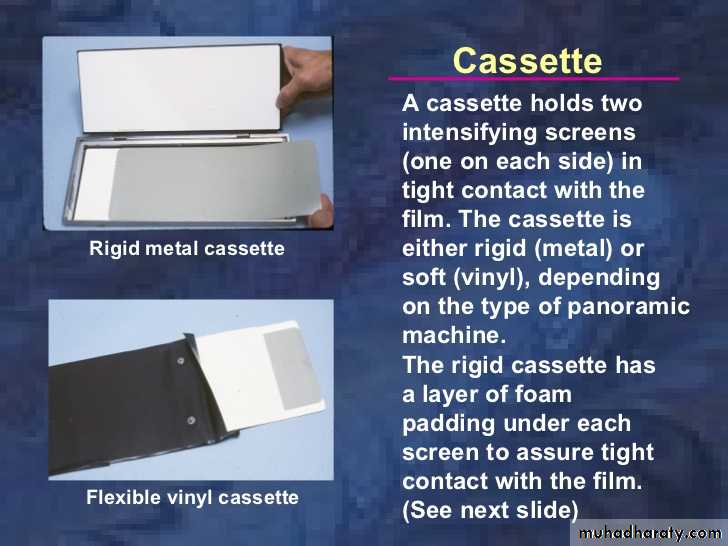
Is used to hold the extra oral film and intensifying screens.
Cassette are available in a variety of sizes that corresponding to the film size and screens.The cassette is may be flexible or rigid.
A rigid cassette is more expensive, last longer and protect the screen from the damage better than the flexible type.
Cassette
16
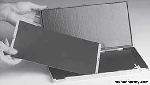
Cassette
Both types of cassette most are light tight in order to:Protect the film from the exposure to the light
To hold the screens in the perfect contact with extra oral film because lack of contact resulted in a loss of image sharpness.
The front cover of the cassette is made of plastic to permit the passage of x-ray beam.
The back cover is constructed from heavy metal to reduce scattered radiation.
The cassette must be marked to oriented the finished radiograph; a metal L or R are attached to the front cover of cassette to indicate the patient side either left or right.
17
• Film is adversely affected by heat, humidity and radiation.
• To prevent film fog the unexposed and unprocessed film should be kept in a cool, dry place and stored in the areas that are shield from source of radiation and not stored in the area where the patient exposed to x-radiation.• To prevent film fog, lead lined or radiation–resistant film dispensers and storage box should be used.
• Film must be used before the labeled expiration date.
Film Storage and Protecting
18
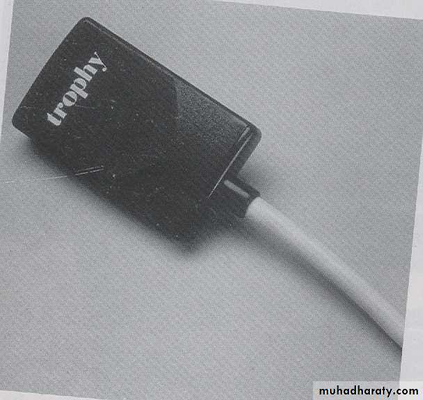
• The Electronic image receptors used for filmless or direct digital radiography creates a latent image.
• An electronic latent image is create when x-ray active electrons contained in the receptor.
• The electronic latent image is transferred to and stored in computer can be converted to a visible image on the monitor or print on the paper
Electronic Image Receptors
19
• Linear array detector: Are used for extra oral film projection such as skull, panoramic and tomographic radiograph
• Area array detectors: Are used in an intra oral projection and bitewing film
Electronic Image Receptors
19