
GIT 4
Third year class
By Dr.Riyadh A. Ali
Department of pathology
TUCOM

Titles
Normal colonic mucosa
Ulcerative colitis
Colonic polyps
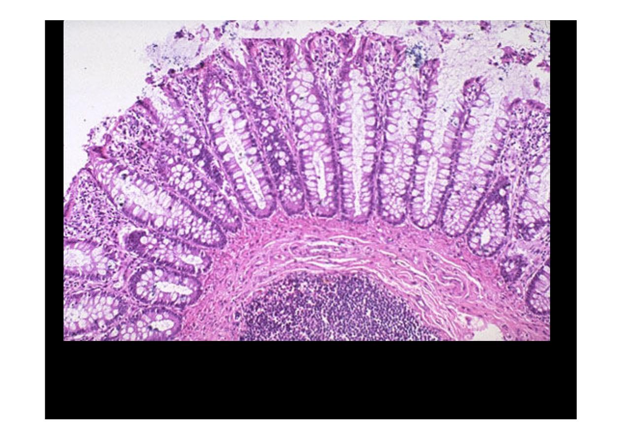
This is
normal colonic mucosa
. Note the crypts that are lined by numerous
goblet cells. In the submucosa is a lymphoid nodule. The gut-associated
lymphoid tissue as a unit represents the largest lymphoid organ of the body.
Ulcerative colitis
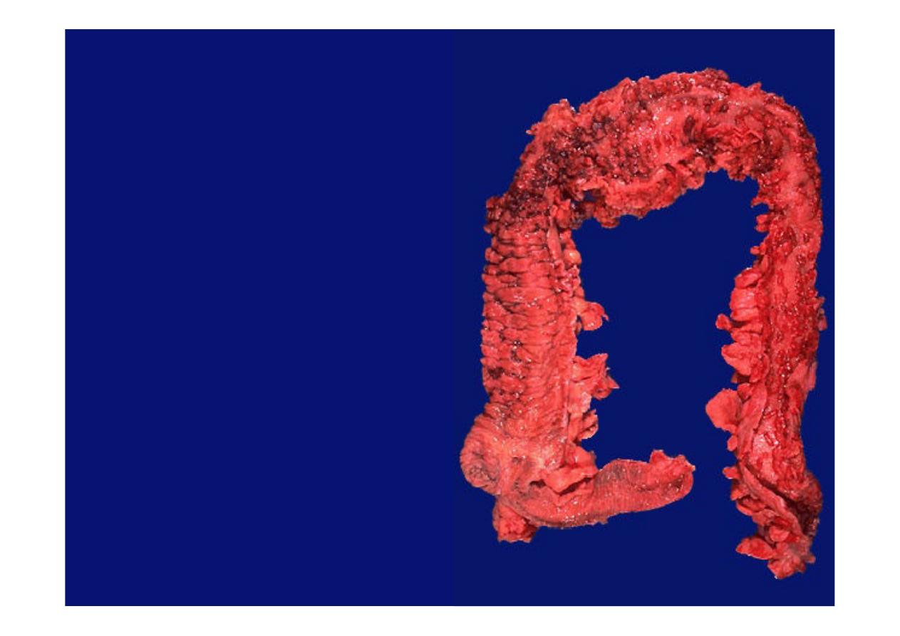
This gross appearance is
characteristic for
ulcerative
colitis
. The most intense
inflammation begins at the
lower right in the sigmoid
colon and extends upward
and around to the ascending
colon. At the lower left is the
ileocecal valve with a portion
of terminal ileum that is not
involved. Inflammation with
ulcerative colitis tends to be
continuous along the mucosal
surface and tends to begin in
the rectum. The mucosa
becomes eroded, as in this
photograph, which shows
only remaining islands of
mucosa called
"pseudopolyps".
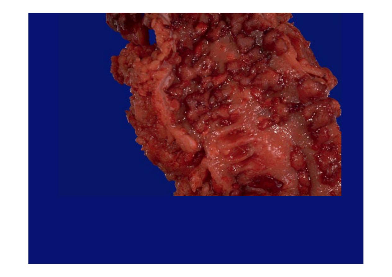
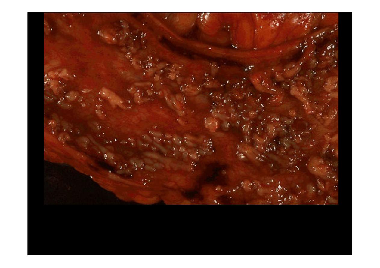
At higher magnification, the pseudopolyps can be seen clearly as raised red
islands of inflamed mucosa. Between the pseudopolyps is only remaining
muscularis.
Ulcerative colitis.
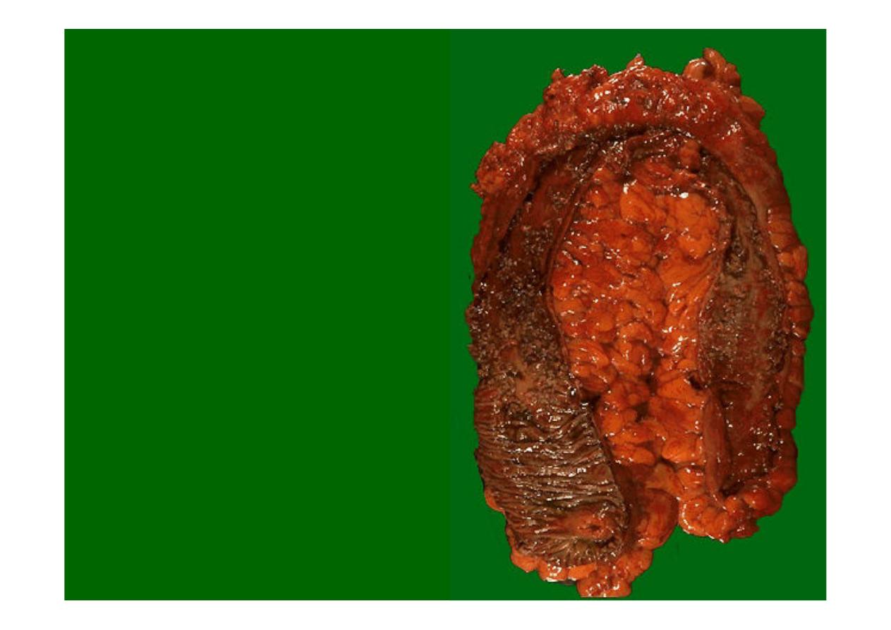
Here is another
example of extensive
ulcerative colitis
.
The ileocecal valve is
seen at the lower left.
Just above this
begins increasing
mucosal
inflammation.
Pseudopolyps are seen here. The remaining mucosa has been ulcerated
away and is hyperemic.
Ulcerative colitis
Microscopically
, the inflammation of
ulcerative colitis
is confined primarily to the
mucosa. Here, the mucosa is eroded by an ulcer that undermines surrounding
mucosa
At higher magnification
, the intense inflammation of the mucosa is seen. The colonic
mucosal epithelium demonstrates
loss of goblet cells
. An exudate is present over
the surface. Both acute and chronic inflammatory cells are present.
Ulcerative colitis
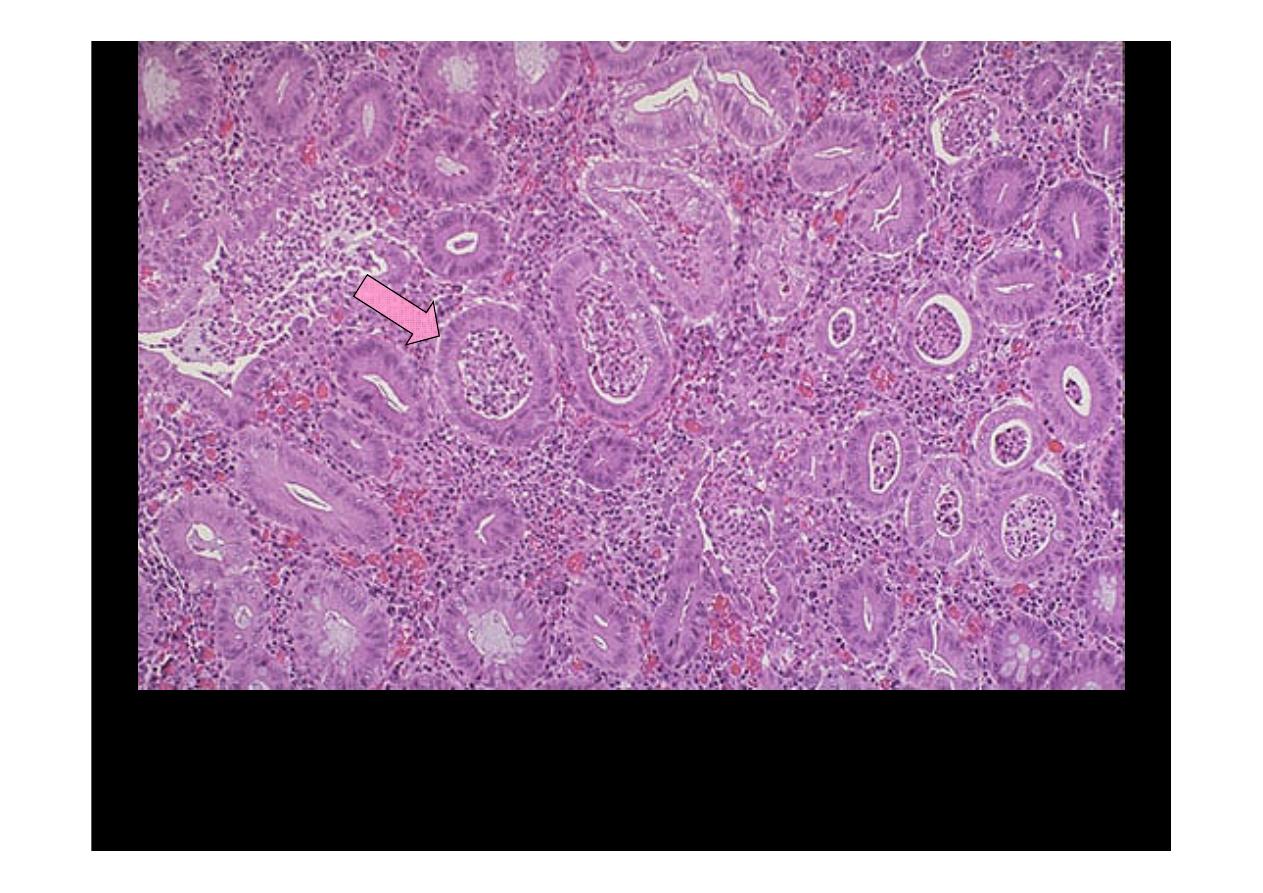
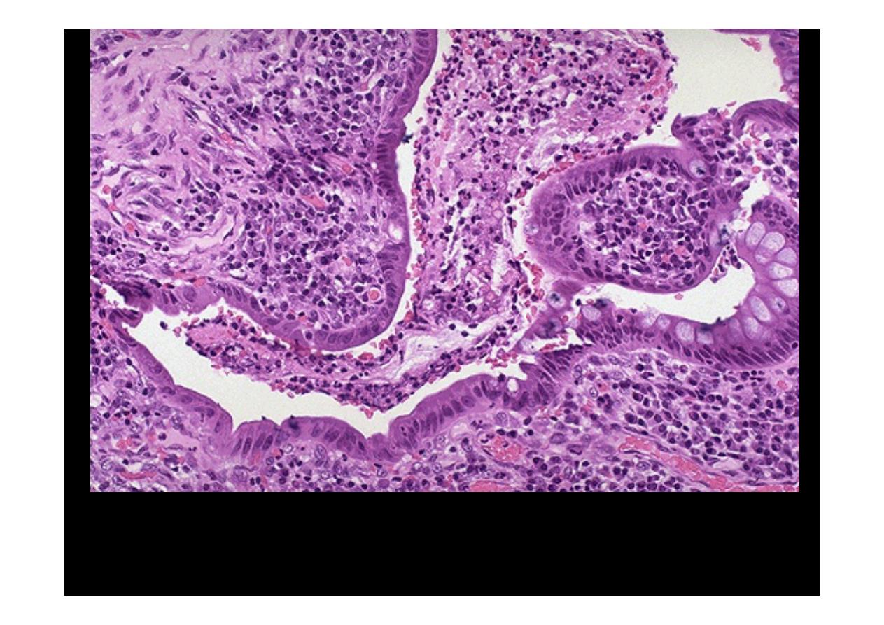
The colonic mucosa of active
ulcerative colitis
shows
"crypt abscesses"
in which
a neutrophilic exudate is found in glandular lumens. The submucosa shows
intense inflammation. The glands demonstrate
loss of goblet cells
and
hyperchromatic nuclei with inflammatory atypia.
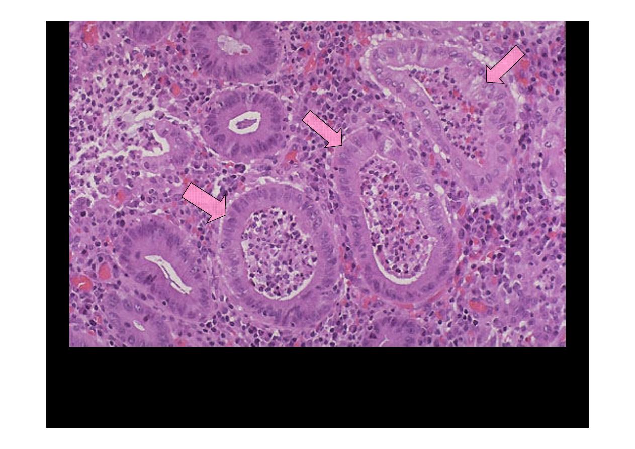
The colonic mucosa of active
ulcerative colitis
shows
"crypt abscesses"
in which a
neutrophilic exudate is found in glandular lumens. The submucosa shows intense
inflammation. The glands demonstrate
loss of goblet cells
and hyperchromatic nuclei
with inflammatory atypia.(higher power)
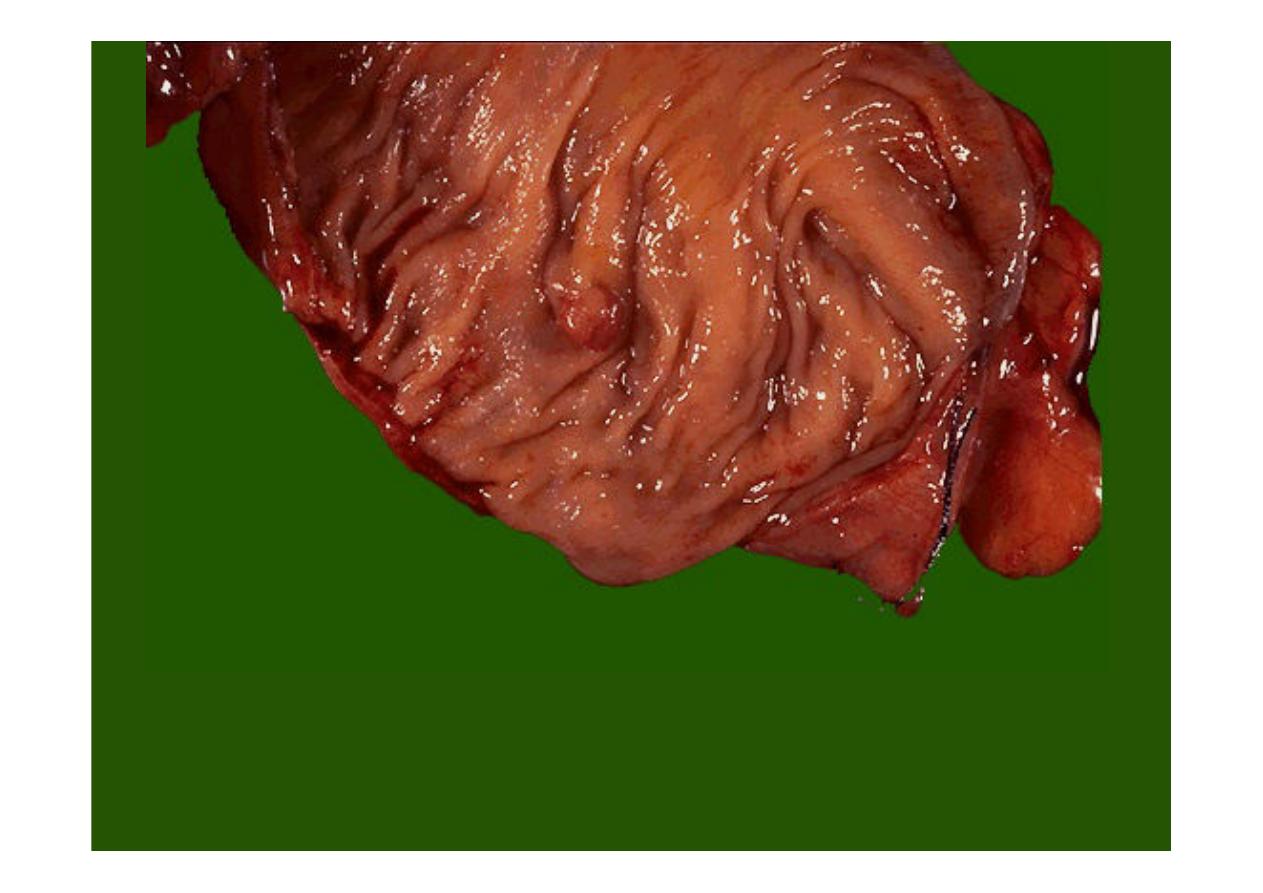
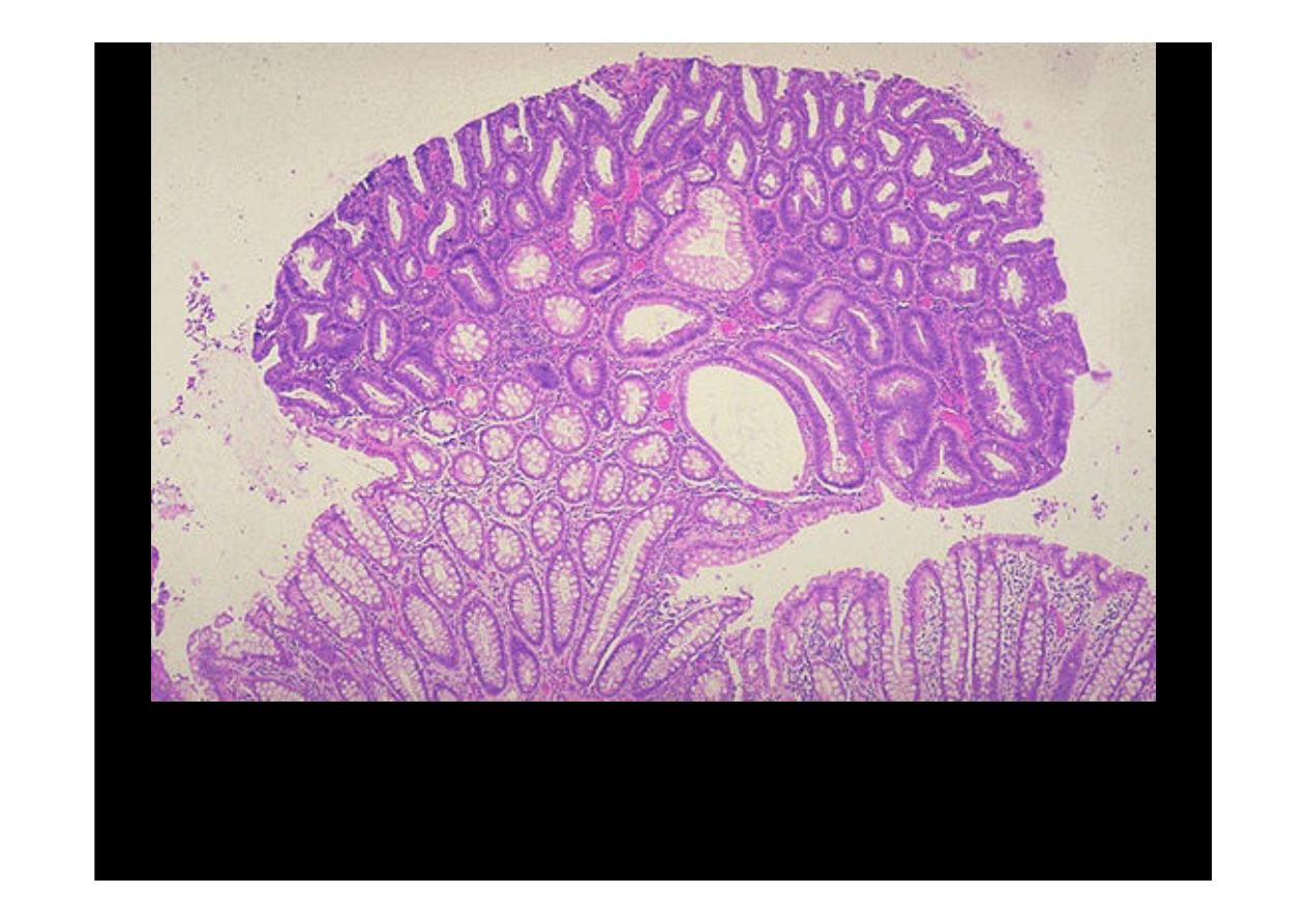
Here is a small
adenomatous polyp
in the middle of the colon seen here. It has
smooth surfaces and is discreet. These are common. Small ones are virtually
always benign. Those larger than 2 cm carry a much greater risk for development of
a carcinoma.
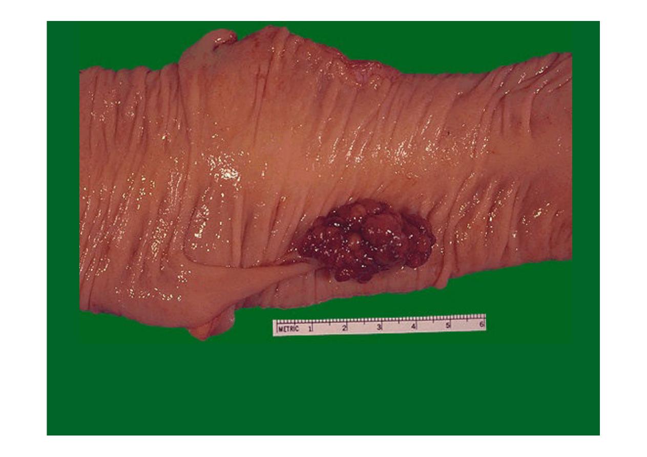
This
adenomatous polyp
has a hemorrhagic surface (which is why they may
first be detected with stool occult blood screening) and a long narrow stalk.
The size of this polyp--above 2 cm--makes the possibility of malignancy more
likely, but this polyp proved to be benign.
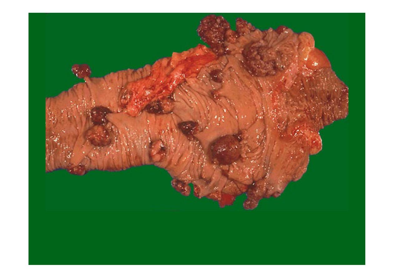
Here are
multiple adenomatous polyps
of the cecum. A small portion of
terminal ileum appears at the right.
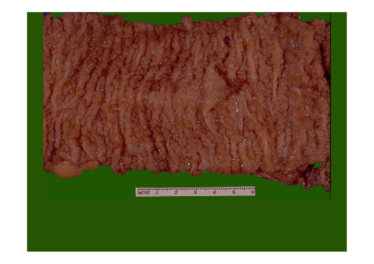
This is
familial polyposis
in which the mucosal surface of the colon is
essentially a carpet of small adenomatous polyps, there is a 100% risk over
time for development of adenocarcinoma.
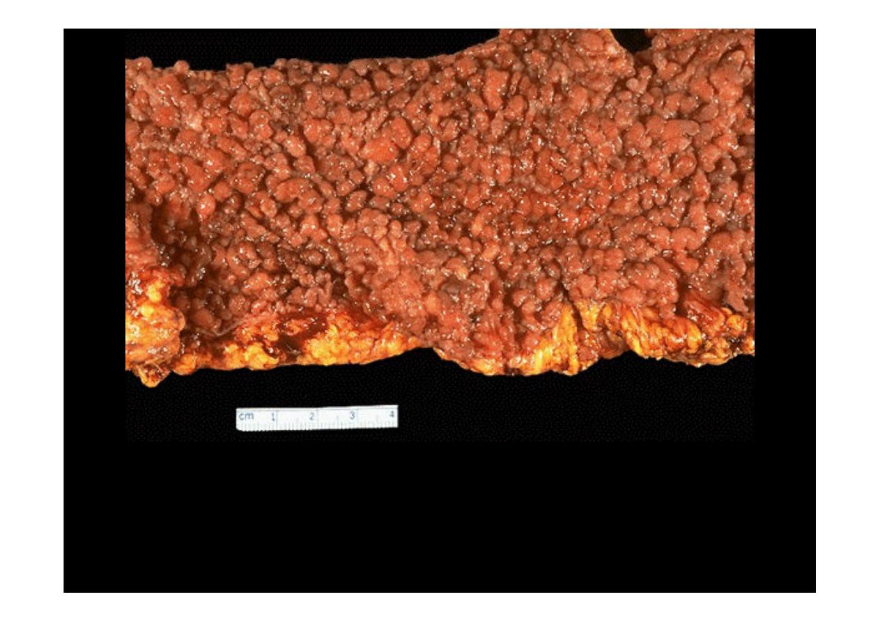
Here is another example of
polyposis
with numerous small polyps covering the
colonic mucosa.
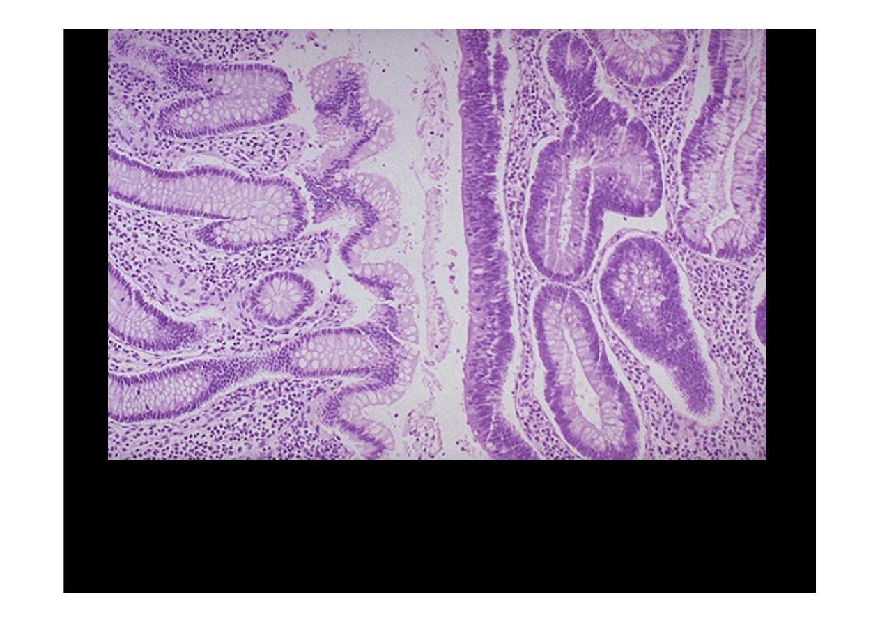
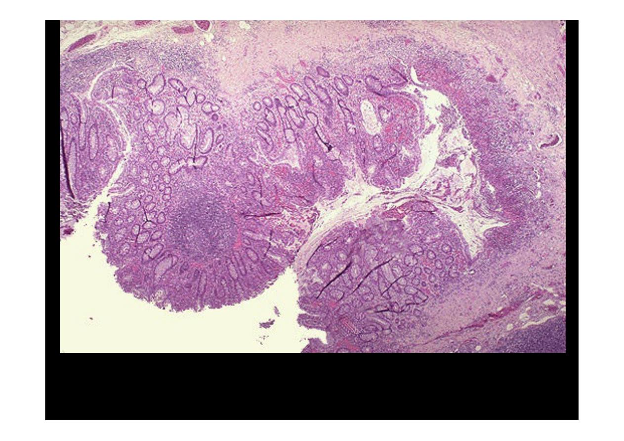
A microscopic comparison of
normal colonic mucosa
on the left and that of an
adenomatous polyp (tubular adenoma)
on the right is seen here. The
neoplastic glands are more irregular with darker (hyperchromatic) and more
crowded nuclei. This neoplasm is benign and well-differentiated, as it still closely
resembles the normal colonic structure.
This is a small
adenomatous polyp (tubular adenoma)
seen microscopically to have
more crowded, disorganized glands than the normal underlying colonic mucosa.
Goblet cells are less numerous and the cells lining the glands of the polyp have
hyperchromatic nuclei. However, it is still well-differentiated and is benign.
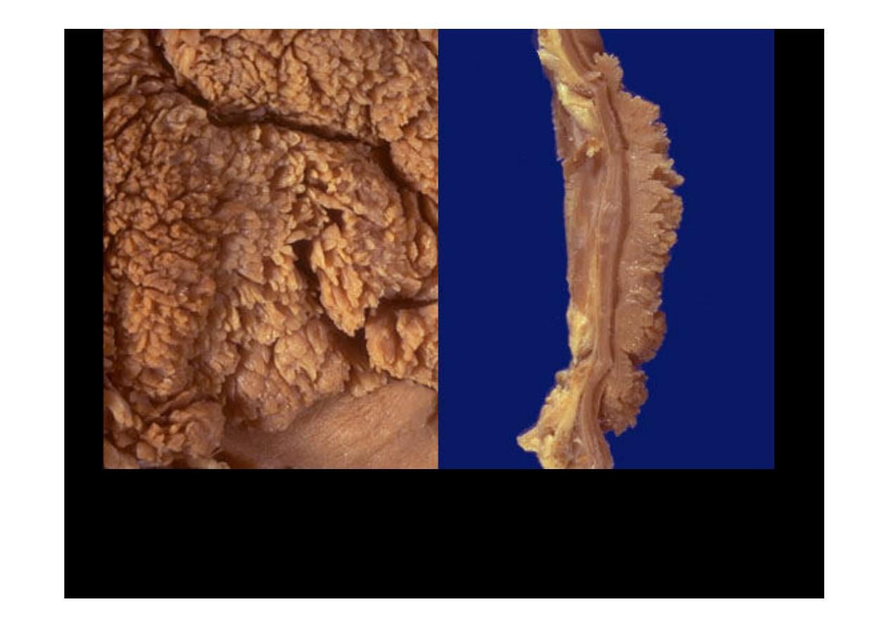
The gross appearance of a
villous adenoma
is shown above the surface at the
left, and in cross section at the right. Note that this type of adenoma is sessile,
rather than pedunculated, and larger than a tubular adenoma (adenomatous
polyp). A villous adenoma averages several centimeters in diameter, and may be
up to 10 cm.
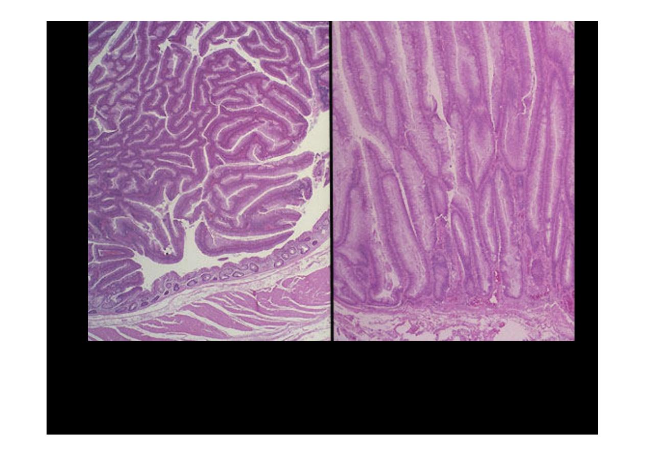
Microscopically, a
villous adenoma
is shown at its edge on the left, and projecting above
the basement membrane at the right. The cauliflower-like appearance is due to the elongated
glandular structures covered by dysplastic epithelium. Though villous adenomas are
less
common
than adenomatous polyps, they are much more likely to have invasive carcinoma
in them (about 40% of villous adenomas).