
Liver Diseases 2
Third Year Class
By Dr.Riyadh A. Ali
Department of Pathology
TUCOM
Articles
• Cholestasis
• Secondary Tumors (Gross only)
• Hepatocellular Carcinoma
Cholestasis
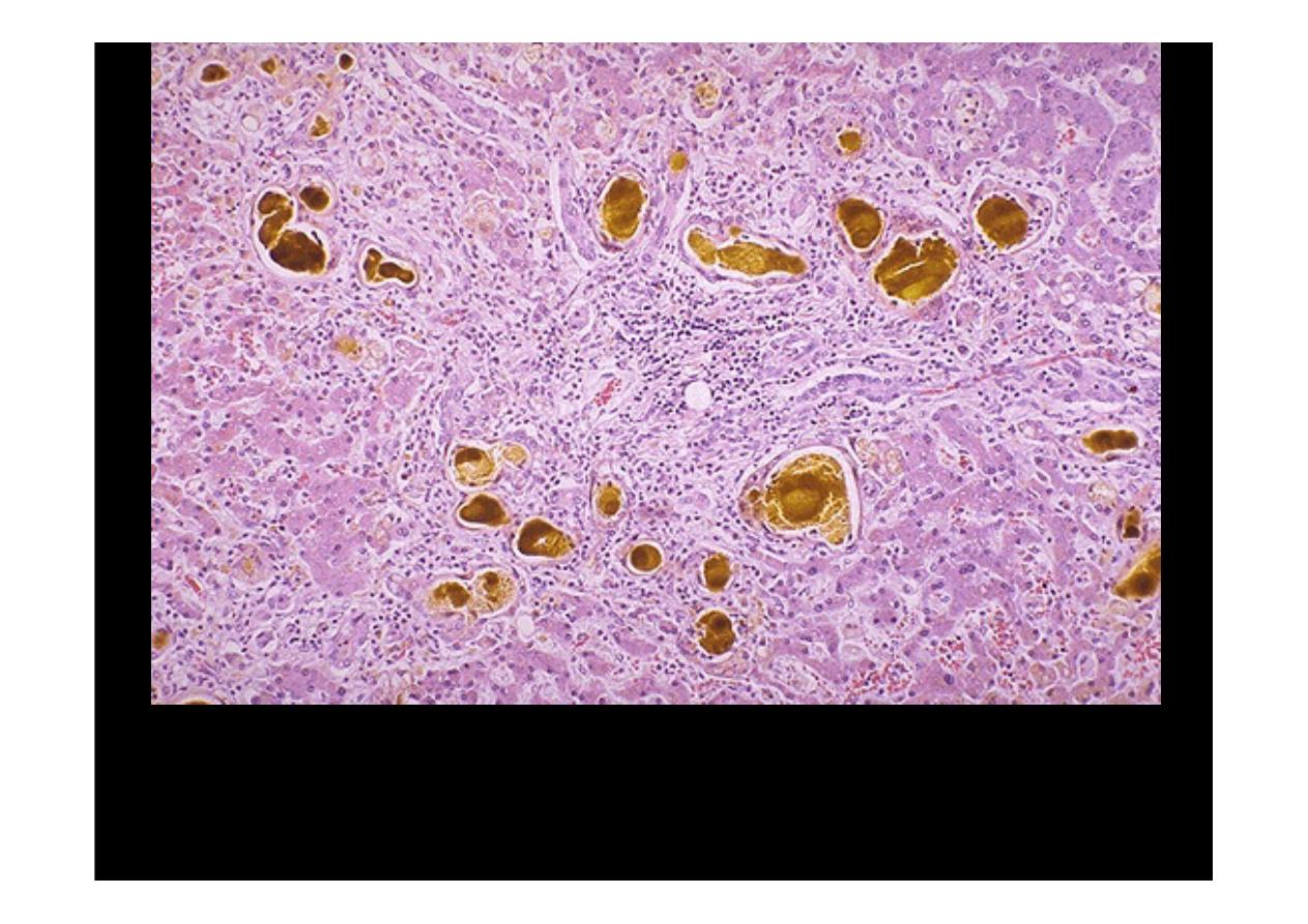
The yellowish-green accumulations of pigment seen here are bile. Most often
this is due to extrahepatic biliary tract obstruction. However, bile may also
accumulate in liver (called
cholestasis
) when there is hepatocyte injury.
Small brown-yellow bile plugs are seen here in canaliculi. The total serum
bilirubin is increased
(the direct bilirubin is greater than indirect )
and the
patient demonstrates jaundice (icterus) by physical examination.
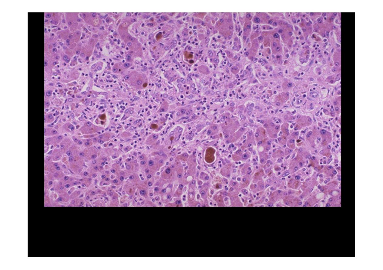
Cholestasis
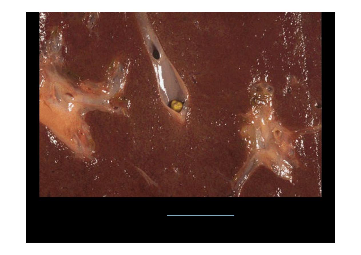
Here is an example of intrahepatic (
Intrahepatic lithiasis
) obstruction with a
small stone in an intrahepatic bile duct. This could produce a localized
cholestasis

Secondary Tumors of
Liver
Gross only
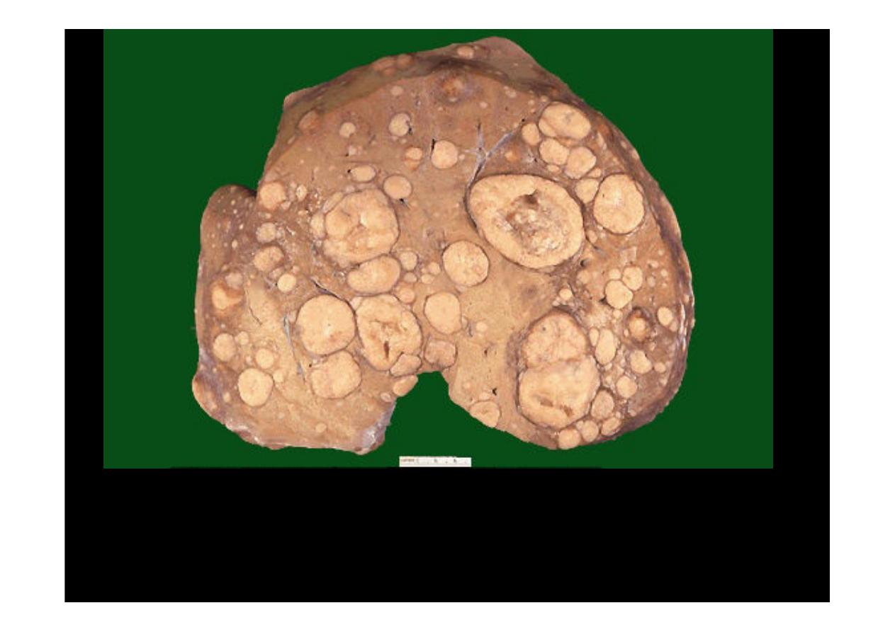
Note the numerous mass lesions that are of variable size. Some of the larger
ones demonstrate central necrosis. The masses are
metastases to the liver
.
The obstruction from such masses generally elevates alkaline phosphatase
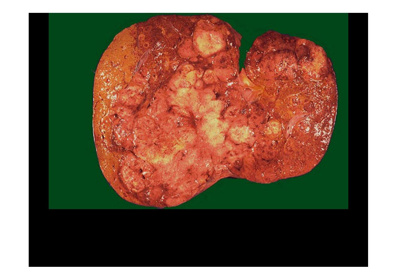
Here is a liver with
metastatic adenocarcinoma
.
Of all neoplasms in the liver,
metastases are the most common
. Here are multiple irregular yellowish masses
distributed haphazardly throughout liver, some show necrosis and destruction of
neighbor liver tissue seen well.
Here are liver metastases from an adenocarcinoma primary
in the colon, numerous mass lesions that are of variable size
scattered throughout the specimen

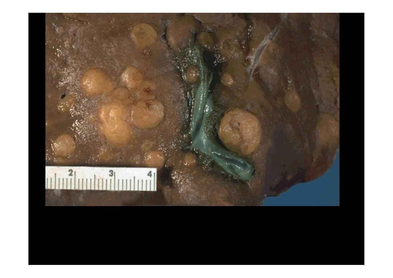
Hepatocellular
Carcinoma
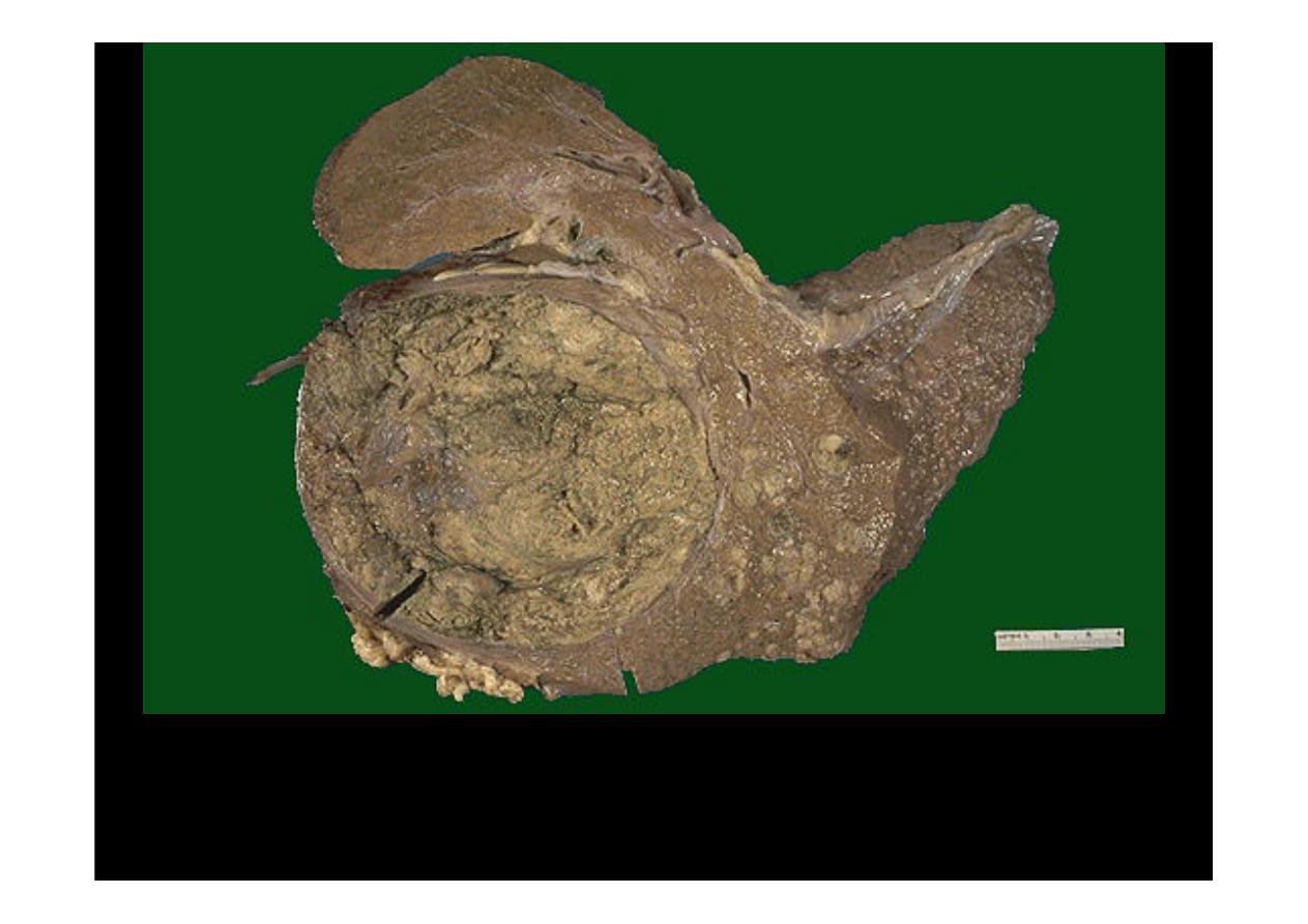
Here is an
hepatocellular carcinoma
. The neoplasm is large and bulky and
has a greenish cast because it contains bile. To the right of the main mass
are smaller satellite nodules. Such liver cancers arise in the setting of
cirrhosis
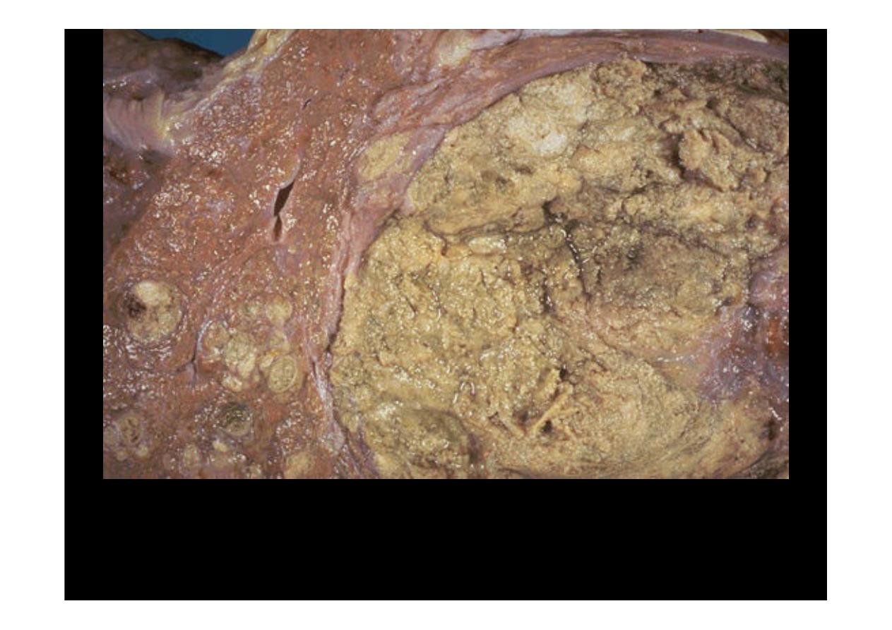
Closer view of previous same tumor, The satellite nodules of this
hepatocellular carcinoma
represent either intrahepatic spread of the tumor
or multicentric origin of the tumor
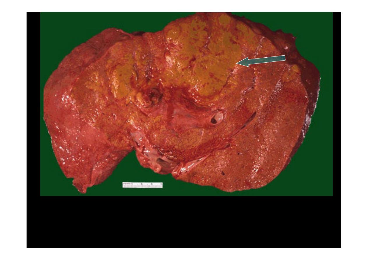
Here is another
hepatocellular carcinoma
with a greenish yellow hue (
arrow
).
One clue to the presence of such a neoplasm is an elevated serum alpha-
fetoprotein. Such masses may also focally obstruct the biliary tract and lead to an
elevated alkaline phosphatase
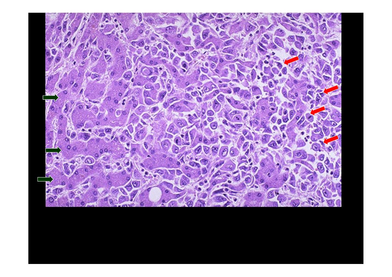
The malignant cells of this
hepatocellular carcinoma
(seen mostly on
the right,
red arrows
) are well differentiated and interdigitate with
normal, larger hepatocytes (seen mostly at the left,
green arrows
)

At the right is an area of necrosis and hemorrhage in this
hepatocellular
carcinoma
. Liver cell carcinomas are very prone to necrosis and
hemorrhage.