
Liver and
Gallbladder Diseases 3
Third Year Class
By Dr.Riyadh A. Ali
Department of Pathology
TUCOM

Articles
• Chronic liver inflammation
• Chronic hepatitis
• Chronic active hepatitis
• Liver congestion
• Chronic cholecystitis
Chronic Liver
Inflammation
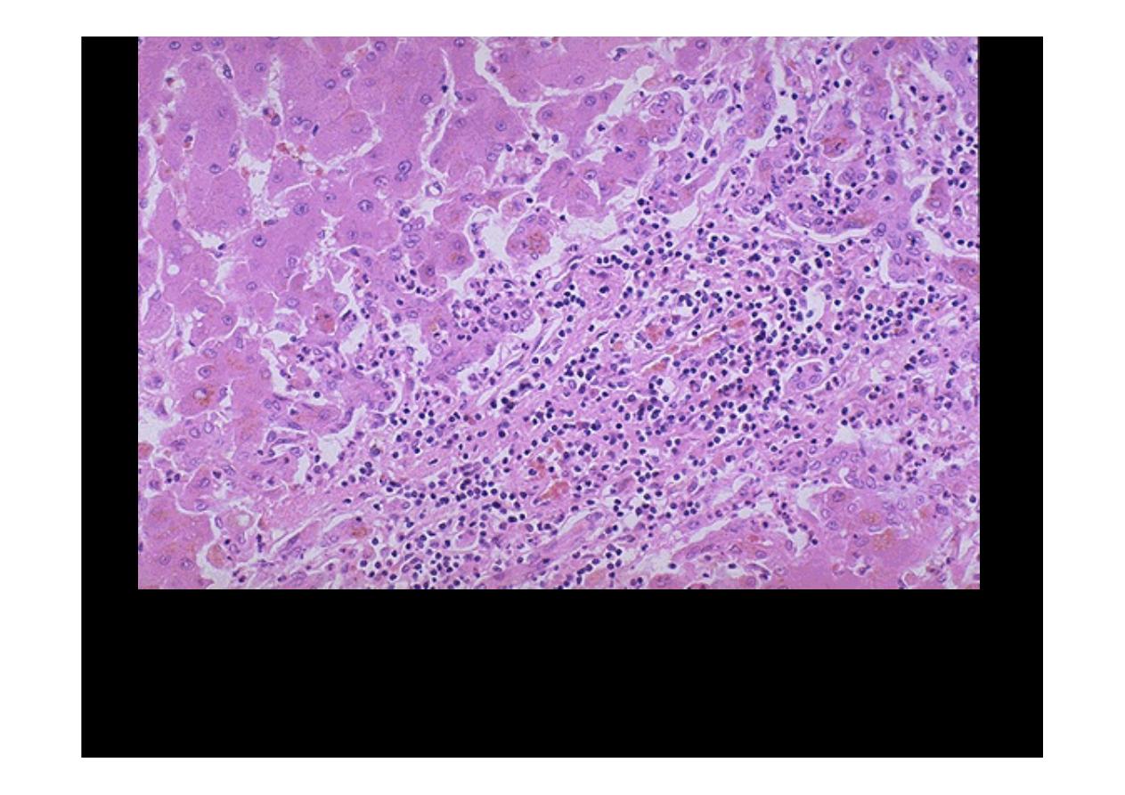
A mononuclear inflammatory cell infiltrate extends from portal areas and
disrupts the limiting plate of hepatocytes which are undergoing necrosis, the
so-called "
piecemeal" necrosis
of
chronic active hepatitis
. In this case, the
hepatitis B surface antigen (HbsAg) and hepatitis B core antibody (HbcAb)
were positive.

Individual hepatocytes are affected by viral hepatitis. Viral hepatitis A rarely leads to
signficant necrosis, but hepatitis B can result in a
fulminant hepatitis with
extensive necrosis
. A large pink cell undergoing "ballooning degeneration" is seen
below the right arrow. At a later stage, a dying hepatocyte is seen shrinking down to
form an eosinophilic "councilman body" below the arrow on the left.
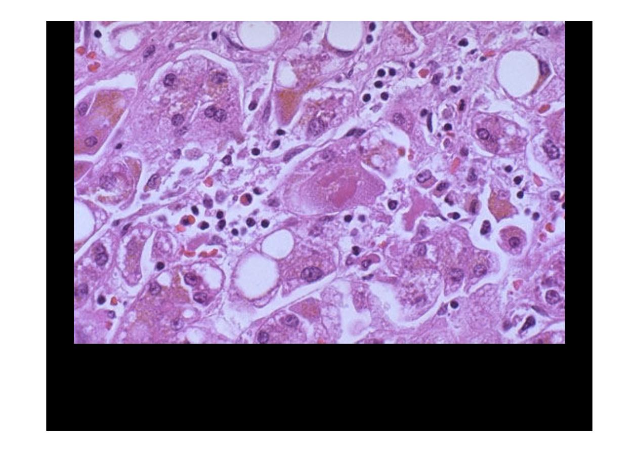
In this case, necrosis and inflammation are prominent.
Chronic hepatitis
This is a case of
viral hepatitis
C which is at a high stage with extensive fibrosis
and progression to macronodular cirrhosis, as evidenced by the large regenerative
nodule at the center right.
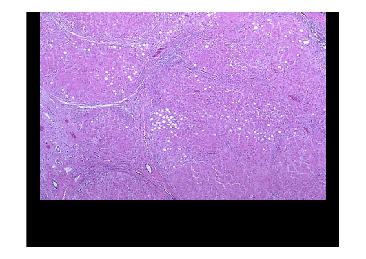
Liver Congestion
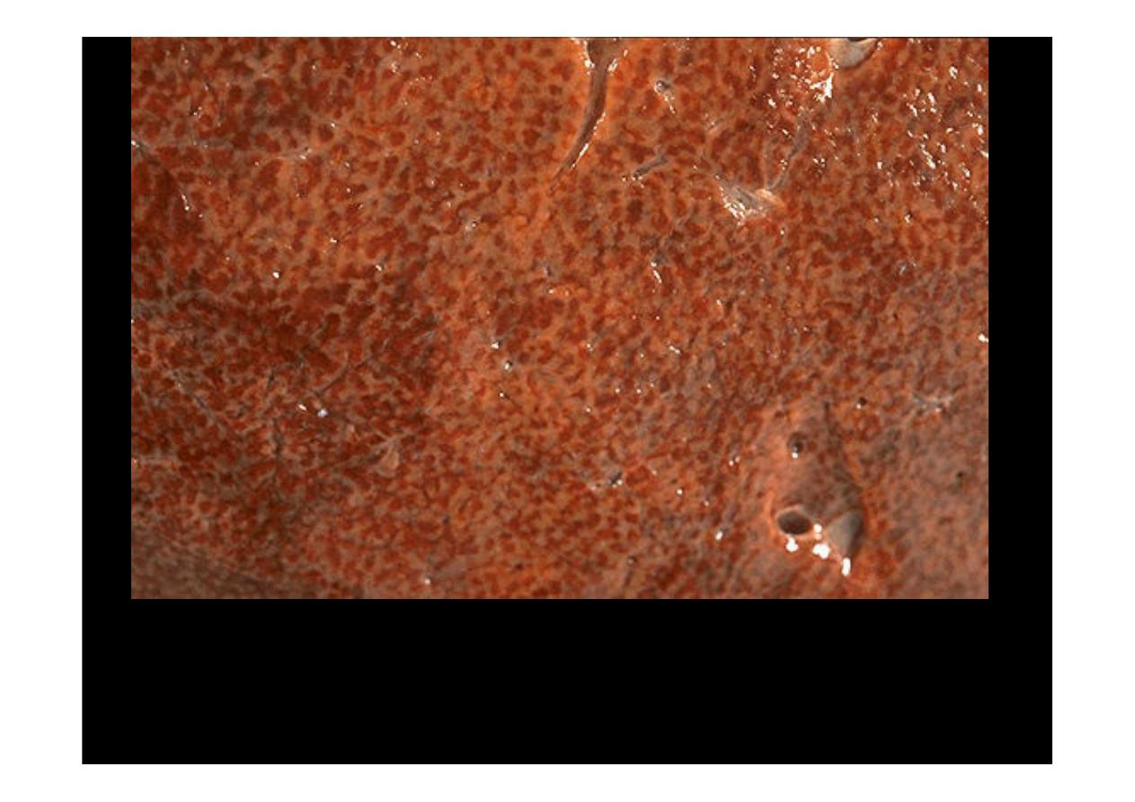
Here is an example of a "
nutmeg
" liver seen with chronic passive
congestion of
the liver
. Note the dark red congested regions that represent accumulation of
RBC's in centrilobular regions

Microscopically, the nutmeg pattern results from congestion around
the central veins, as seen here. This is usually due to a "right sided"
heart failure.
liver congestion
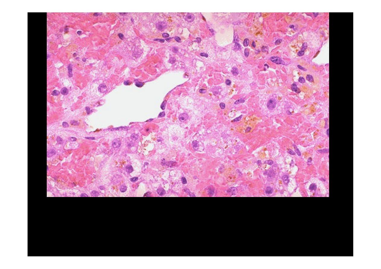
If the passive
congestion
is pronounced, then there can be centrilobular
necrosis, because the oxygenation in zone 3 of the hepatic lobule is not
great. The light brown pigment seen here in the necrotic hepatocytes
around the central vein is lipochrome.
Chronic
Cholecystitis
Chronic
Cholecystitis
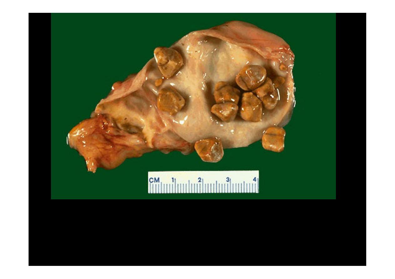
Yellow tan faceted gallstones are present. The gallbladder shows
evidence of
chronic cholecystitis
because the mucosa is tan and the
wall and surface are pale, suggesting collagenization as a result of
scarring with chronic inflammation.
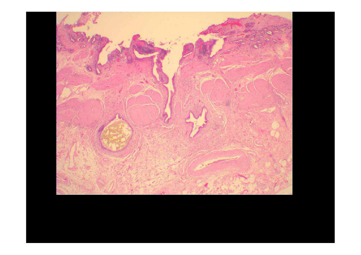
Chronic inflammation gallbladder. Lymphocytes inflammatory cells and
Rokitansky's-Aschoff sinuses usually present and may have associated bile
granulomas.
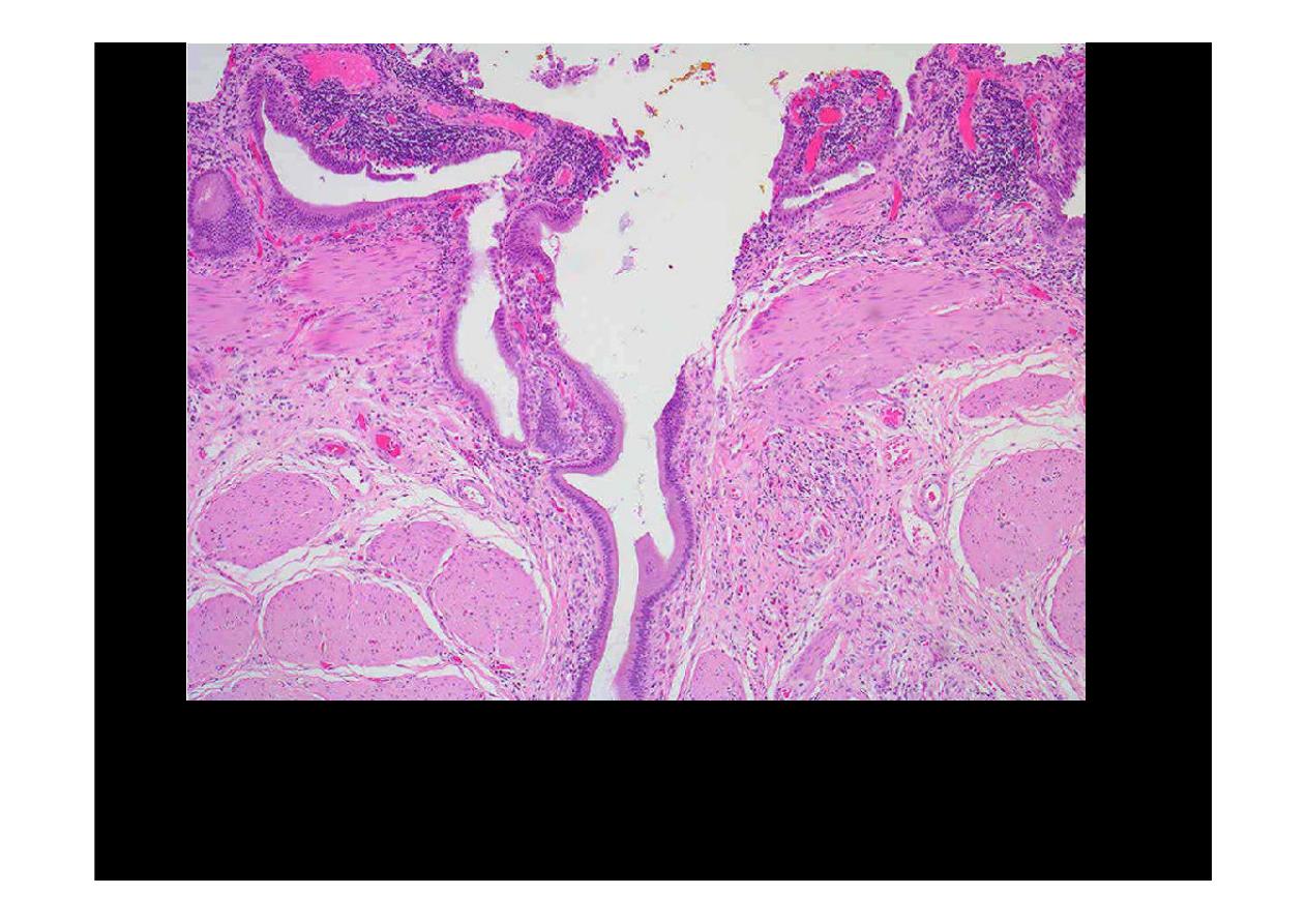
Chronic inflammation with increased lymphocytes and formation
Rokitansky's-Aschoff sinuses (diverticula with increased smooth
muscle, related to chronic increase in lumenal pressure) usually
present.