
Lung Diseases 2
Third Year Class
By Dr.Riyadh A. Ali
Department Of Pathology
TUCOM
Articles
• Pneumonia
• Lung Abscess
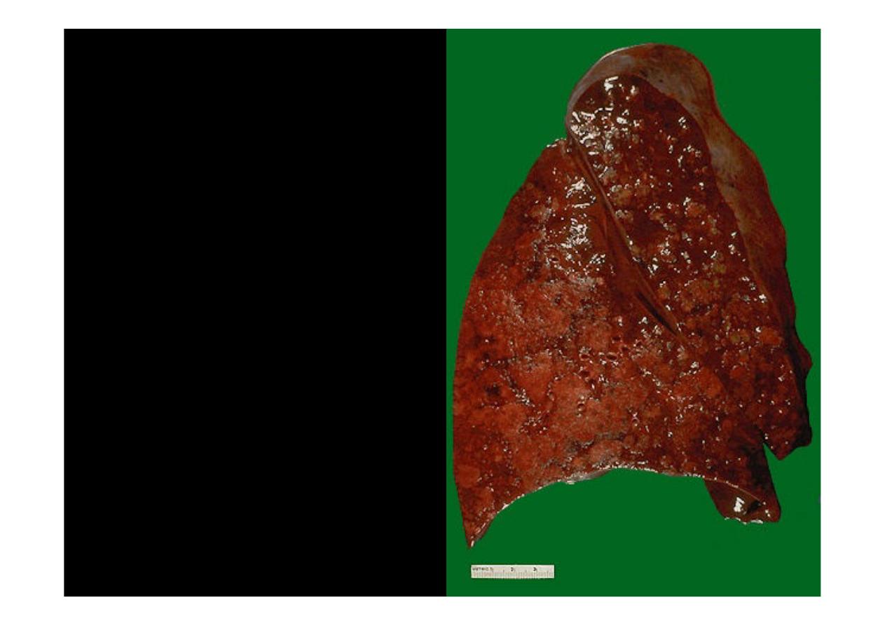
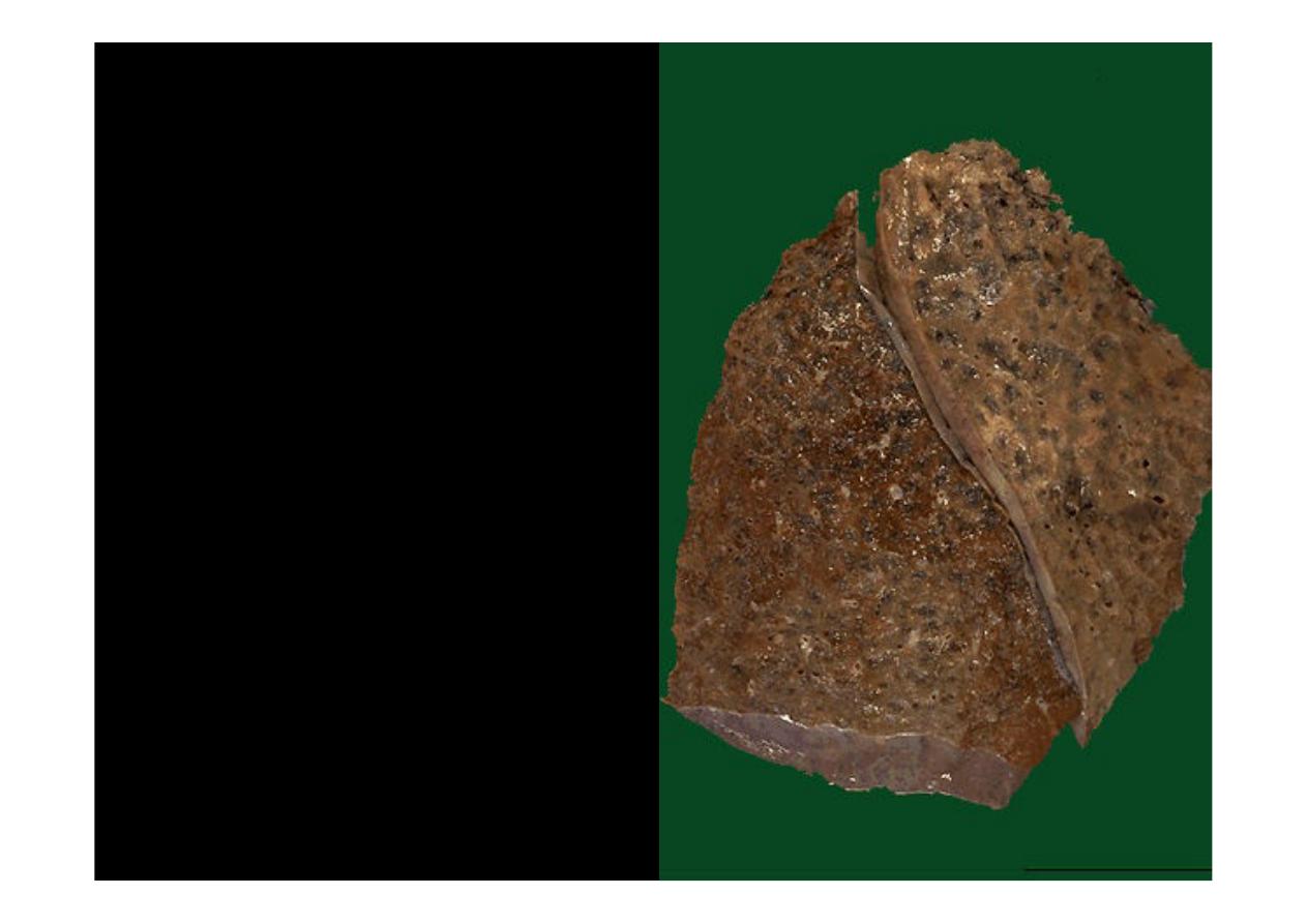
The cut surface of this lung
demonstrates the typical
appearance of a
bronchopneumonia
with areas
of tan-yellow consolidation, that
characterized by patchy areas
of pulmonary consolidation.
These areas become almost
confluent in the left lower lobe
on the bottom left of the
photograph. The areas of
consolidation are firmer than the
surrounding lung. Remaining
lung is dark red because of
marked pulmonary congestion
bronchopneumonia
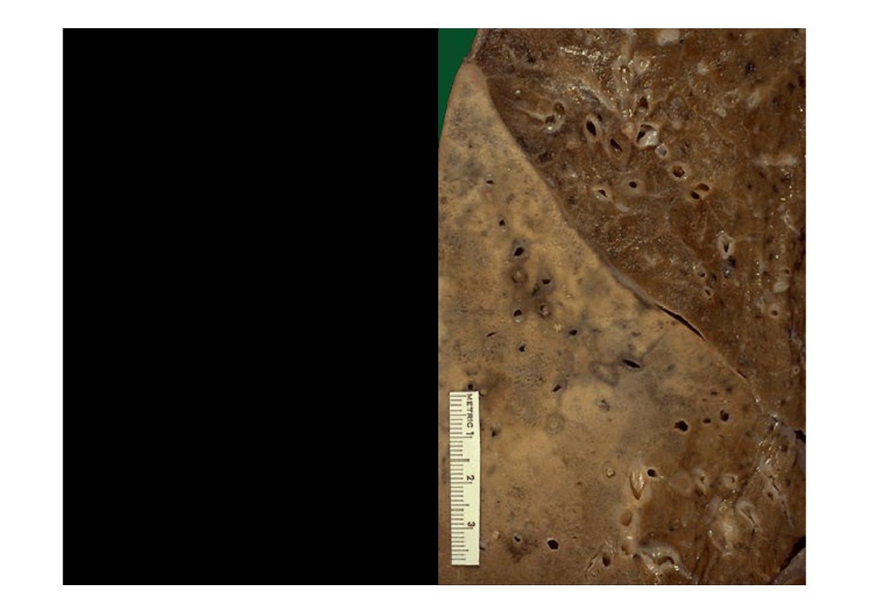
. The
lighter areas that appear to
be raised on cut surface from
the surrounding lung are the
areas of consolidation of the
lung, the pattern of patchy
distribution of a
bronchopneumonia is seen.
The consolidated areas here
very closely match the
pattern of lung lobules
(hence the term "lobular"
pneumonia)
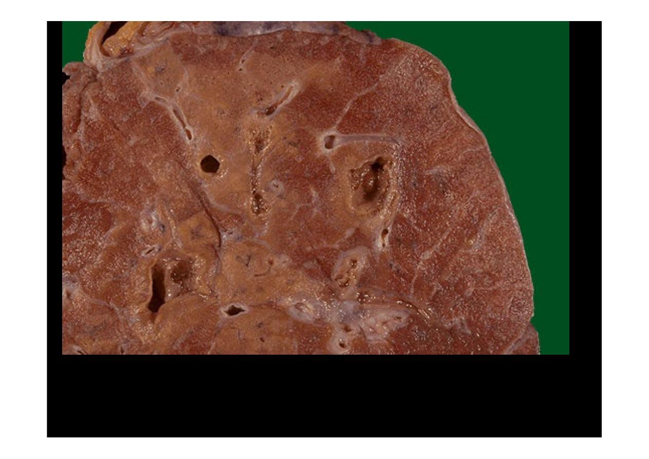
This
bronchopneumonia
is more subtle, but there
are areas of lighter tan
consolidation. The hilum is
seen at the lower right with
radiating pulmonary
arteries and bronchi
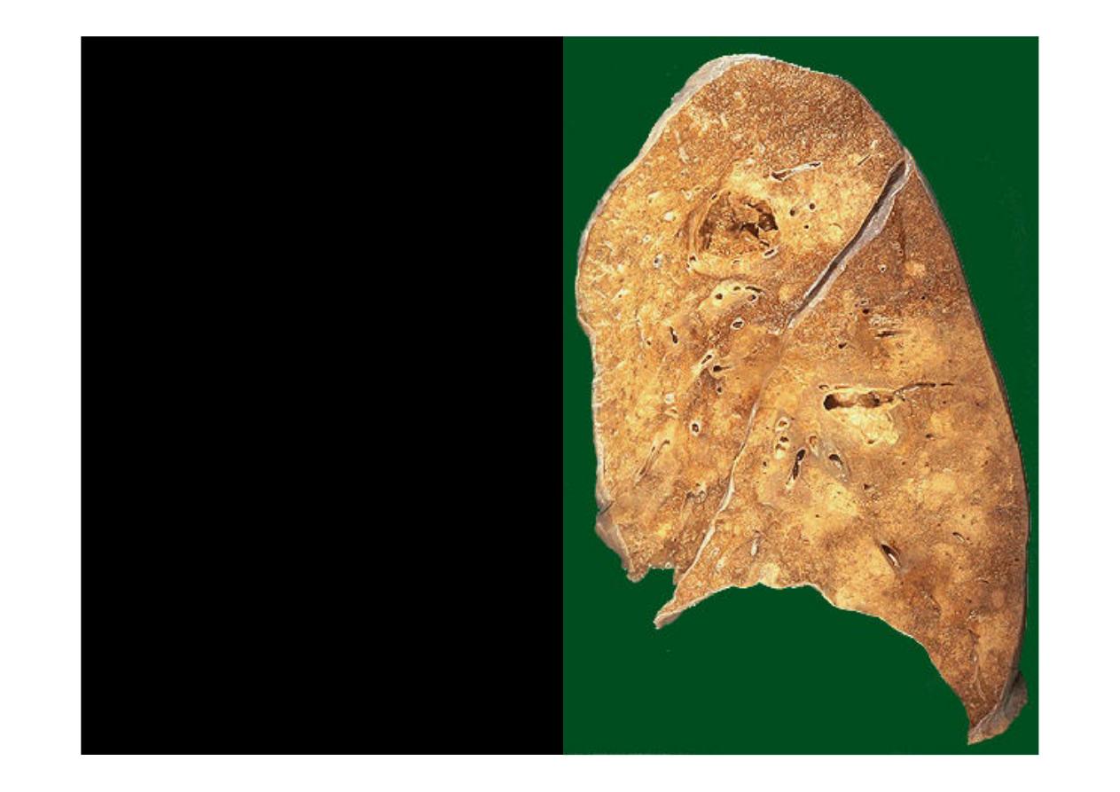
This is a
lobar pneumonia
in
which consolidation of the
entire left upper lobe has
occurred
lobar pneumonia
demonstrates the distinct
difference between the
upper lobe and the
consolidated lower lobe
that appear fainter in color
and harder
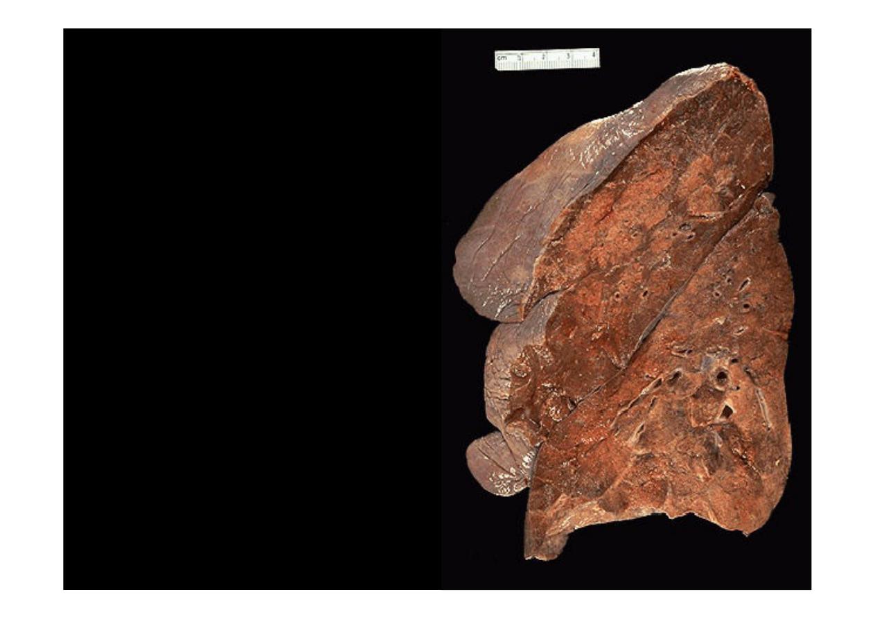
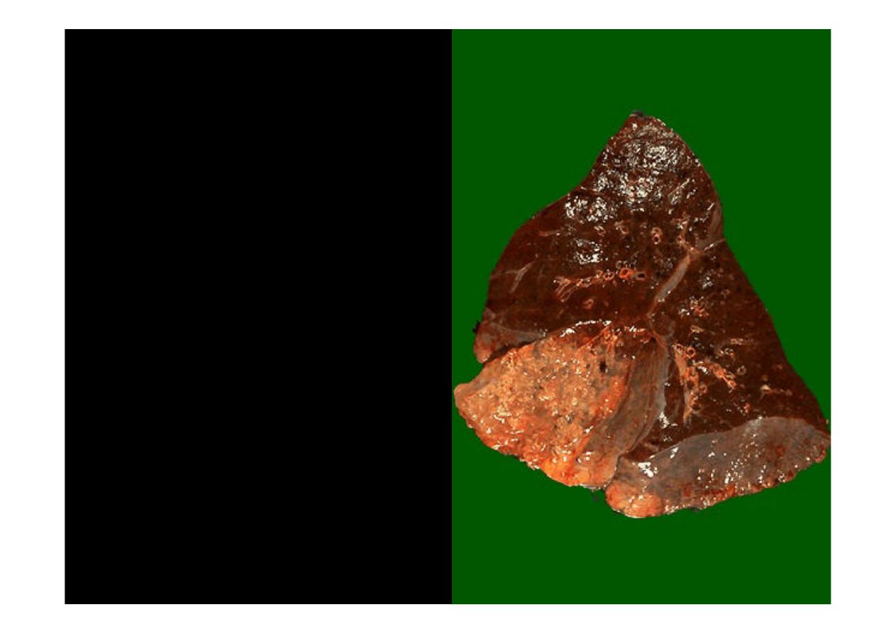
two
lung abscesses
, one
in the upper lobe and one
in the lower lobe of this left
lung. An abscess is a
complication of severe
pneumonia, most typically
from virulent organisms
such as S. aureus
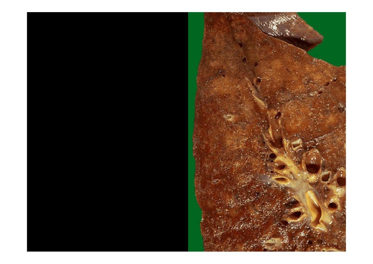
This is an
abscessing bronchopneumonia
in which several abscesses with
irregular, rough-surfaced walls are seen within areas of tan consolidation
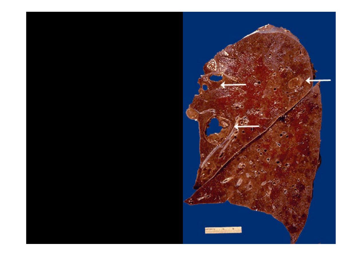
The white arrows mark areas of
abscess
formation in the upper
lobe of this lung. The liquefactive
necrosis of an abscess is apparent,
because the purulent contents are
draining out to leave a cavity. On
a chest radiograph, the liquefied
central contents of an abscess can
appear as an "air-fluid level".
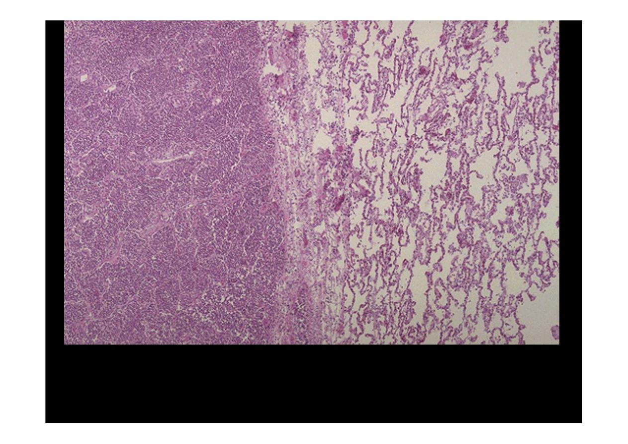
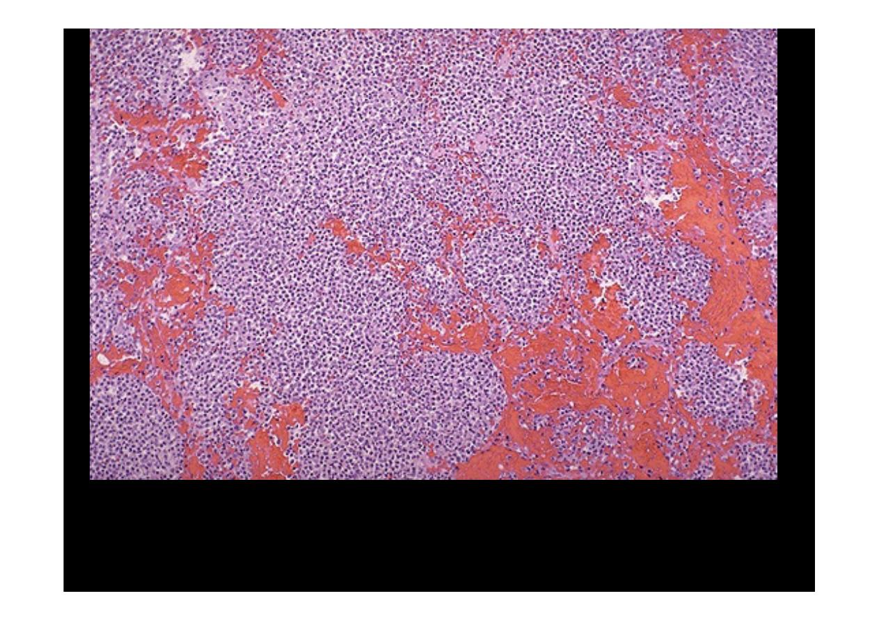
At the left the alveoli are filled with a neutrophilic exudate that corresponds
to the areas of consolidation seen grossly with the
bronchopneumonia
At higher magnification can be seen a patchy area of alveoli that are filled
with inflammatory cells. The alveolar structure is still maintained.
pneumonia
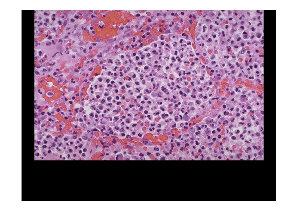
At high magnification, the alveolar exudate of mainly neutrophils is seen. The
surrounding alveolar walls have capillaries that are dilated and filled with RBC's.
Such an exudative process is typical for bacterial infection. This exudate gives rise
to the productive cough of purulent yellow sputum seen with
bacterial pneumonias
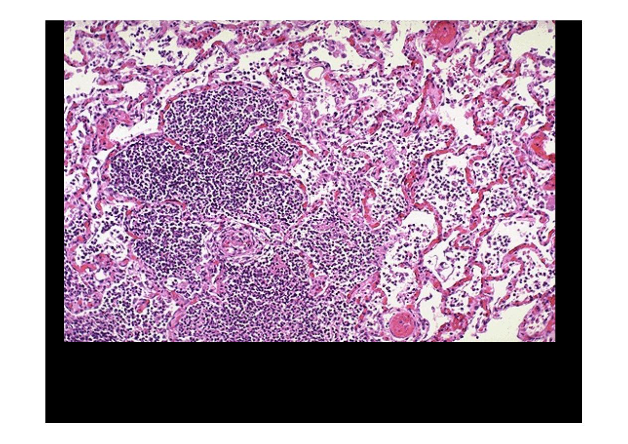
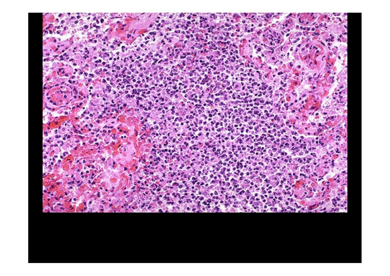
More virulent bacteria and/or more severe
pneumonias
can be associated
with destruction of lung tissue and hemorrhage. Here, alveolar walls are no
longer visible because there is early abscess formation. There is also
hemorrhage.
At higher magnification, early
abscessing pneumonia
is shown. Alveolar walls
are not clearly seen, only sheets of neutrophils
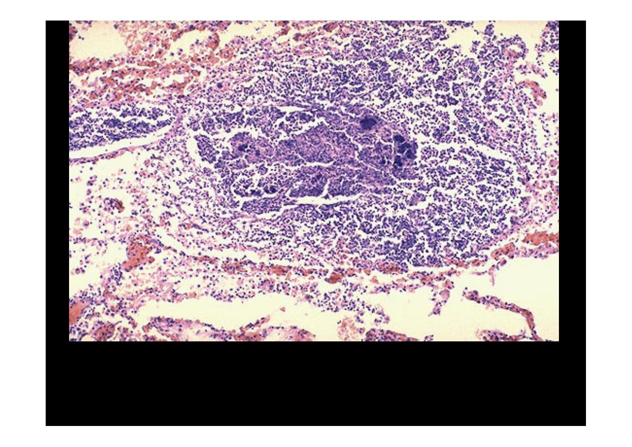
This more focal
abscess
containing a neutrophilic exudate as well as dark
blue bacterial colonies suggests aspiration or hematogenous spread of
infection to the lung
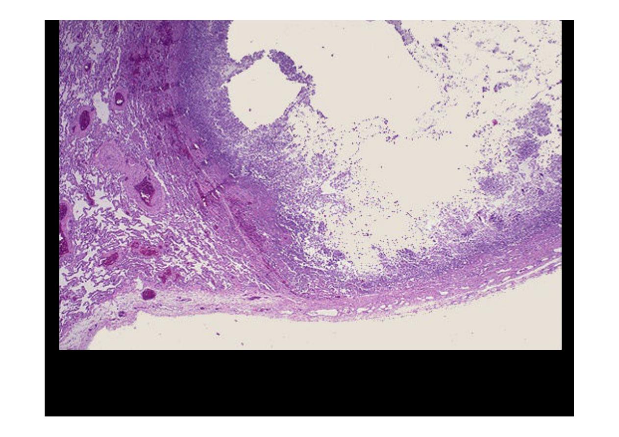
At low magnification, a
lung abscess
is seen on the right bordered by a thin
wall of organizing granulation tissue. The center of the abscess contains
neutrophils and necrotic debris. Often, the contents are liquid,
Almost the entire
middle lobe of this
right lung is involved
by a
chronic abscess
as seen here on
section. The area of
abscess is yellow tan,
and it was very firm