By
Dr.Amer salih

Diseases of nasal septum
Nasal septal diseases can be categorized as congenital
and acquired . the later one is significantly more
common than the former.
Anatomy of nasal septum.
The
nasal
septum
composed
from
columella
,
membranous between columella and anterior part of
cartilaginous septum , cartilaginous part and finally the
bony part .

CONGENITAL
LESIONS
OF
NASAL SEPTUM
1- Sincipital encephalocele. Brain tissue that is
trapped in the nose during embryologic development
usually result in either a type of sincipital encephalocele
or nasal glioma . sincipital encephalocele has a direct
connection to the intracranial cavity so this connection
must be obliterated to avoid potential future meningitis
and abcess formation.
2- Nasal glioma . It is also consist of disorganized
trapped brain tissue but unlike encephalocele don’t has
connection with intracranial cavity and unlike the
intracranial glioma don’t has neoplastic potential.

3-
Teratomas,dermoids
and
epidermoids
.
Incomplete regression of the embryonic frontonasal
diverticula with trapping of nonglial tissue may result in
the formation of teratomas , dermods or epidermoids.
Teratomas contain ectoderm, mesoderm and endoderm ,
whereas
epidermoid and dermoid are composed
exclusively from ectoderm.
4- Choanal atresia and stenosis. Choanal atresia
occurs in one of 8000 live births and is commonly
associated with syndromes and systemic anomalies.it
divides in to bony and membranous types , usually the
bony type is more common the membranous type.

Diagnosis;
1- clinical examination. This is done by nasal
examination by anterior rhinoscopy and nasal
endoscopy.
2- CT scan and MRI.
Treatment.
Intranasal resection of the lesion or by endoscopic
surgery.in case of choanal atresia , the membranous
type can be treated by forceful introduction of
nasogastric tube , while the bony type treated by
surgical drilling and widening of bone closing the
posterior choana.

Acquired lesions
1- Wegener Granulomatosis:- It is
a type of
granulomatous lesion affecting the upper and lower
respiratory tract . The Advanced type Wegener
granulomatosis may lead to destructive lesions of the
hard palate , sinonasal-oral fistuala or complete nasal
septal destruction . Usually treated by surgical
resection
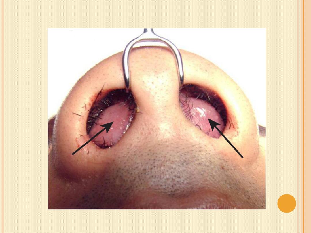
2-
Septal
haematoma.
It
is
occur
due
to
subperichondrial blood collection in the nasal septum.,
due to nasal trauma ,more common in children
,presented with bilateral nasal swelling.

Causes
1. nasal trauma. Due to a fracture or dislocation of the
cartilaginous septum, they mostly occur in the anterior
septum.
2. septal surgery.
3. blood disorders.
Clinical features
usually the patient presented with bilateral nasal
obstruction.
The hematoma appears as cystic swelling bluish red in
color protrudes into the vestibules and completely
obstructs the nasal entrance on both sides.

Tapping revealed blood, early diagnosis is important.
delay diagnosis and treatment lead to
abscess
formation and septal cartilage necrosis.
Treatment;
Urgent surgical drainage within 24 to72 hours and
antibiotics.

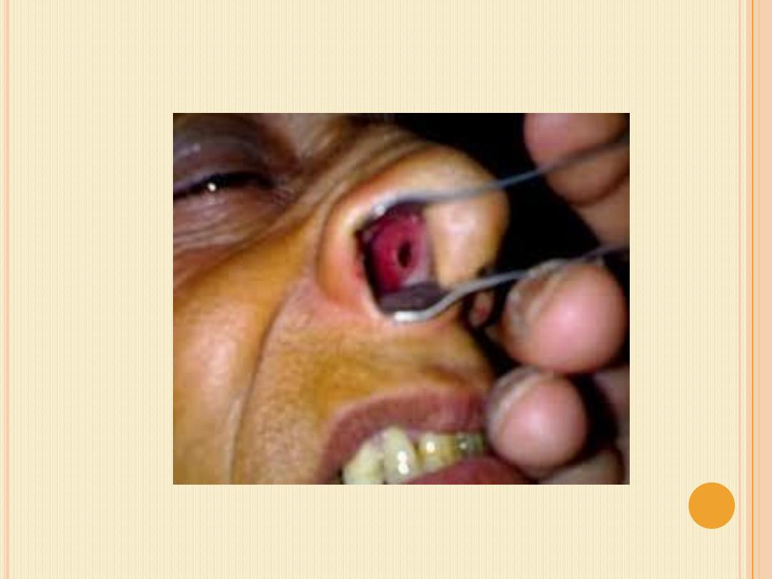
3- Septal abscess . Subperichondrial or subperiosteal
Collection of pus in the nasal septum.
Etiology
1. It follows delay drainage of septal haematoma 3-5 days.
2. Nasal infection( vestibulitis).
3. Acute ethmoiditis and sphenoiditis
Clinical features .
There are fever, nasal obstruction and throbbing pain over
bridge of the nose and tenderness. Aspiration reveal pus.
Treatment :
Drainage
of abscess and immediate reconstruction of the
septum with good antibiotics cover .

Complications ;
1- Saddle nose.
2- Septal perforation.
3- Cavernous sinus thrombosis.
4- Maxillary hypoplasia.
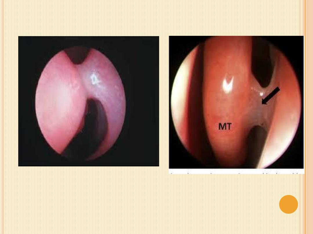
4.Septal perforation. It a hole or fissure in the
cartilaginous part of nasal septum.

Etiology
1. Trauma; most common cause like
*septal cautery for epistaxis.
*nose picking and piercing
*septal surgery
. * complication of septal abscess
2. local use of cocaine.
3. Infection . TB. and syphilis cause posterior bony
perforation.
4.
malignancies
melanoma
,
adenocarcinoma
,
sequamous cell carcinoma.
5. Inflammatory ; sarcoidosis and wegener's granuloma,

Clinical features;
1- whisling noise.
2- crustation and bloody discharge.
3- difficulty breathing.
4- nasal pressure and discomfort
Treatment.
*majority are asymptomatic and require no specific
treatment.
*symptomatic perforation
Non-surgical treatment ; includes
* nasal douches alkaline ,normal saline.
*Ointment.
*Obturator (Button )
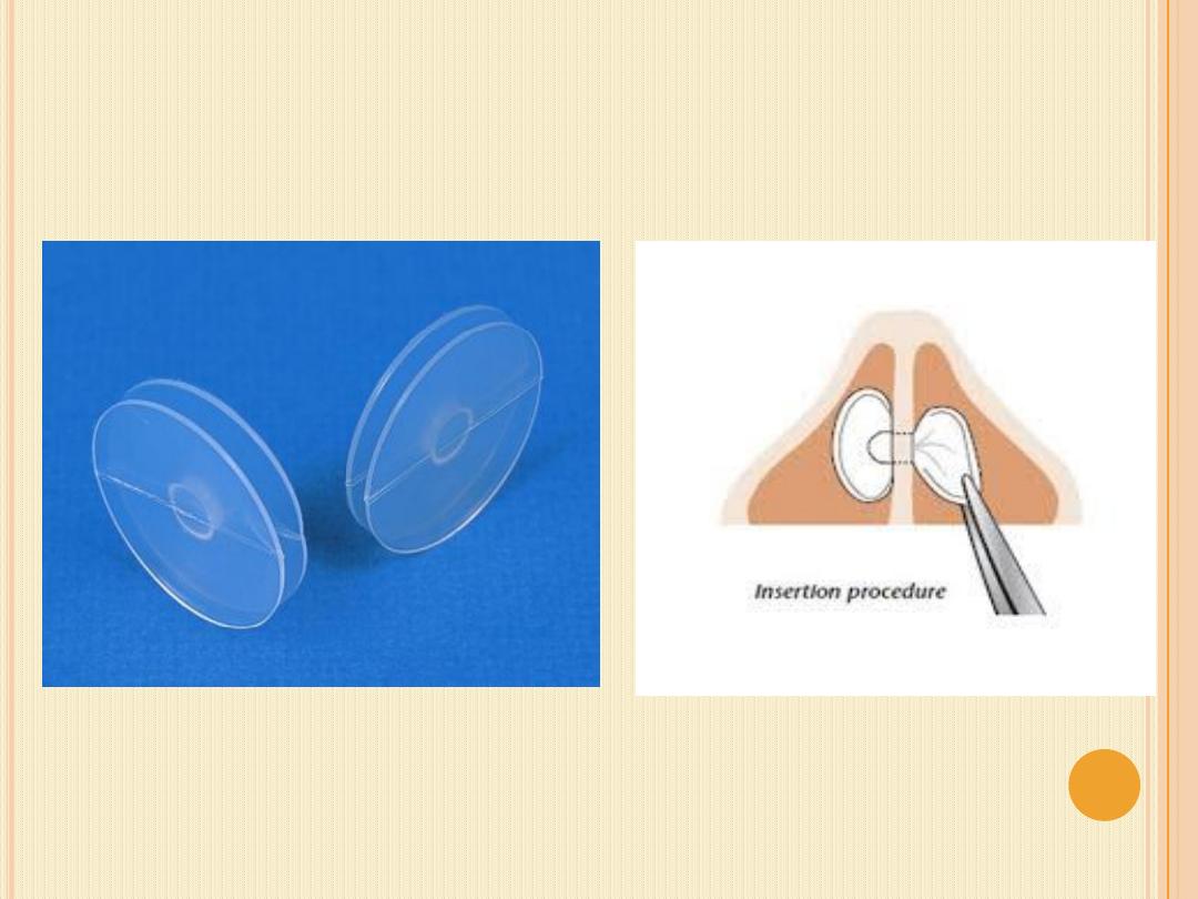

*Surgical treatment ; for small to medium hole can be closed
by surgery.
*local nasal flap buccal mucosal,;*free graft composite
autograft.
5-Intranasal adhesion(spike).
this is
occur between turbinate with septum or turbinate with
lateral wall
Causes;
1. Nasal surgery ( septoplasty , polypectomy , FESS).
2. Nasal packing
3. fracture nose.
4. Forign body.
Treatment
. remove of synechia , and Insertion of
splint to prevent recurrence.

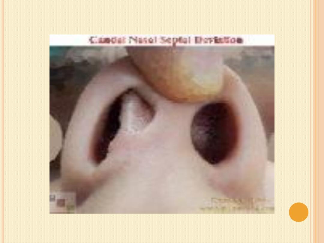
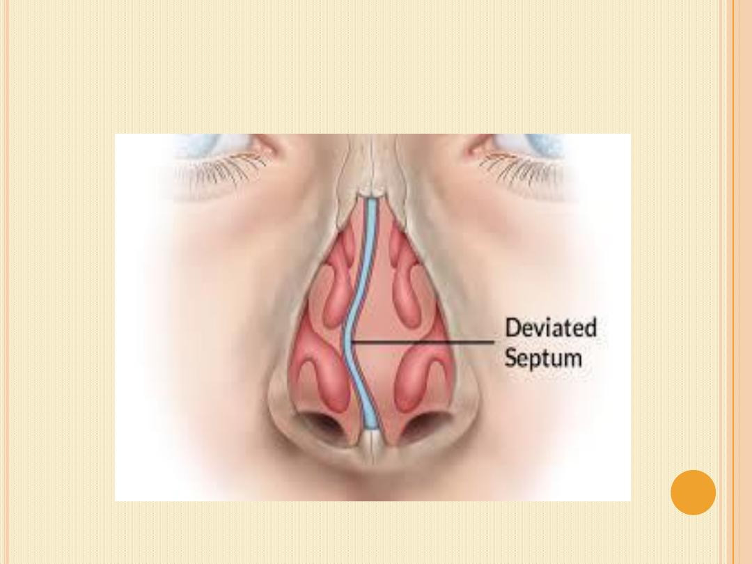
6-Septal deviation .
It is physical disorder of
the nose involving displacement of nasal septum . Some
displacement is common , affecting 80% of people , mosly
without their knowledge.
Causes;
1- Impact trauma , such as by blow to the face.
2- Congenital.Like abnormal posture intrauterine or
compression of the nose during childbirth.
3- Genetic connective tissue disorders like marfan
syndrome and Ehlers-Danlos syndrome.
4- Secondery to compression by mass like nasal polyp
and tumor.
5- Racial .like in Caucasian.

Clinical features.
1- Nasal obstruction . may be unilateral or bilateral
some the sever form associated with sleep apnea.
2- Headache and facial pain . It is due to sinusitis or
compression on the nerve cause sphenopalatine
neuralgia and anterior ethomoid neuralgia.
3- Anosmia.
4- Epistaxis.

Diagnosis;
1- History.
2- Examination. By anterior rhinoscopy and endoscopy
that show the following types
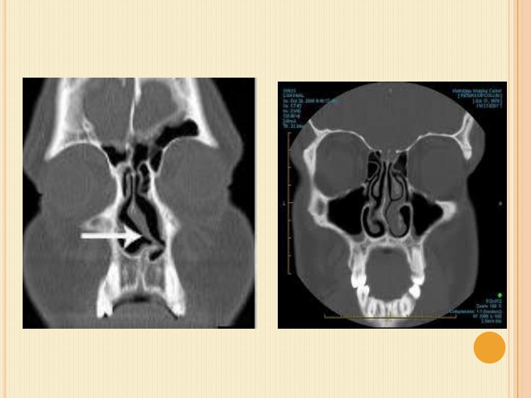
1- C-shape
2- S-shape
3- Caudal dislocation.
4- Spur.
3- Imaging . By x-ray and Ct scan to show the deviation
and complications

Treatment.
1- Asymptomatic need no treatment.
2- Symptomatic.
1- Medical treatment for sinusitis and
partial
relieve of obstruction
2- septoplasty by straitening the deviated part and
remove the severely deviated bone and cartilage.
3- Conservative septoplasty . This is done in children
to
prevent
future
complication
like
maxillary
hypoplasia.

Complications of septoplasty.
1- Septal perforation.
2- Septal abscess and septal hematoma.
3- Scarring and crustation inside the nose.
4- Adhesion between the septum and lateral nasal wall.
5- Saddle nose and dropped nasal tip.
6- Incomplete correction and persistence of nasal
symptoms.
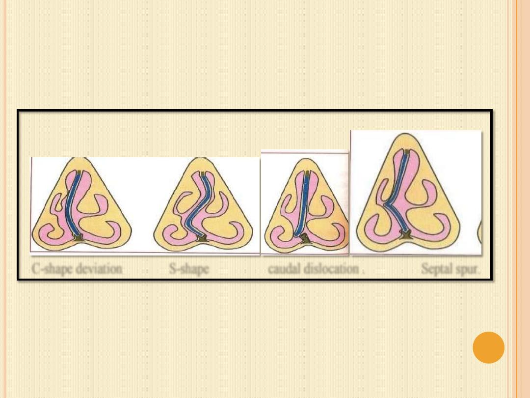
C-shape deviation S-shape caudal dislocation . Septal spur.