
Dental X-ray Image Characteristics
VISUAL CHARACTERISTICS

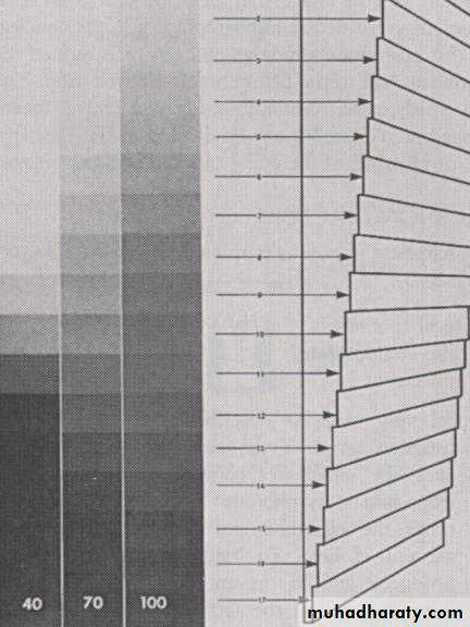
DENSITY
:
The over all degree of blackness or darkness of an dental radiography also density refers to the degree of optical transmission of light rays through a processed film.
When dental radiograph is viewed against a light source, the relative transparency of areas on the radiograph depends on the distribution of black silver particles in the emulsion.
Dark area represent heavier deposit of black silver particles.
INFLUENSING FACTORS
1- Exposure Factors Include:Millamperage: An increase in millamperage produce more x-ray that expose the film, if the millamperage is increased, the film density increased and the radiograph appears darker.
Operating kilovoltage peak: Increasing the kVp increase the density by increasing the mean energy of the x-ray beam and producing higher energy x-ray, if the kVp is increase the film density increase and the film appears darker.
Exposure time : An increase in exposure time increase film density by increasing the total number of x-ray that reach the film surface, if exposure time is increased, more x-ray reach the film and film density increase and film appears darker.
2- Patient Parameters Include:
Subject thickness: Larger patients with greater tissue thickness will require larger exposure time or increased mA setting.Object density: Object density affects film density, denser object such as bone and enamel will attenuate more x-ray in the x-ray beam that they let fewer x-ray pass though metallic filling to strike the film and thus interact with silver halide, consequently those area will be radiopaque or white on the film.
3- Source–Film Distance: The density of a film also depends on the amount of radiation reaching the film or the distance of the film from the source to the film. The inverse square law state that the x-ray beam varies inversely with the square of distance from the source. Thus we change the length from 8 inch to 16 inch the intensity of x-ray beam reach the film would be 1\4 and this affect density and must be compensate by increasing exposure time.
4- Development Condition: Film density may also increase by prolonging the development time during processing or increase the temperature of developer.
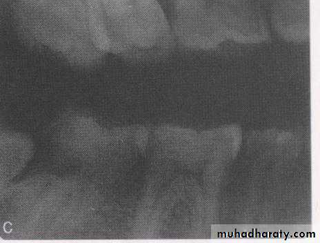
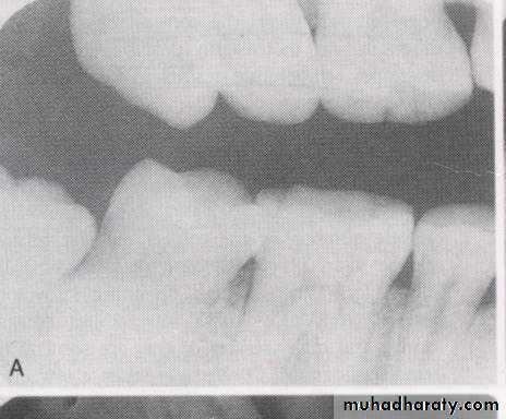
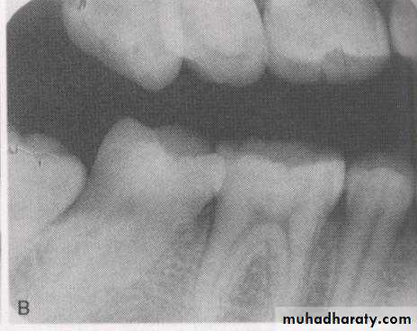
CONTRAST
Is the difference in the degree of blackness between adjacent area on dental radiograph. Densities on the radiograph are not simply black and white but encompass multiple shade of gray called scale of contrast.
High contrast image (short scale contrast) would be black and white with few gray shades.
Low contrast image (long scale contrast) usually contain a wide range of shades of gray .INFLUENCING FACTORS
Only one exposure factors has direct influencing on the contrast of radiograph.
Operating Kilo Voltage Peak: Increasing the kilovolt affects film contrast increasing the mean or average energy of x-ray and by producing higher energy x-rays, x-ray with higher energy are better able to penetrate tissue as a result more variation in tissue density are record on the film and appears as a varying shade of gray. A higher kilo voltage produce a film with decreased or low contrast and radiograph exhibit many shade of gray..
GEOMETRIC CHARACTERISTICS
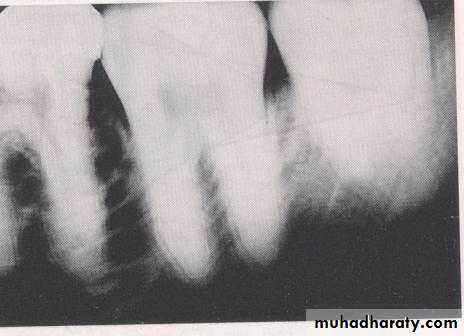
SHARAPNESS: Sharpness also known as detail, resolution or definition refers to the capability of the x-ray film to reproduce the distinct outlines of an object or in the other word to how well the smallest details of an object are reproduced on a dental radiograph.INFLUENCING FACTORS
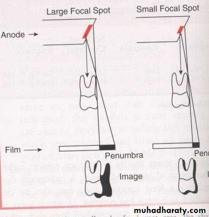
Focal Spot Size: The smaller the focal spot area the sharper the image appears, the larger the focal spot area the greater the loss of image sharpness.
Film Composition: The composition of the film emulsion influence sharpness. Sharpness is relative to the size of the crystals found in the film emulsion, the emulsion of faster film contains larger crystals that produce less image sharpness, where as slower film contains smaller crystals that produce more image sharpness. Unsharpness occurs because the large crystal do not produce object out lines as small crystal.
Movement
: Movement influence film sharpness, loss of image sharpness occurs if either the film or patient moves during x-ray exposure.
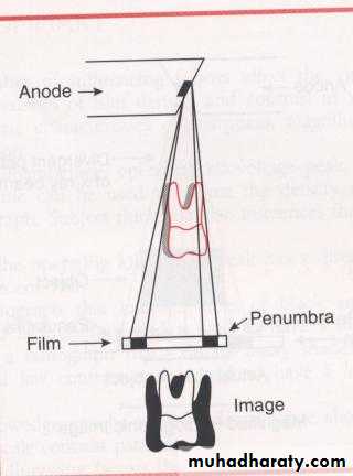
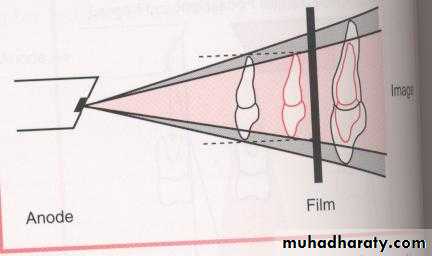
MAGNIFICATION: Image magnification refers to the radiographic image that appears larger than the actual size of the object. Magnification results from divergent path of x-ray beam.
INFLUNCING FACTORS
Target–Film Distance
: Also known as source to film distance is the distance between the x-rays (focal spot of tungsten target) and the film. The target film distance is determined by length of the position indicating device, when a longer PID is used more parallel rays from the middle of x-ray beam strike the object rather than the diverging x-rays from the periphery of the beam, as a result a longer PID and target film distance result in less image magnification .
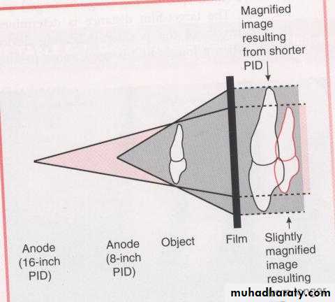
Object–Film Distance : Object film distance is the distance between the object being radiographed and dental x-ray film, the teeth and film should be as closed as possible, the closer the proximity of the tooth to the film, the less image enlargement will be on the film.
DIASTORTION: dimensional distortion of radiographic image is a variation in the true size and shape of the object being radiographed. A distorted image dose not have the same size and shape as the object being radiographed. Distortion result from the unequal magnification of different parts of the same object. Distortion results from improper film alignment or angulations of x-ray beam .
INFLUENCING FACTORS
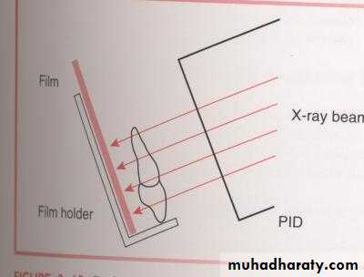
Object–Film Alignment: In order to minimize dimensional distortion the object and film must be parallel to each other if the object and film are not parallel an angular relation ship result. An angular relation ship produce a variation of distances between the teeth and film that result in distorted image.
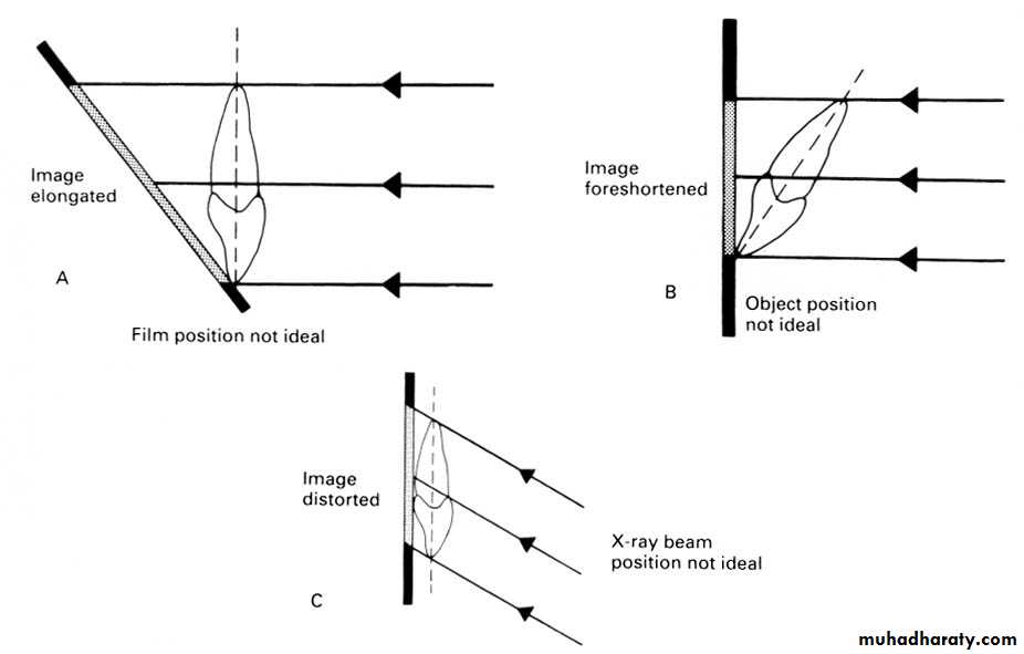
X-ray Beam Angulations: To minimize dimensional distortion the x-ray beam must be direct perpendicular to the tooth and the film in order to recorded the adjacent structures in their true spatial relationships.
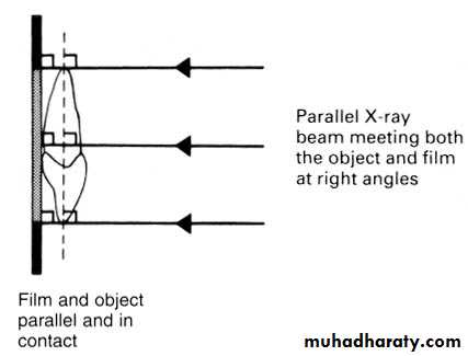
Ray Beam Characteristics
The ideal X-ray beam used for imaging should be:• Sufficiently penetrating, to pass through the patient and react with the film emulsion and produce good contrast between the different shadows .
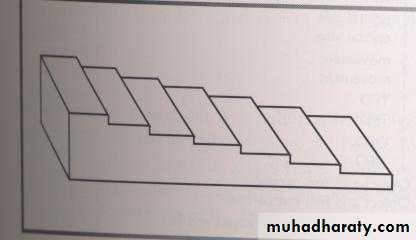
• Parallel, i.e. non-diverging, to prevent magnification of the image.
• Produced from a point source, to reduce blurring of the edges of the image, a phenomenon known as the penumbra effect