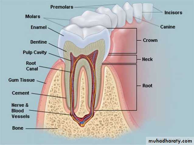
NORMAL TOOTH ANATOMY
ENAMEL: Enamel is the most radiopaque of any tissue seen radiographically and cover the coronal portion of the tooth.

DENTIN: Dentin is found beneath the enamel layer of a tooth and surrounds the pulp cavity, the dentin appears radiopaque, but lighter than enamel.

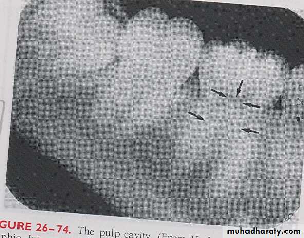
PULP CAVITY: The pulp cavity consist of a pulp chamber and pulp canals, its contain blood vessels, nerve and lymphatic and appears relatively radiolucent on dental radiograph.

LAMINA DURA: Laminadura is the wall of the tooth socket that surrounds the roots of tooth, on periapical radiograph the laminadura appears as dense radiopaque line that surround the root of a tooth.

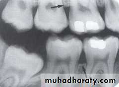
ALVEOLAR CREST: The alveolar crest is the most coronal portion of alveolar bone found between teeth. On dental radiograph the alveolar crest appears radiopaque and is typically located 1.5-2mm below the cementoenamel junction

PERIODENTAL LIGAMENT SPACE: The Periodontal ligament space is the space between the roots of the tooth and laminadura, the pdl space contain connective tissue fibers, blood vessels and lymphatic. on dental radiograph the pdl space appears as a thin radiolucent line around the root of a tooth.
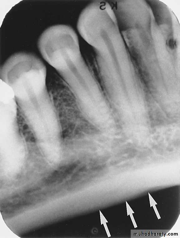
Cortical bone, also referred to as compact bone, is the dense outer layer of bone. Cortical bone resists the passage of the x-ray beam and appears radiopaque on a dental image. The inferior border of the mandible is composed of cortical bone and appears radiopaque
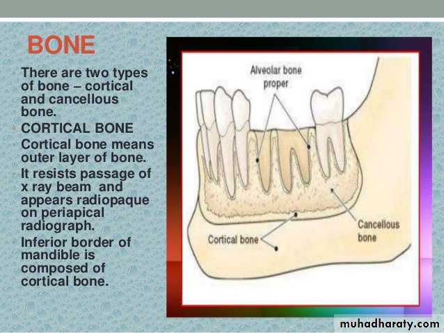
CANCEllOUS
BONE
The cancellous bone (also called trabecular bone or spongiosa) lies between the cortical plates in both jaws. It is composed of thin radiopaque plates and rods (trabeculae) surrounding many small radiolucent pockets of marrow.
BONY LANDMARKS OF MAXILLA
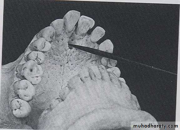
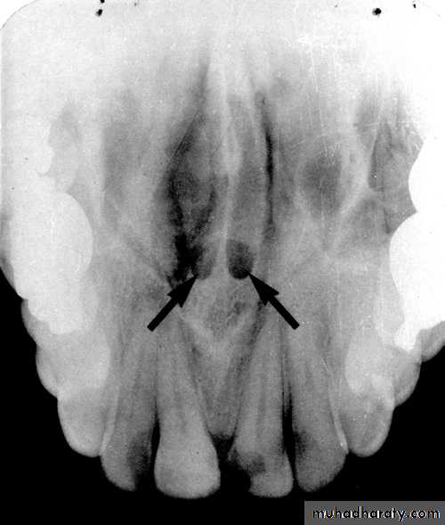
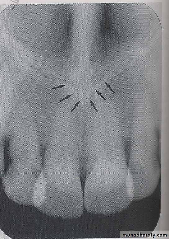
INCISIVE FORAMEN: The incisive foramen is an opening in the bone that is located at the midline of the anterior portion of hard palate. Radiographically the incisive foramen appears as a small ovoid or round radiolucent area located between root of maxillary central incisor.

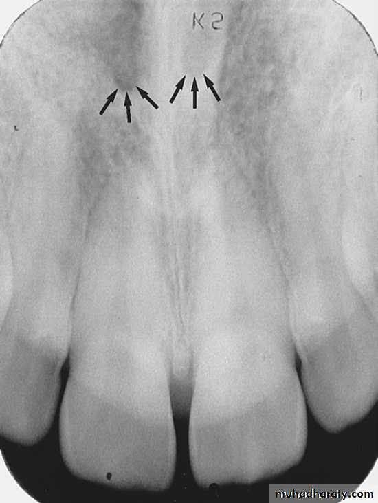
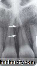
Superior Foramina of Incisive Canal
The superior foramina of the incisive canal are two tiny openings or holes in bone that are located on the floor of the nasal cavity. The superior foramina are the openings of two small canals that extend downward and medially from the floor of the nasal cavity. These two small canals join together to form the incisive canal and share a common exit, the incisive foramen.Appearance. On a maxillary periapical image, the superior foramina appear as two small, round radiolucencies located superior to the apices of the maxillary central incisors

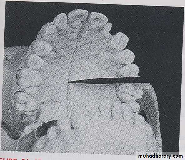
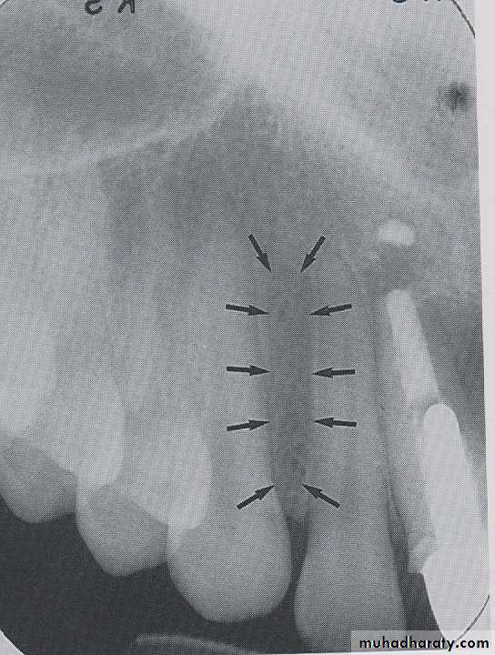
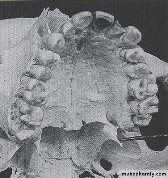
MEDIAN PALATEL SUTURE: The median palatal suture is the immovable joint between two palatine process of maxilla. Radiographically the suture appears as thin radiolucent line between the central incisor, the median palatal suture bounded in both side by dense cortical bone that appear radiopaque.
LATERAL FOSSE
:
The lateral fosse is a smooth depressed area of maxilla located between canine and lateral incisor. Radiographically the lateral fosse appears as a radiolucent area between the maxillary canine and lateral incisor.
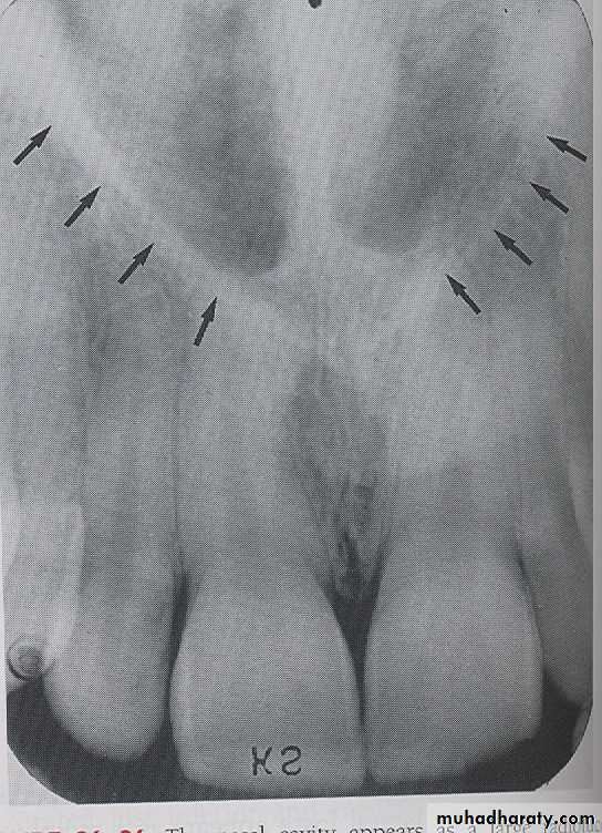
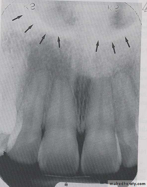
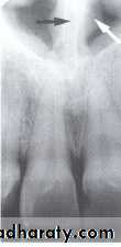
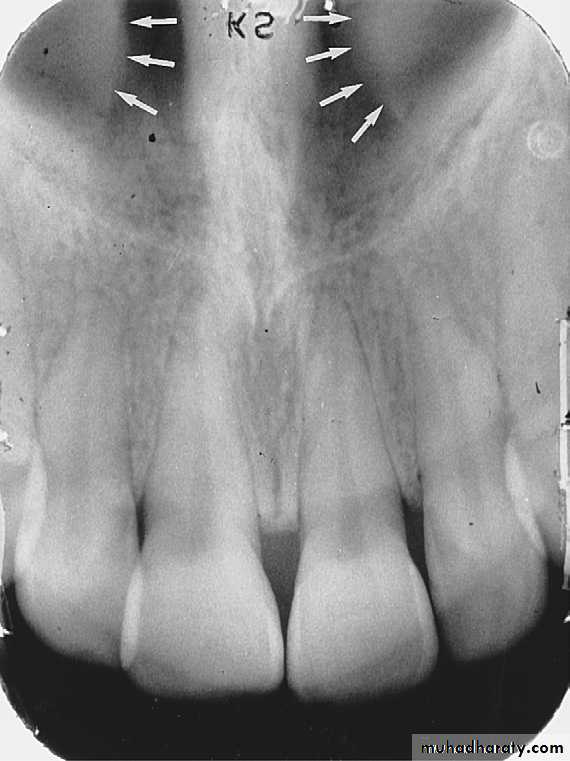
NASAL CAVITY: Is a pear-shaped compartment of bone located superior to the maxilla. On a maxillary perapical radiograph the nasal cavity appears as a large radiolucent area above the maxillary incisor.
NASAL SEPTUM:
The nasal septum is a vertical bony wall that divides the nasal cavity into right and left nasal fosse. Radiographically nasal septum appears as a vertical radiopaque partition that divide the nasal cavity.
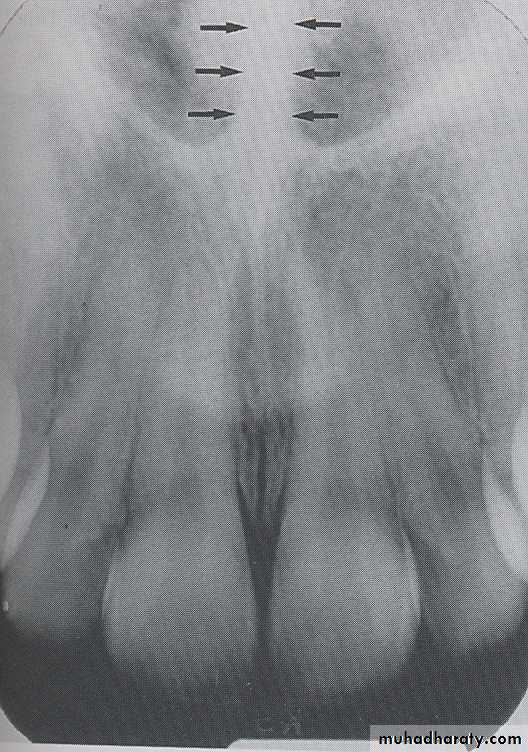
ANTERIOR NASAL SPINE
: Is a sharp projection of the maxilla located at the anterior portion of nasal cavity. Radiographically the anterior nasal spin appears as a V shaped radiopaque area located at the intersection of the floor of nasal cavity and the nasal septum.
Inferior Nasal Conchae
Inferior nasal conchae are plates of bone that extend from the lateral walls of the nasal cavity. Inferior nasal conchae are seen in the lower lateral portions of the nasal cavity.Radiographically, inferior nasal conchae appear as a diffuse radiopaque mass or projection within the nasal cavity
NOSE
The soft tissue of the tip of the nose is frequently seen in projections of the maxillary central and lateral incisors, superimposed over the roots of these teeth. The image of the nose has a uniform, slightly opaque appearance with a sharp border.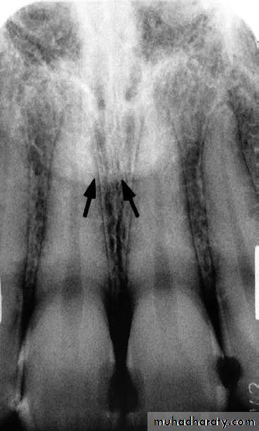
MAXILLARY SINUS:
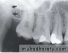
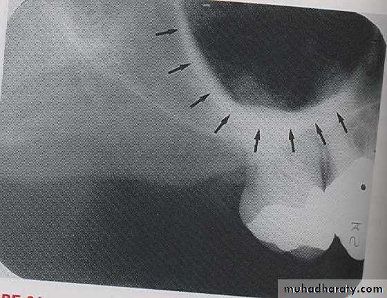
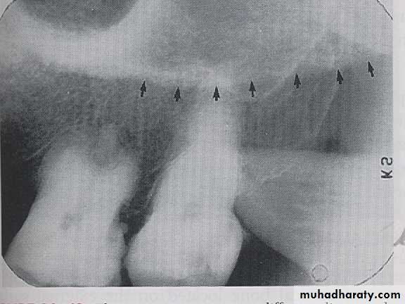
The maxillary sinus are paired cavities or compartments of bone located within the maxilla. The maxillary sinus are located above the maxillary permolars and molars teeth. Radiographically the sinus appears as a radiolucent area located above the apices of maxillary premolars and molars. The floor of sinus is composed of dense cortical bone and appear as radiopaque.
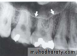
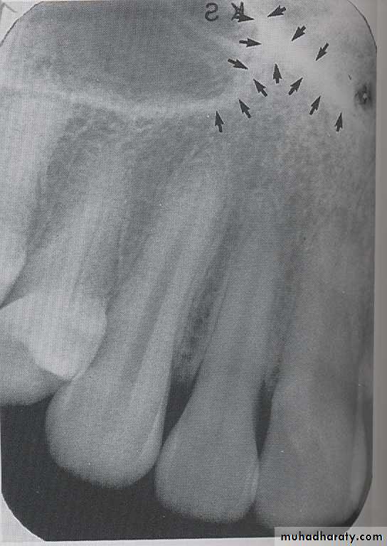
SEPTA WITHIN THE MAXILLARY SINUS:
Septa are bony wall that divided the maxillary sinus into compartment. Radiographically the septa appear as a radiopaque line within the sinus.
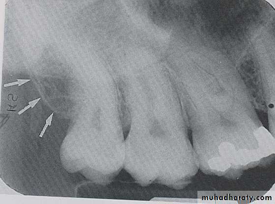
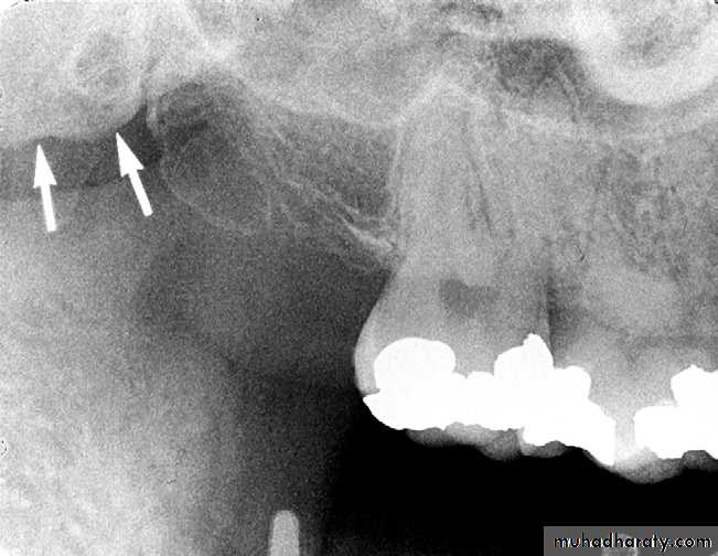
Nutrient Canals within Maxillary Sinus
Nutrient canals may be seen within maxillary sinuses. Nutrient canals are tiny, tubelike passageways through bone, which contain blood vessels and nerves that supply maxillary teeth and interdental areas. On a maxillary periapical image, a nutrient canal appears as a narrow radiolucent band bounded by two thin radiopaque lines.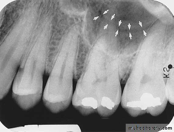
INVERTED Y
: The inverted Y refers to the intersection of the maxillary sinus and the nasal cavity. Radiographically the inverted Y appears as a radiopaque upside-down Y formed by intersection of the lateral wall of nasal fosse and anterior border of maxillary sinus both the lateral wall of nasal cavity and anterior border of maxillary sinus are composed of dense cortical bone and appears as a radiopaque band. The inverted Y is located above the maxillary canine.
MAXILLARY TUBEROSITY
:
Maxillary tuberosity is a round prominence of bone that extended posterior to the third molar region. On periapical radiography maxillary tuberosity appears as a radiopaque bulge distal to third molar region.
Pterygoid Plates
Pterygoid plates lie immediately posterior to the tuberosity of the maxilla. The image of these two plates is extremely variable, and they do not appear at all on many intraoral radiographs of the third molar area. When they are apparent, they almost appear as single radiopaque homogeneous shadow without any evidence of trabeculationHAMULUS:
The hamulus also known as the hamular process is a small hook like projection of bone extending from medial pterygoid plate of sphenoid bone it locater posterior to maxillary tuberosity region. Radiographically, the hamulus appears as a radiopaque hook like projection posterior to maxillary tuberosity area.
ZYGOMATIC PROCESS OF MAXILLA
:
The zygomatic process of maxilla is a bony projection of maxilla that articulates with the zygoma. On periapical radiography process appears as a J–U shaped radiopacity located superior to the maxillary first molar.
ZYGOMA: The zygoma or cheek bone articulates with the zygomatic process of maxilla. On maxillary periapical radiograph the zygoma appears as a diffuse radiopaque band extending posterior from the zygomatic process of maxilla.
BONY LANDMARKS OF THE MANDIBLE
SYMPHYSIS
Radiographs of the region of the mandibular symphysis in infants demonstrate a radiolucent line through the midline of the jaw between the images of the forming deciduous central incisors. This suture usually fuses by the end of the first year of life, after which it is no longer radiographically apparent.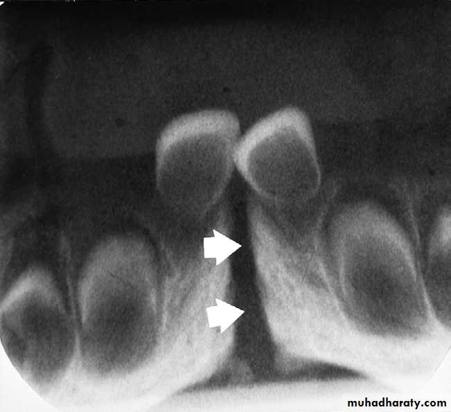
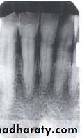
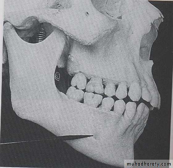
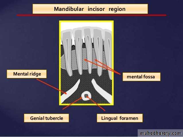
GENIAL TUBERCLES
:
The genial tubercle are tiny bumps of bone that serve as the attachment sites for the genioglossus and geniohyoid muscles, its located on lingual aspect of the mandible. On mandible periapical radiograph the genial tubercle appears as a ring shaped radiopacity below the apices of the mandibular incisor.
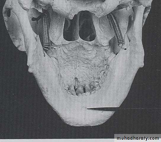
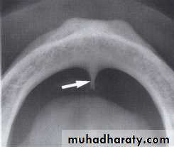
LINGUAL FORAMEN
:
The lingual foramen is a tiny opening or hole in the bone located on the internal surface of mandible, its located near the midline and surrounded by genial tubercle. Radiographically, the lingual foramen appears as small radiolucent dot located inferior to the apices of mandibular incisor.
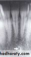
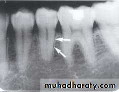
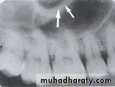
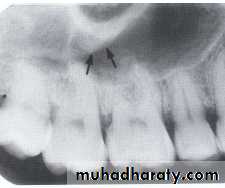
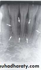
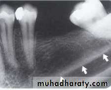
NUTRIENT CANALS
:
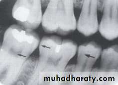
Nutrient canals carry a neurovascular bundle and appear as radiolucent lines of fairly uniform width. They are most often seen on mandibular periapical radiographs running vertically from the inferior dental canal directly to the apex of a tooth or into the inter dental space between the mandibular incisors.
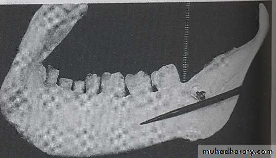
MENTAL RIDGE : Is a linear prominence of cortical bone located on the external surface of anterior portion of mandible. The mental ridge extends from premolar region to the midline. Radiographically the mental ridge appears as a thick radiopaque band extends from premolar regions to incisor region.
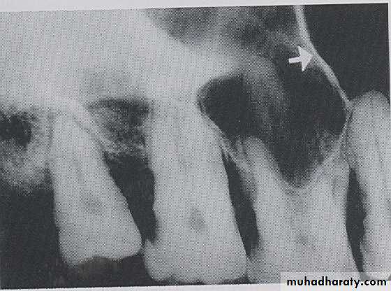
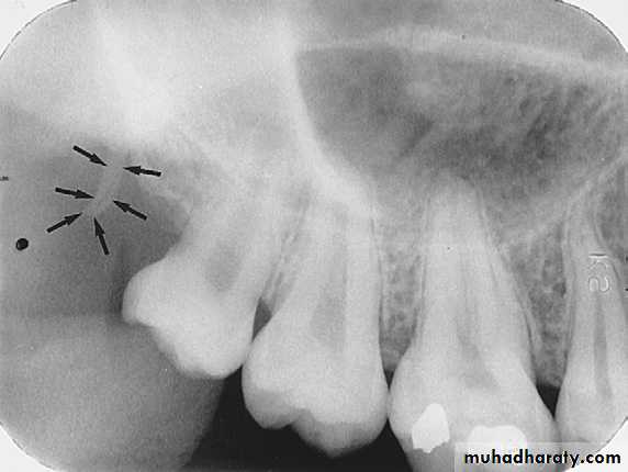
MENTAL FOSSE:
The mental fosse is a scooped out depressed area of bone located on the external surface of anterior portion of mandible, its located above the mental ridge in the mandible incisor region. On periapical radiograph, the mental fosse appears as a radiolucent area above the mental ridge.
MENTAL FORAMEN:
The mental foramen is an opening or hole in the bone located on the external surface of mandible in the region of mandibular premolars. Radiographically, the mental foramen appears as small ovoid or round radiolucent area located in the apical region of mandibular premolars.
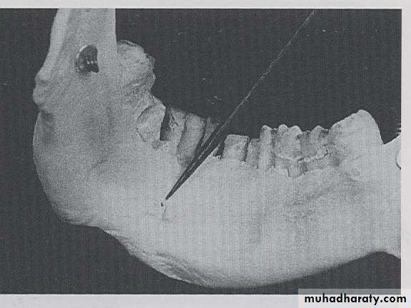
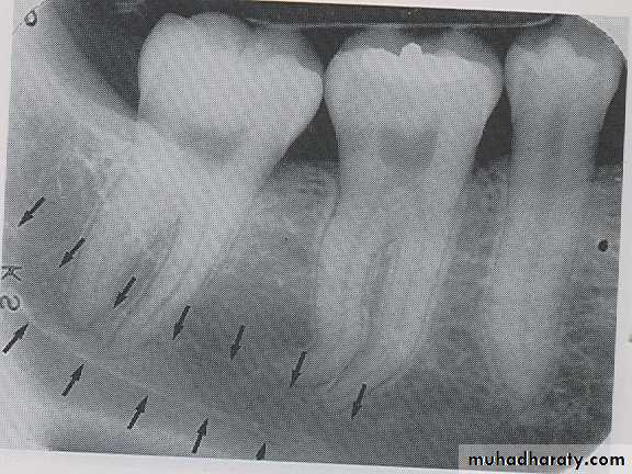
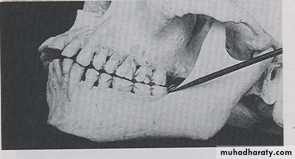
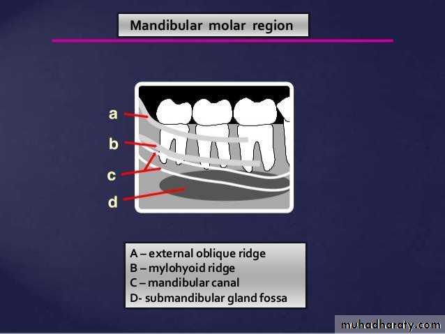
MYLOHYOID RIDGE
: The mylohyoid ridge is a linear prominence of bone located on the internal surface of mandible, its extended from the molar region downward and forward toward the lower border of mandibular symphysis and serves as the attachment site for the mylohyoid muscles. Radiographic appearance, mylohyoid ridge appears as a dense the radiopaque band that extends downward and forward from molar region.
SUB MANDIBULAR FOSSE:
Submandibular fosse is a scooped–out depressed area of bone located on the internal surface of mandible inferior to mylohyoid ridge. On periapical radiography the submandibular fosse appears as radiolucent area in the molars region below the mylohyoid ridge.
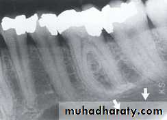
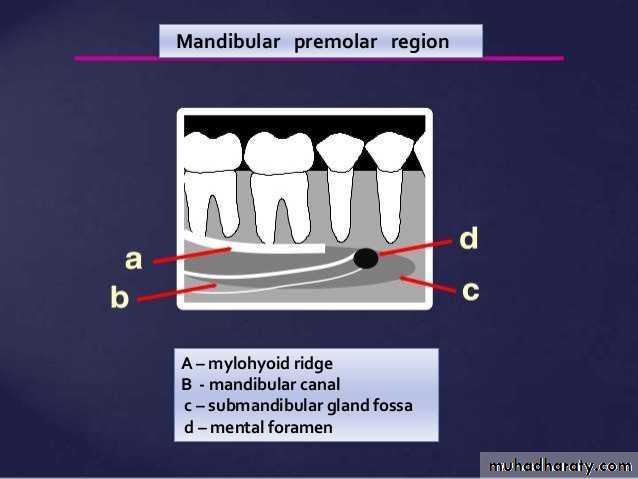
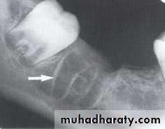
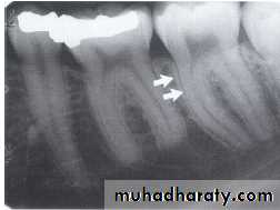
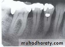
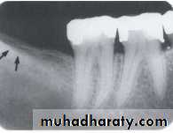
MANDIBULAR CANAL:
The mandibular canal is a tube like passage way through the bone that travels the length of the mandible, its extends from the mandibular foramen to the mental foramen and houses the inferior alveolar nerve and blood vessels. On radiograph the mandibular canal appears as a radiolucent band outlined by two thin radiopaque lines that represent the cortical wall of the canal.
INTERNAL OBLIQUE RIDGE:
The internal oblique line is linear prominence of bone located on internal surface of mandible that extended downward and forward from the ramus. Radiographically the line appears as radiopaque band that extends from the ramus of the mandible.
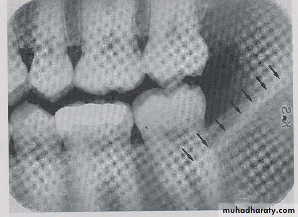
EXTERNAL OBLIQUE LINE :
The external oblique line is linear prominence of bone located on external surface of the body of mandible, the anterior end of ramus ends in the external oblique line. Radiographically the ridge appears as radiopaque band extending downward and forward from the anterior border of the ramus, its ends in the mandibular third molar region.
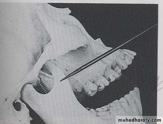
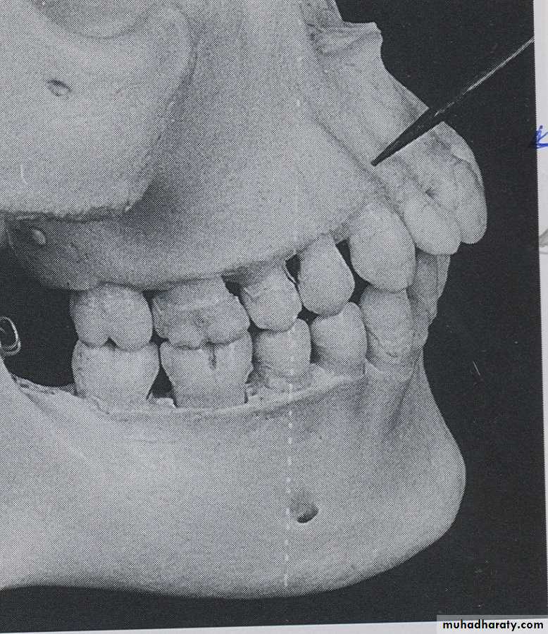
CORONOID PROCESS:
The coronoid process is a marked prominence of bone on the anterior ramus of mandible. Radiographically the coronoid process is not seen on mandibular periapical radiography but dose appears on maxillary periapical film as triangular radiopacity superimposed over the maxillary tuberosity region.