
1
Lecture (1)
د.قاسم امير تاج الدين
x-ray
Definition
X rays are a type of radiation used in imaging and therapy that uses short
most substances except heavy metals
Electromagnetic radiation of wavelengths ranging between 5.0 × 10
−6
and 5.0 × 10
−4
μm
X-rays are produced when fast-moving electrons with sufficient energy strike a target.
Most of the electron energy is converted to heat, but a very minute amount —less than
1 per cent —is converted to x-rays
Spectrum of Electromagnetic Radiations. X-rays resemble visible light rays very
closely but have the distinguishing feature that their wave lengths are very short
Feature of x-ray :
• No mass
• No charge
• Travel at the speed of light
• Short wave length
• High energy
• Source of x-ray from the electron shells
x-ray production
:
uses a high voltage to accelerate
to a high velocity. The high velocity electrons
collide with a metal target, the
, creating the x-rays.
In medical x-ray tubes the target is usually

2
the insert is evacuated from air because :
• Prevent oxidation of the cathode filament
• Decrease resistance against electron movement
Advantage of oil in x-ray tube :
• Shock absorber
• Heat absorber
Two different processes give rise to radiation of X-ray
frequency. In one process radiation is emitted by the high-
speed electrons themselves as they are slowed or even
stopped in passing near the positively charged nuclei of the
anode material. This radiation is often called
brehmsstrahlung [Ger.,=braking radiation]. In a second
process radiation is emitted by the electrons of the anode
atoms when incoming electrons from the cathode knock
electrons near the nuclei out of orbit and they are replaced
by other electrons from outer orbits. The
frequencies given off with any particular anode material
thus consists of a continuous range of frequencies emitted
in the first process, and superimposed on it a number of
sharp peaks of intensity corresponding to discrete
frequencies at which X rays are emitted in the second
process. The sharp peaks constitute the X-ray line
spectrum for the anode material and will differ for
different materials
.

3
X-radiation (composed of X-rays) is a form of
, corresponding to
15
Hz to 30 × 10
18
Hz) and energies in the range 120 eV
to 120 keV. They are shorter in wavelength than
languages, X-radiation is called Röntgen radiation after one of its first
investigators, Wilhelm Conrad Röntgen.
X-rays, or
rays, are electromagnetic waves in which periodically
variable electric and magnetic fields are
to the direction of propagation. Thus they are identical in nature with
visible light and all the other types of radiation that constitute the
electromagnetic spectrum. In general, x-rays are generated as the result of
energy transitions of atomic electrons caused by the bombardment of a
material of high atomic weight by high-energy electrons.
The range of x-rays in the electromagnetic spectrum, as excited in x-ray
tubes by the bombardment of
high accelerating potential, overlaps the
100 nanometers on the long-wavelength side, and the shortest-wavelength
limit moves downward as voltages increase. An accelerating potential of
10
9
volts, now readily generated, produces a
−15
m (10
−6
nm). An average wavelength used in research is 0.1 nm, or about 1/6000
the wavelength of yellow light. See also
X rays pass easily through air and soft tissue of the body. When they
encounter more dense material, such as a
, bone, or a metal
fragment, they are stopped. Diagnostic x rays are performed by
positioning the part of the body to be examined between a focused beam
of x rays and a plate containing film. This process is painless. The greater
the density of the material that the x rays pass through, the more rays are
absorbed. Thus bone absorbs more x rays than muscle or fat, and tumors
may absorb more x rays than surrounding tissue. The x rays that pass

4
through the body strike the photographic plate and interact with silver
molecules on the surface of the film.
Once the film plates have been processed, dense material such as bone
shows up as white, while softer tissue shows up as shades of gray, and
airspaces look black. A radiologist, who is a physician trained to interpret
diagnostic x rays, examines the pictures and reports to the doctor who
ordered the tests. Plain film x rays normally take only a few minutes to
perform and can be done in a hospital, radiological center, clinic, doctor's
or
's office, or at
with a portable x-ray machine.
X ray, invisible, highly penetrating
of much shorter wavelength (higher frequency)
than visible light. The wavelength range for X rays is from
about 10
−8
m to about 10
−11
m, or from less than a billionth
of an inch to less than a trillionth of an inch; the
corresponding frequency range is from about 3 × 10
16
Hz
to about 3 × 10
19
Hz (1 Hz = 1 cps).
X rays are also produced in a highly evacuated glass bulb,
called an X-ray tube, that contains essentially two
electrodes—an anode made of platinum, tungsten, or
another heavy metal of high melting point, and a cathode.
When a high voltage is applied between the electrodes,
streams of electrons (cathode rays) are accelerated from
the cathode to the anode and produce X rays as they strike
the anode
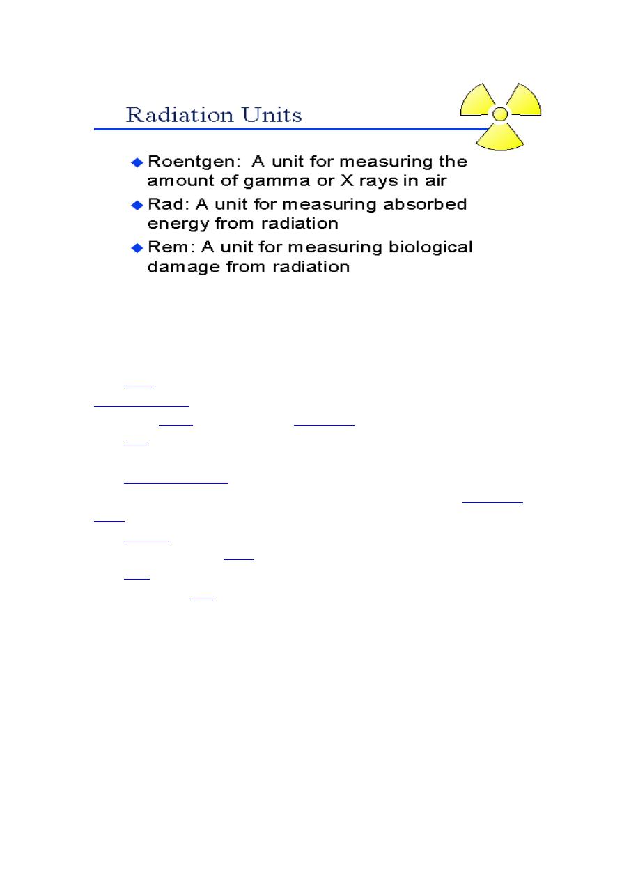
5
(Gy) which has units of (J/kg), is the SI unit of
which is the amount of radiation required to
of energy in 1
is the (obsolete) corresponding traditional unit, equal to
0.01 J deposited per kg. 100 rad = 1 Gy.
The
is the measure of the biological effect of
radiation on human tissue. For X-rays it is equal to the
(Sv) is the SI unit of equivalent dose, which for X-
is the traditional unit of equivalent dose. For X-rays it
is equal to the
or 0.01 J of energy deposited per kg. 1 sievert
= 100 rem

6
CT scan
Definition
Computed tomography (CT) an imaging method that uses x-
rays to create cross-sectional pictures of the body.
Alternative Names
CAT scan; Computed axial tomography (CAT) scan
What is computed tomography?
Computed tomography, commonly known as a CT scan, uses X-
rays and computers to produce images of a cross-section of the
body. The patient must lie as still as possible as the table moves
through the large, donut-shaped scanning device. Movement
could blur
In conventional x-rays, a beam of energy is aimed at the body
part being studied. A plate behind the body part captures the
variations of the energy beam after it passes through skin, bone,
muscle, and other tissue. While much information can be
obtained from a regular x-ray, specific detail about internal
organs and other structures is not available
With computed tomography scan (also called CT or CAT scan),
the x-ray beam moves in a circle around the body. This allows
for many different views of the same organ or structure, and
provides much greater detail. The x-ray information is sent to a
computer which interprets the x-ray data and displays it in two-
dimensional form on a monitor
How the Test is Performed
You will be asked to lie on a narrow table that slides into the
center of the CT scanner. Depending on the study being done,
you may need to lie on your stomach, back, or side.
Once inside the scanner, the machine's x-ray beam rotates
around you. (Modern "spiral" scanners can perform the exam in
one continuous motion.