Extra oral radiography
As the term extraoral suggests, an extraoral radiograph is one that is placed outside the mouth during x-ray exposure. It is used to image large areas of the skull or jaws. Many types of extraoral films exist; such films are primarily used in orthodontics and oral surgery.Uses of extraoral film include :
(1) evaluation of large areas of skull and jaws;
(2) evaluation of growth and development;
(3) evaluation of impacted teeth;
(4) detection of diseases, lesions,and conditions of the jaws;
(5) examination of the extent of large lesions;
(6) evaluation of trauma;
(7) evaluation of TMj area.
The use of special equipment, including x-ray unit, screen film, intensifying screens ,grid, and cassette, is necessary in extraoral radiography. The most common extraoral film is the panoramic radiograph.
Panoramic radiograph
The panoramic radiograph allows the dental professional to view a large area of the maxilla and mandible on a single film, in it both the film and tube head are connected and rotate simultaneously around the patient during exposure; Rotational centers allow the image layer to conform to elliptical shape of dental arches. The number and location of the rotational centers influence the size and shape of the focal trough.Focal trough(image layer) :
Is a three-dimensional curved zone in which structures are clearly demonstrated on a panoramic radiograph. Structures within the focal trough appear reasonably well defined, whereas structures outside the focal trough appear blurred.
Types o f panoramic x –ray machines :
Double-center rotation machines have two Rotational centers one for the right and one for the left side of the jaws.
Triple-center rotation machines have three centers of rotation and create an un interrupted radiographic image of the jaws.
Moving-center rotation machines rotate around a continuously moving center that is similar to the arches creating an un interrupted image of the jaws.
Each panoramic x-ray unit has a focal trough that designed to accommodate the average jaw. Each manufacturer provides specific instructions about patient positioning to ensure that the teeth are positioned within the focal trough. The quality of the resulting panoramic radiograph depends on the positioning of the patient's teeth within the focal trough and how closely the patient's jaws conform to the focal trough designed for the average jaw.
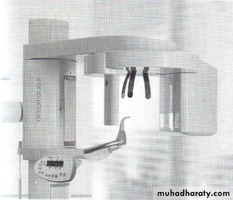
The main components of the panoramic unit include the following
1. X-ray tube head
2. Head positioner
3. Exposure controls
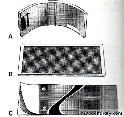
Film: Screen film is used in panoramic radiography. it sensitive to light emitted from intensifying screen it is placed between two intensifying screens in a cassette holder. When the cassette holder is exposed to x-rays, the screens convert the x-ray energy into light, which in turn exposes the screen film. The film used in panoramic radiography is available in two sizes: 5 x 12 inch and 6 x 12 inch.
Focal trough
Intensifying screens:
There are two basic types of intensifying screens: calcium tungstate and rare earth. Calcium tungstate screens emit blue light, and the rare earth screens emit green light. Rare earth screens require less x-ray exposure than calcium tungstate screens and are considered " faster."Consequently rare earth screens are recommended in panoramic radiography because there is less radiation exposure for the patient.Cassette:
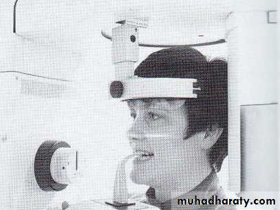
The cassette is a device that is used to hold the extra oral film and intensifying screen. Midsagittal plane :An imaginary line that divides the patient's face into right and left sides) perpendicular to the floor .The patient's head must not be tipped or tilted; if the midsagittal plane is not positioned perpendicular to the floor, a distorted image results on the panoramic radiograph.
Frankfort plane : An imaginary plane that passes through the top of the ear canal an
the bottom of the eye socket . When the Frankfort plane is parallel to the floor, the occlusal plane is positioned at the correct angle.
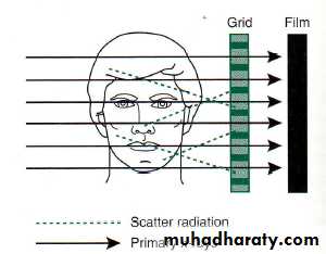
Grid :A grid is a device used to reduce the amount of scatter radiation that reaches an extraoral film during exposure. Scatter radiation causes film fog and reduces film contrast. A grid can be used to decrease film fog and increase the contrast of the radiographic image.
Ghost Image: A ghost image is a radiopaque artifact seen on a panoramic film that is produced when a radiodense object is penetrated twice by the x-ray beam.
A ghost image resembles its real counterpart and is found on the opposite side of the film; it appears indistinct, larger, and higher than its actual counterpart. For example, a ghost image of a hoop earring appears on the opposite side of the film as a radiopacity that is larger and higher than the real hoop earring. Also it appears blurred in both a horizontal and a vertical direction.
Large hoop earrings (1) , ghost images (2).
Uses of the panoramic radiograph includeThe panoramic radiograph is typically used to supplement bite-wing and periapical
films and is not a substitute for intraoral films. It should not be used to evaluate
caries, periodontal disease, or periapical lesions but it's uses :
(1) evaluation of impacted teeth;
(2) evaluation of eruption patterns and growth and development;
(3) detection of diseases, lesions, and conditions of the jaws;
(4) examination of extent of large lesions;
(5) evaluation of trauma.
Advantages of Panoramic Radiography
1. Field size :The panoramic radiograph includes coverage of the entire maxilla and mandible. More anatomic structures can be viewed on a panoramic film than on a complete intraoral radiographic series. In addition, lesions and conditions of the jaws that may not be seen on intraoral films can be detected on a panoramic radiograph.
2. Simplicity: Exposure of a panoramic radiograph is relatively simple and requires a minimal amount of time and training for the dental radiographer.
3. Patient cooperation: The exposure of a panoramic radiograph is readily accepted by the patient .Because there is no discomfort involved. For example, children who may not be able to tolerate intraoral projections may find it easier to sit still during the exposure of a panoramic radiograph
4. Minimal exposure: A Panoramic radiograph results in minimal radiation exposure for the patient.
Disadvantages of Panoramic Radiography
Image quality :The images seen on a panoramic radiograph are not as sharp as those on intraoral radiographs because of the intensifying screens ,As a result, the panoramic radiograph cannot used to evaluate dental caries, periodontal disease, or periapical lesions.
Focal trough limitations. Objects of interest that are located outside the focal trough
are not seen.
Distortion : A certain amount of magnification, distortion, and overlapping is present
on a panoramic radiograph, even when proper technique is used.
Equapment cost : The cost of a panoramic x-ray unit is relatively high compared with
the cost of an intraoral x-ray unit.
Before preparing the patient for exposure of a panoramic film, the following tasks
must be completed: preparation of cassette, infection control procedures, selection
of exposure factors, adjustment of panoramic machine for patient height and
proper alignment of movable parts, and loading of cassette into cassette carrier.
After preparing the equipment, the dental radiographer must prepare the patient
by explaining radiographic procedure, placing lead apron on patient, and removing
all radio dense objects from head and neck region.