
Dr.Majeed M.Al-Hamammi
lectures
4
th
year
Medical college
Thi- Qar 2019-2020

Respiratory disease
•
Pulmonary medicine
•
Pulmonology
•
Respiralogy

Objectives
• At the end of this lecture we should be Know
and familiar with
Respiratory physiology
Respiratory anatomy
-Respiratory symptoms.
-Respiratory signs.
-Investigation of respiratory diseases.

Respiratory disease
Is responsible for a major burden of morbidity
and mortality, and conditions such as
• tuberculosis,
• pandemic influenza
• pneumonia
are the most important conditions in world
health terms.

• The increasing prevalence of allergy, asthma
and chronic obstructive pulmonary disease
(COPD) .
• By 2025, smokers world-wide is anticipated to
increase to 1.5 billion.

FUNCTIONAL ANATOMY AND
PHYSIOLOGY
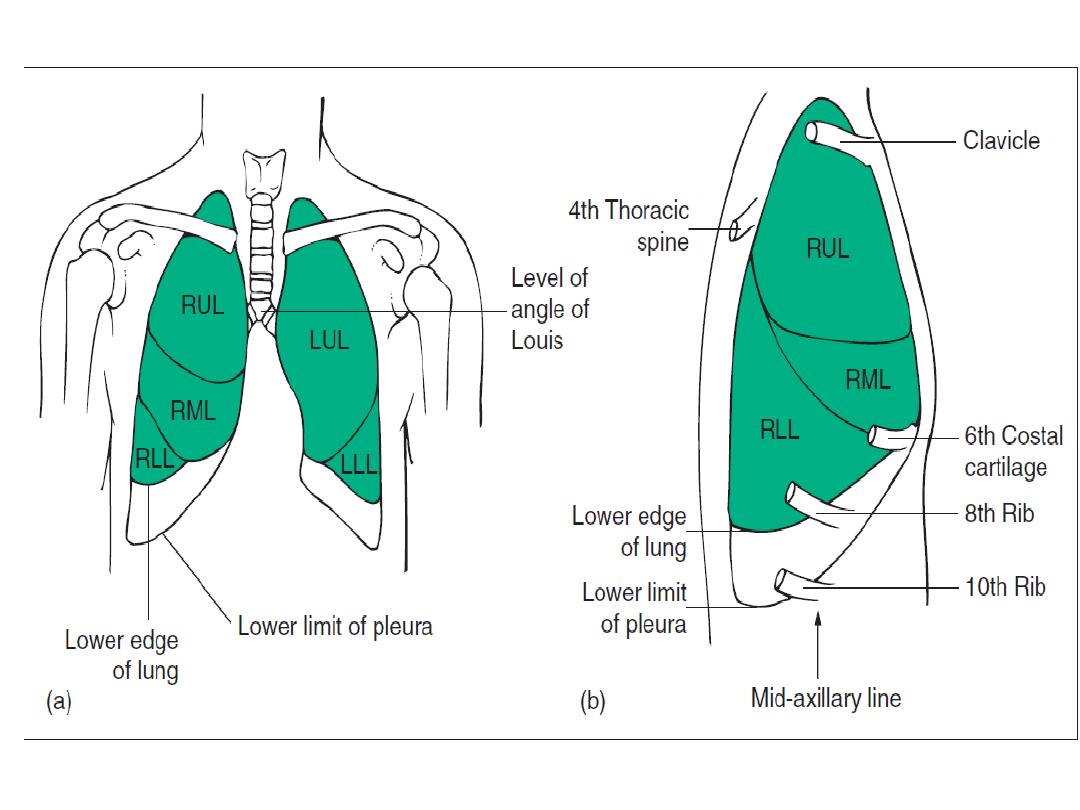
• The lungs occupy the upper two-thirds of the
bony thorax, bounded medially by the spine,
the heart and the mediastinum and inferiorly
by the diaphragm.

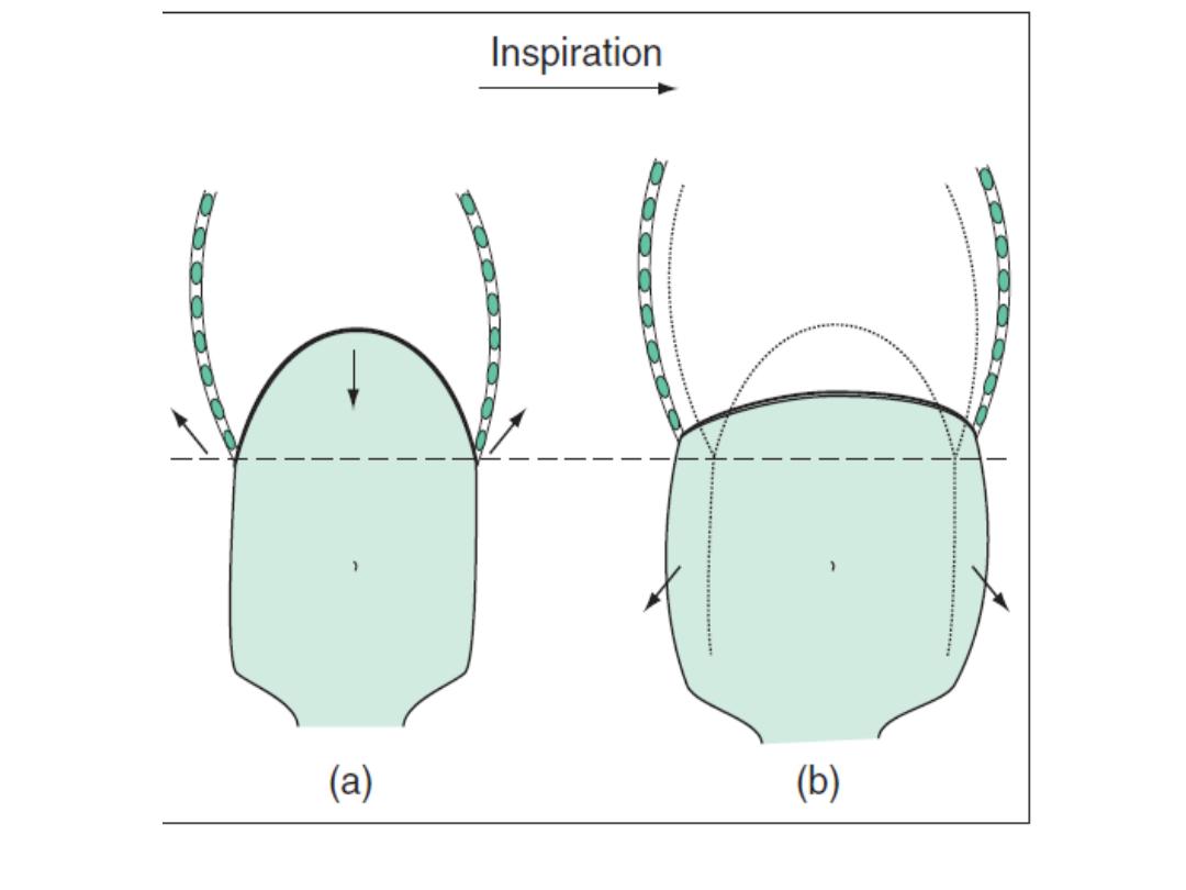
Inspiration
• downward contraction of the dome-shaped
diaphragm .
• contraction of the external intercostal
muscles.

Expiration
• largely passive, driven by elastic recoil of the
lungs.
• The increased demand in inspiration and
expiration operate accessory muscles.

The conducting airways
• from the nose to the alveoli connect the
external environment with the extensive, thin
and vulnerable alveolar surface.
• In the glottis and trachea, obstruction by
foreign bodies and tumours.
• in the third-generation respiratory, very slow
flow rates.

Control of breathing
• The respiratory motor neurons in the medulla
oblongata sense the pH of the cerebrospinal fluid (CSF)
and are indirectly stimulated by a rise in arterialPCO2.
• The carotid bodies sense hypoxaemia but are mainly
activated by arterial PO2 values below 8 Kpa (60
mmHg). They are also sensitised to hypoxia by raised
arterial PCO2..
• Muscle spindles in the respiratory muscles sense
changes in mechanical load.
• Cortical influences can override the automatic control
of breathing.

Ventilation/perfusion matching and the pulmonary
circulation
• Gravity determines the distribution of ventilation
and blood flow in the lungs.
• Hypoxia constricts pulmonary arterioles
• Hypercapenia dilates bronchi.
• Lung disease
which disturb the physiological
matching of regional ventilation and perfusion,
causing respiratory failure .
• Diseases that destroy or thicken the alveolar
capillary membrane (e.g. emphysema or fibrosis)
can impair gas diffusion directly.
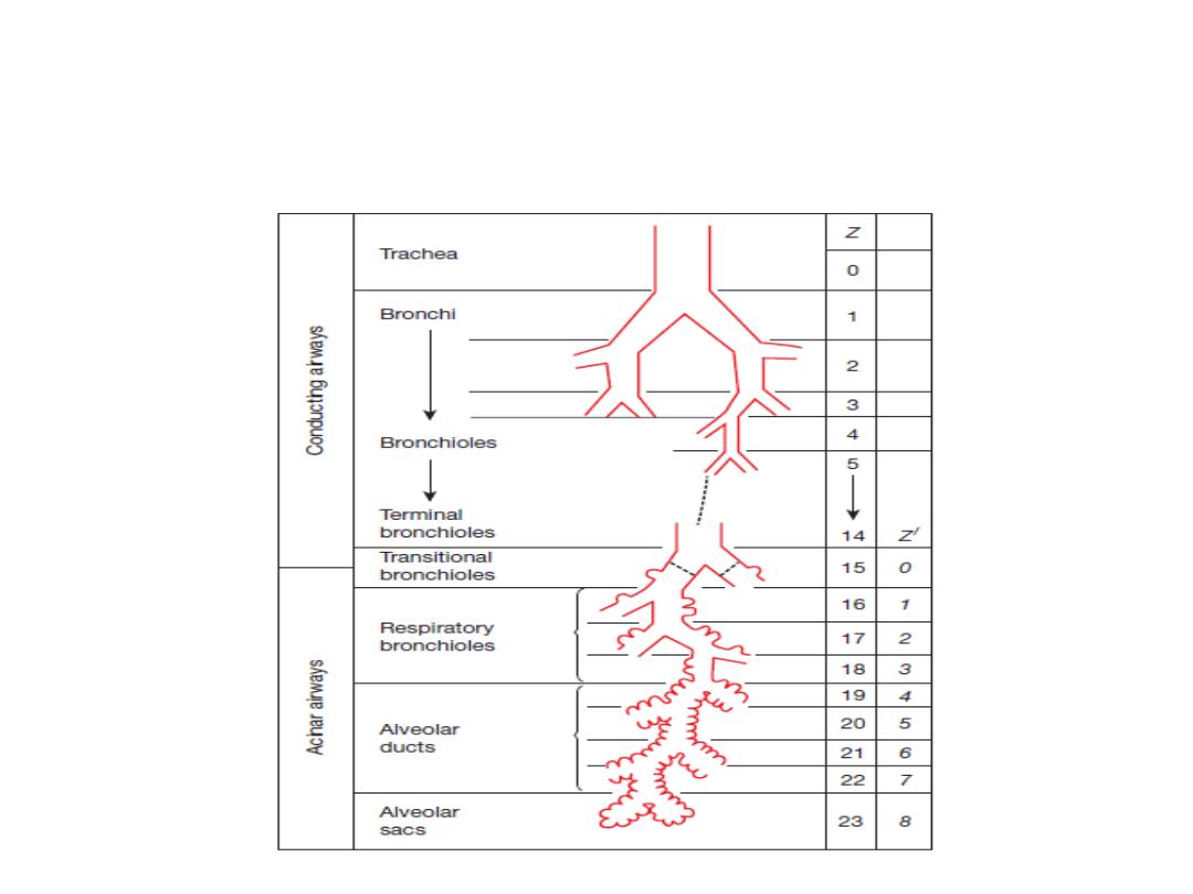
Model of airway branching in human lung by regularized
dichotomy from trachea (generation z = 0) to alveolar ducts and sacs
(generations 19–23). The first 14 generations are purely conducting; transitional
airways (generation 15) lead into the acinar airways with alveoli
that branch over 8 generations (z′).
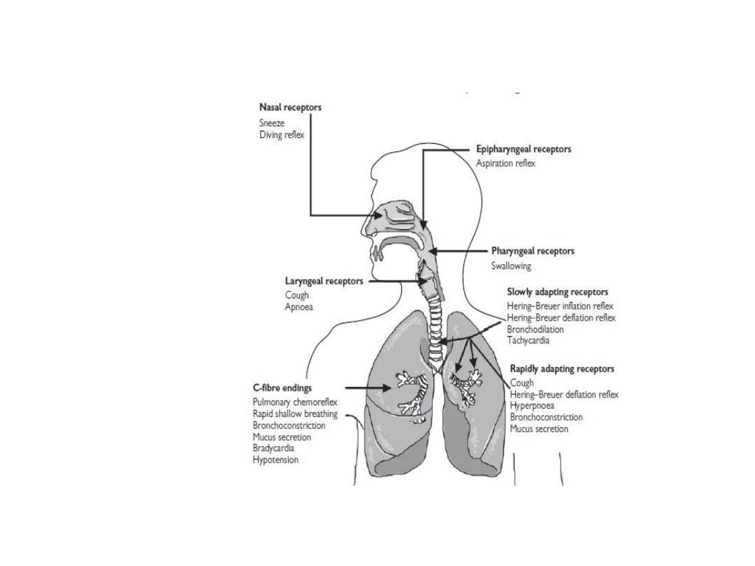
Location of major upper andlower airway receptors
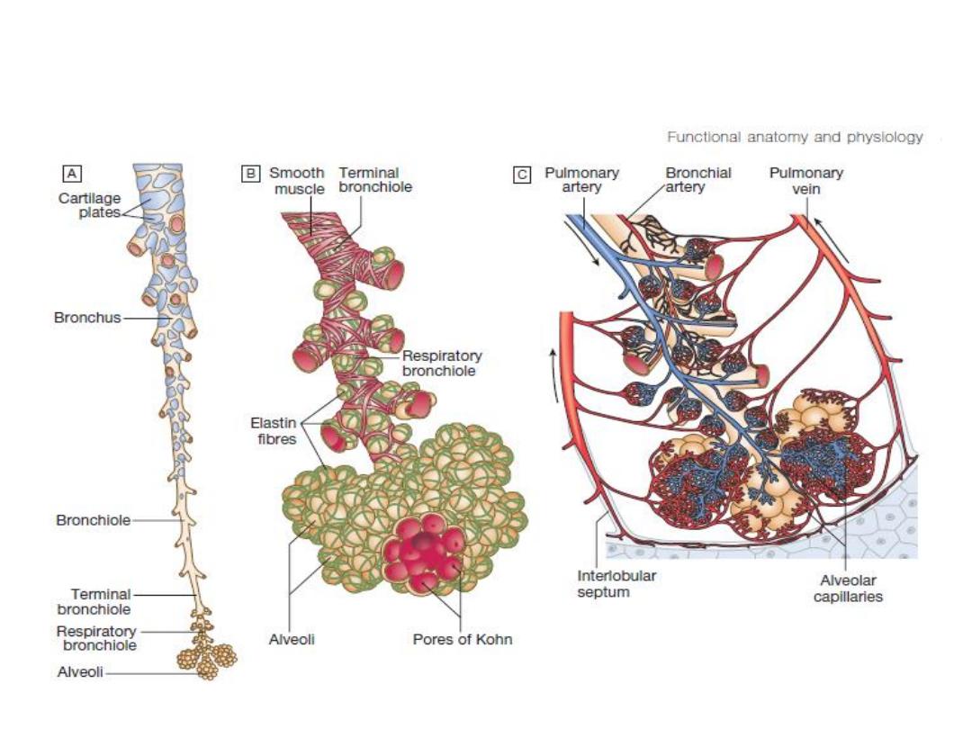
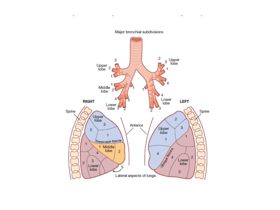
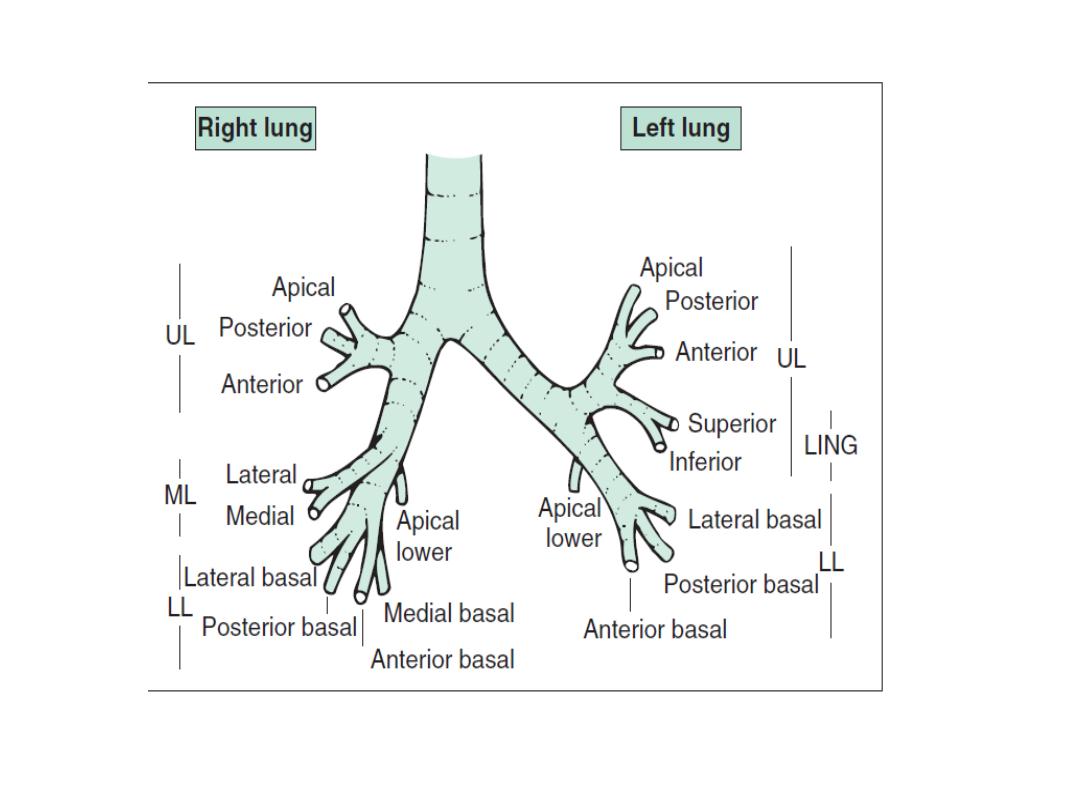
Functional anatomy of the lungs

Lung defences
• Upper airway defences
• nasal hairs.
• the columnar ciliated epithelium.
• cough.
• The larynx.

Lower airway defences
Non specific defences
• mucociliary escalator.
• Airway secretions contain an array of
antimicrobial peptides.
Macrophages.
Adaptive immune defence
• Lung dendritic cells.
• CD4 T-helper
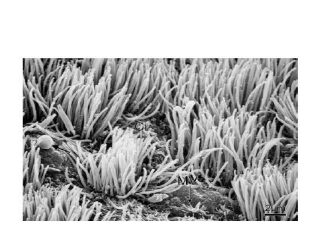
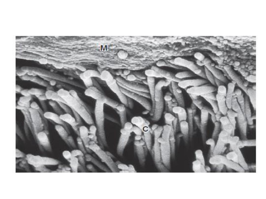
Surface view of bronchiolar epithelium shows tufts of
cilia (Ci) forming on individual ciliated cells and microvilli (MV) on
other cells. Note secretion droplet in process of release from goblet
cell (arrow).
Mucociliary escelator

PRESENTING PROBLEMS IN RESPIRATORY
DISEASE
Cough
• The most frequent symptom of respiratory
disease.
• Sputum production is common
• Acute less than 3 weeks.
• Subacute 3 -8 weeks.
• Chronic more than 8 weeks.

Acute transient cough
• Viral lower respiratory tract infection.
• post-nasal drip resulting from rhinitis or sinusitis,
• aspiration of a foreign body,
• laryngitis
• pharyngitis.
Cough occurs in the context of more serious diseases,
pneumonia.
• Aspiration.
• Congestive heart failure.
• pulmonary embolism.
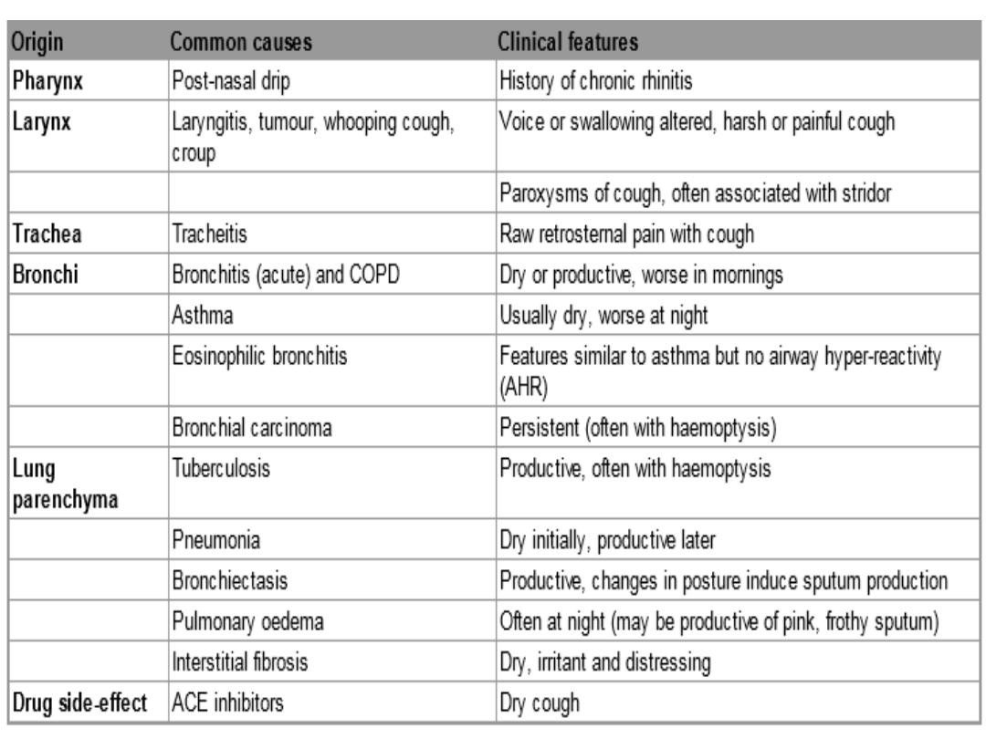

chronic cough
1. cough-variant asthma.
2. post-nasal drip secondary to nasal or sinus
disease.
3. gastro-oesophageal reflux with aspiration.
4. angiotensin-converting enzyme (ACE)
inhibitors .
5. Bordetella pertussis infection in adults .
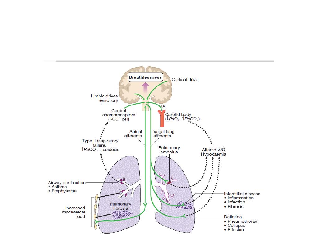
Respiratory stimuli contributing to breathlessness. Mechanisms by which disease can
stimulate the respiratory motor neurons in the
medulla. Breathlessness is usually felt in proportion to the sum of these stimuli.
Further explanation is given on page 543. (V / Q = ventilation/perfusion
match)

Breathlessness
Pathophysiology :
• Respiratory diseases can stimulate breathing and
dyspnoea by:
• stimulating intrapulmonary sensory nerves .
• increasing the mechanical load on the respiratory
muscle.
• causing hypoxia, hypercapnia or acidosis,
stimulating chemoreceptors.
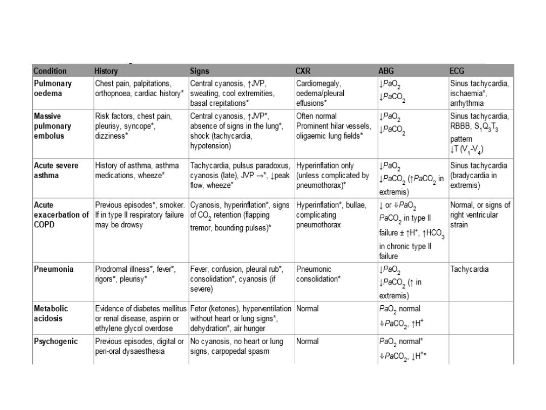
Differential diagnosis of acute breathlessnss
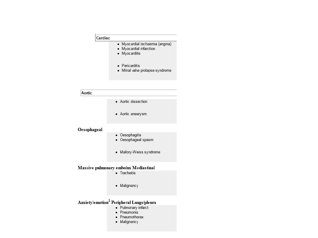
Chest pain :differential diagnosis

Haemoptysis
Coughing up blood.
Many episodes of haemoptysis remain
unexplained even after full investigation

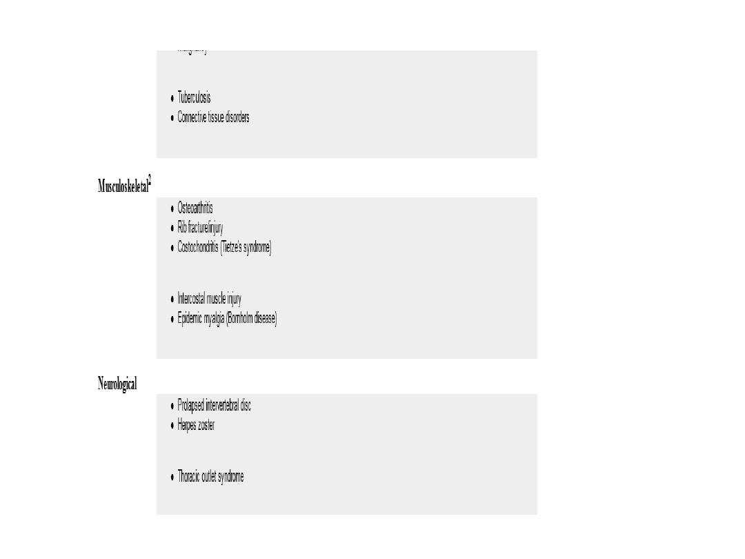
Causes of haemoptysis
Bronchial disease
Carcinoma
Bronchiectasis
Acute bronchitis
Others.
Parenchymal disease
• Tuberculosis
• Others.

• Lung vascular disease
• Pulmonary infarction
• Goodpasture's syndrome
• Others.
• Cardiovascular disease
• Acute left ventricular failure.
• Mitral stenosis .
• Others.
• Blood disorders
• Leukaemia
• Others

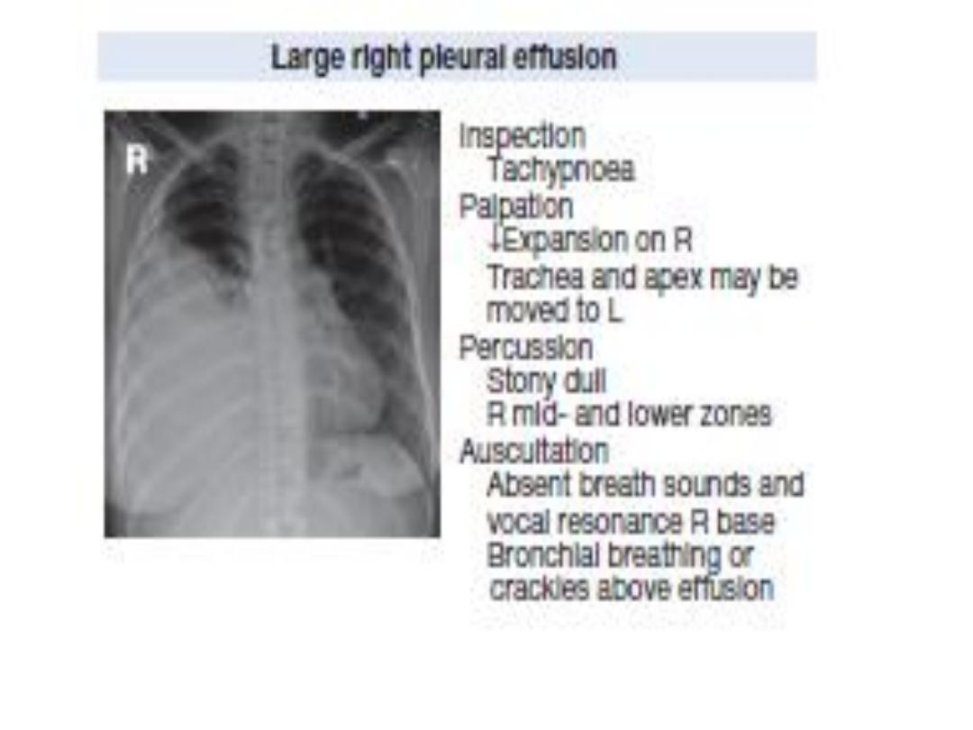
pleural effusion
• Causes of pleural effusion Common causes
Pneumonia ('para-pneumonic effusion')
• Tuberculosis
• Pulmonary infarction*
• Malignant disease
• Cardiac failure*
• Subdiaphragmatic disorders (subphrenic
abscess, pancreatitis etc.)

Uncommon causes
• Hypoproteinaemia*
(nephrotic syndrome, liver failure, malnutrition)
• Connective tissue diseases* (particularly systemic lupus
erythematosus (SLE) and rheumatoid arthritis)
• Acute rheumatic fever
• Post-myocardial infarction syndrome
• Meigs' syndrome (ovarian tumour plus pleural effusion)
• Myxoedema*
• Uraemia*
• Asbestos-related benign pleural effusion
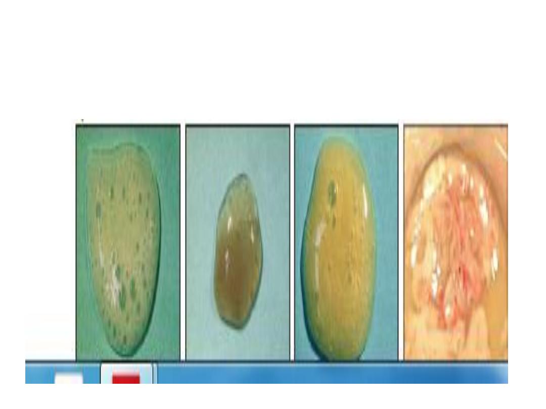
Sputum
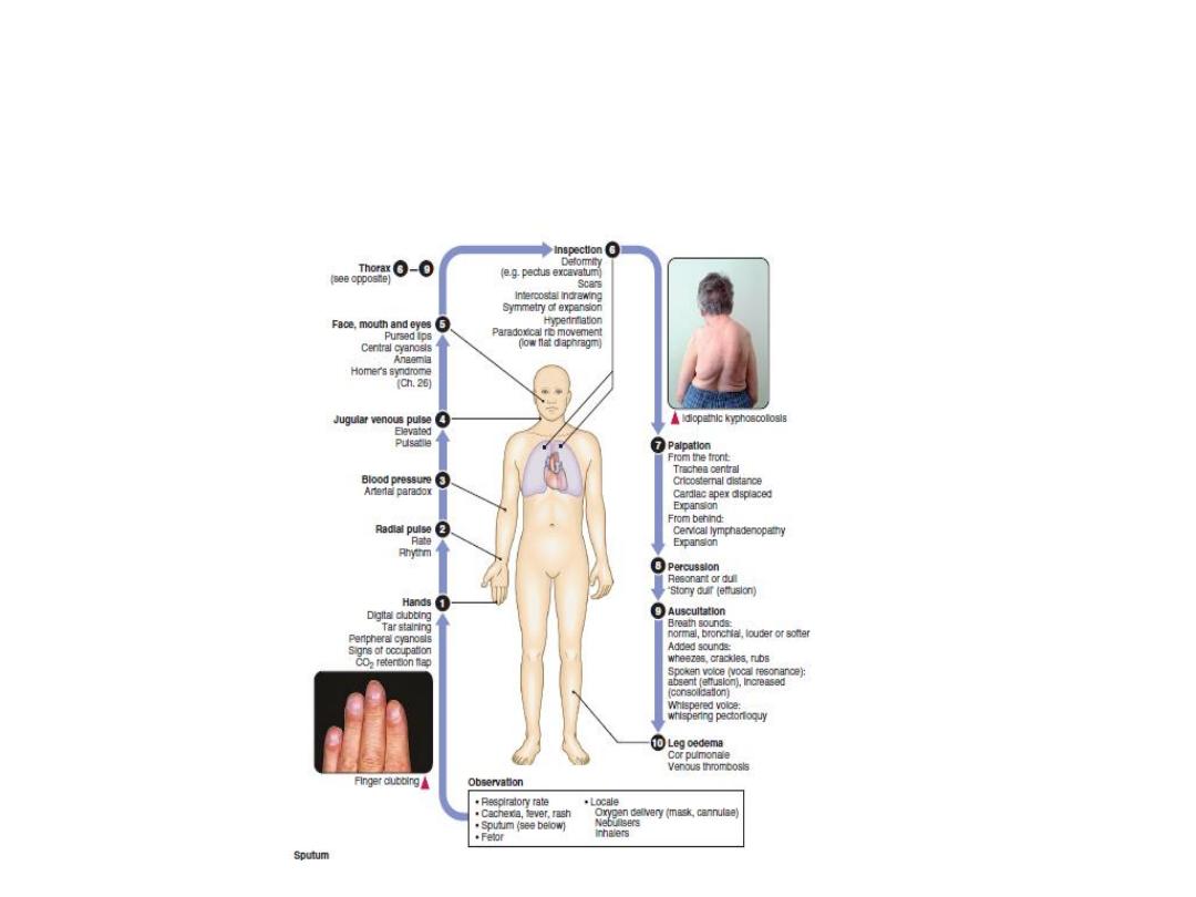
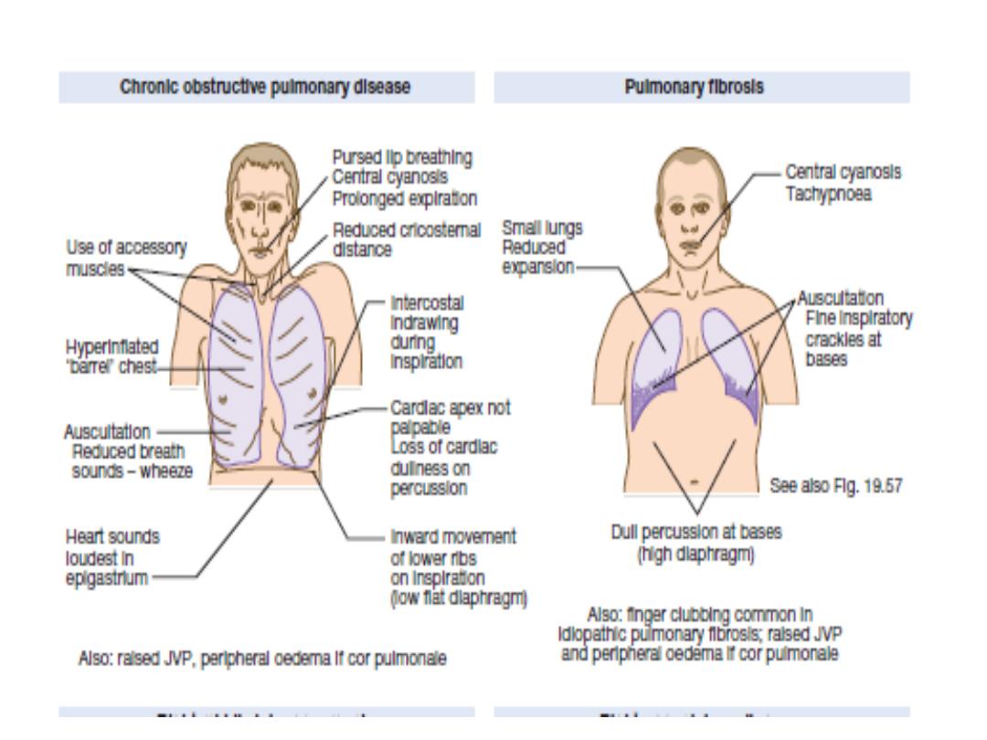
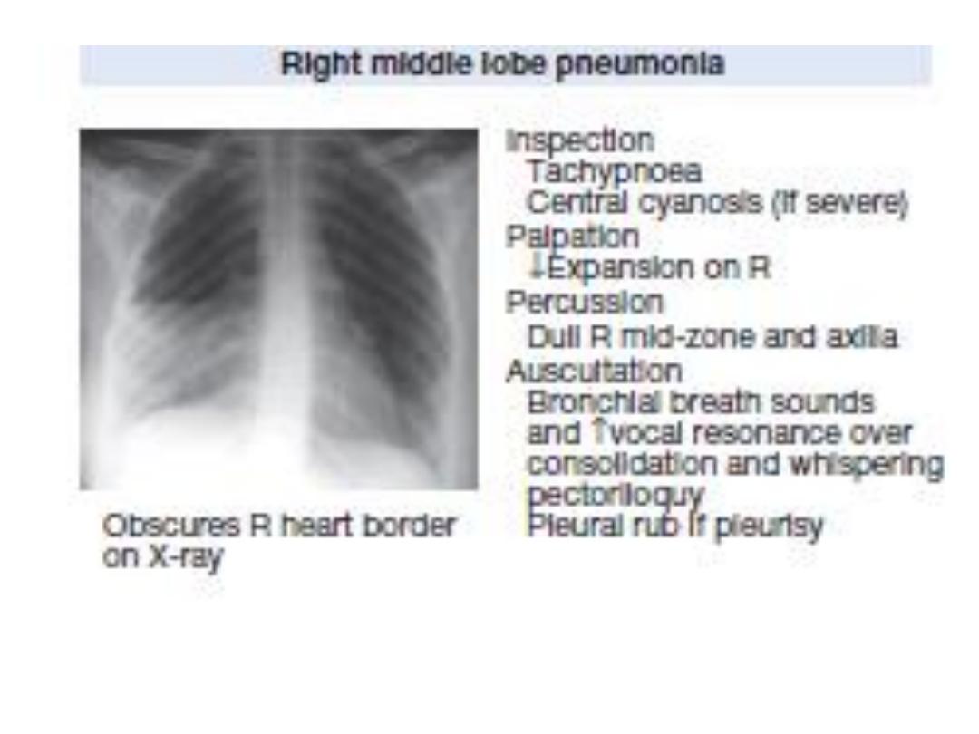
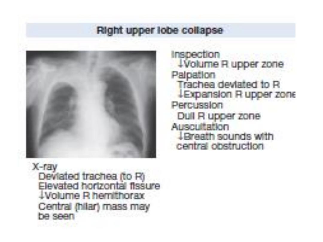
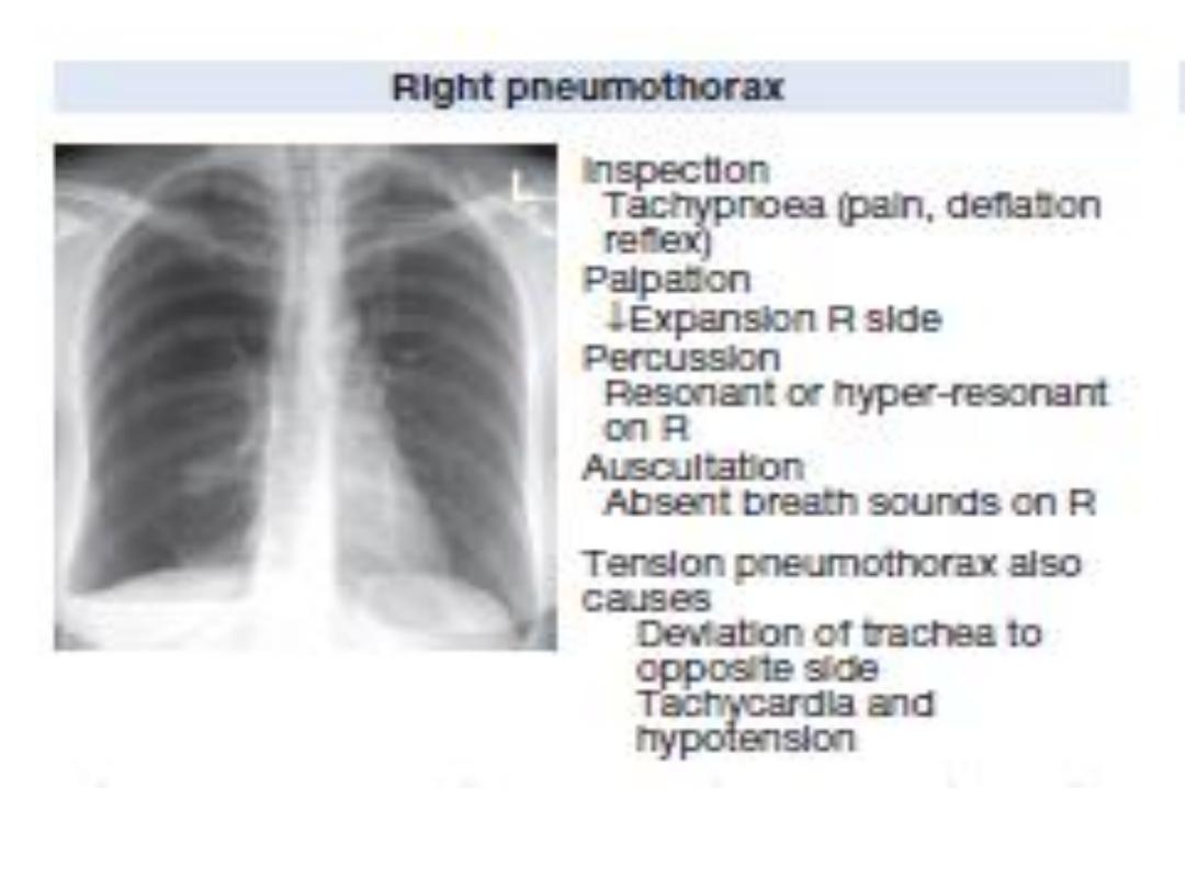
Signs in respiratory disease

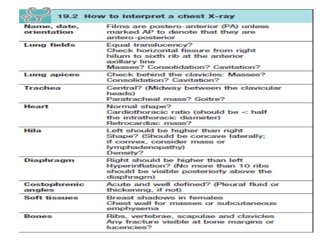
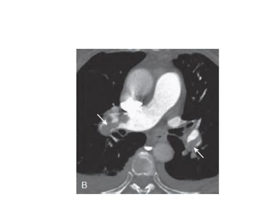
INVESTIGATION OF RESPIRATORY DISEASE
• Imaging
• The 'plain' chest X-ray
• A postero-anterior (PA) film
• lateral film.
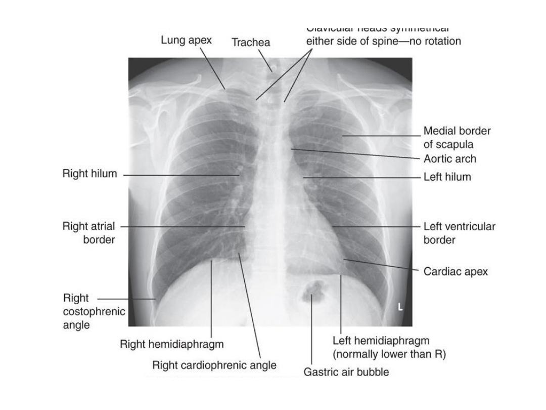
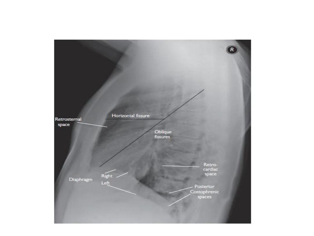
Normal lateral CXR

Computed tomography (CT)
CT provides detailed images of the pulmonary
parenchyma, mediastinum, pleura and bony
structures .
• High-resolution CT (HRCT)
• CT pulmonary angiography (CTPA)
Positron emission tomography (PET)
The radiotracer taken up by malignant tissue.
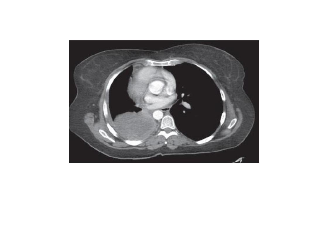
Computed tomography (CT)

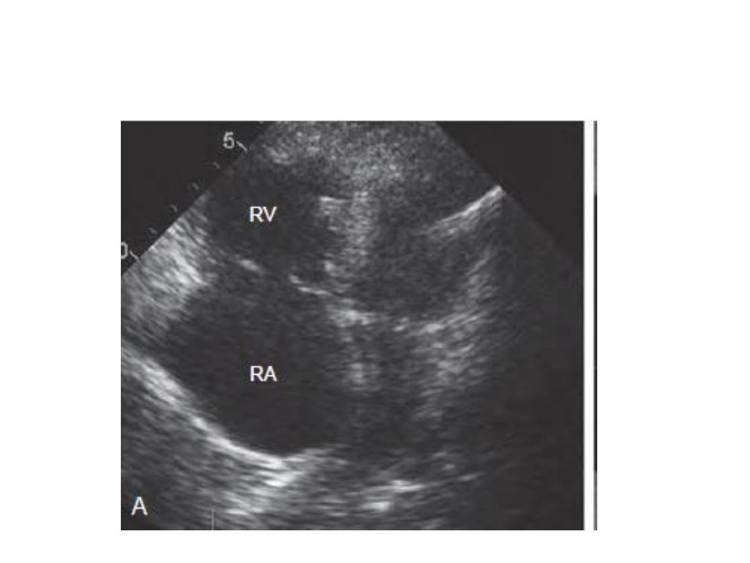
Ultrasound
Ultrasound is sensitive at detecting pleural fluid
.
• pleural biopsy.
• guide needle biopsy.
• Endobronchial ultrasound .
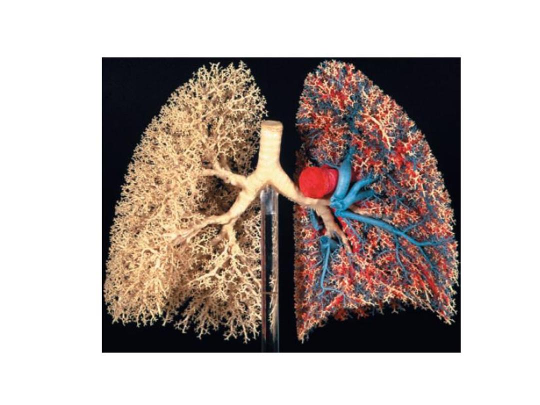

A resin cast of the human airway tree shows the dichotomous
branching of the bronchi from the trachea and the systematic
reduction of airway diameter and length with progressive branching. In
the left lung the pulmonary arteries (red) and veins (blue) .
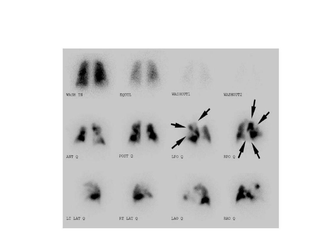
Ventilation-perfusion imaging
Pulmonary angiography
Echocardiography

Endoscopic examination
• Laryngoscopy
Bronchoscopy

Assessment of the mediastinum
• mediastinoscope
• Endobronchial ultrasound (EBUS)
• endoscopic ultrasound (EUS).

• Investigation of pleural disease
• The pleural biopsy using an
• (1)Abram's needle
(2)core biopsy guided by either ultrasound or
CT.
• Thoracoscopy.

Skin tests
• The tuberculin test.
• Skin hypersensitivity tests.
Immunological and serological tests
Microbiological investigations
Histopathological and cytological
examination .
Cytological examination

Immunological and serological tests
• The pneumococcal antigen.
• Influenza viruses can be detected in throat swab
samples.
• Legionella, Mycoplasma, Chlamydia or viruses)
antibody titres may eventually.
• hypersensitivity pneumonitis Precipitating
antibodies .
• Total levels of immunoglobulin E (IgE), and
levels of IgE

Respiratory function testing
Respiratory function tests are used to aid
diagnosis.
• assess functional impairment.
• monitor treatment or progression of disease.
Forced expiratory volume (FEV
1
) and forced
vital capacity (FVC)
• Flow/volume loops
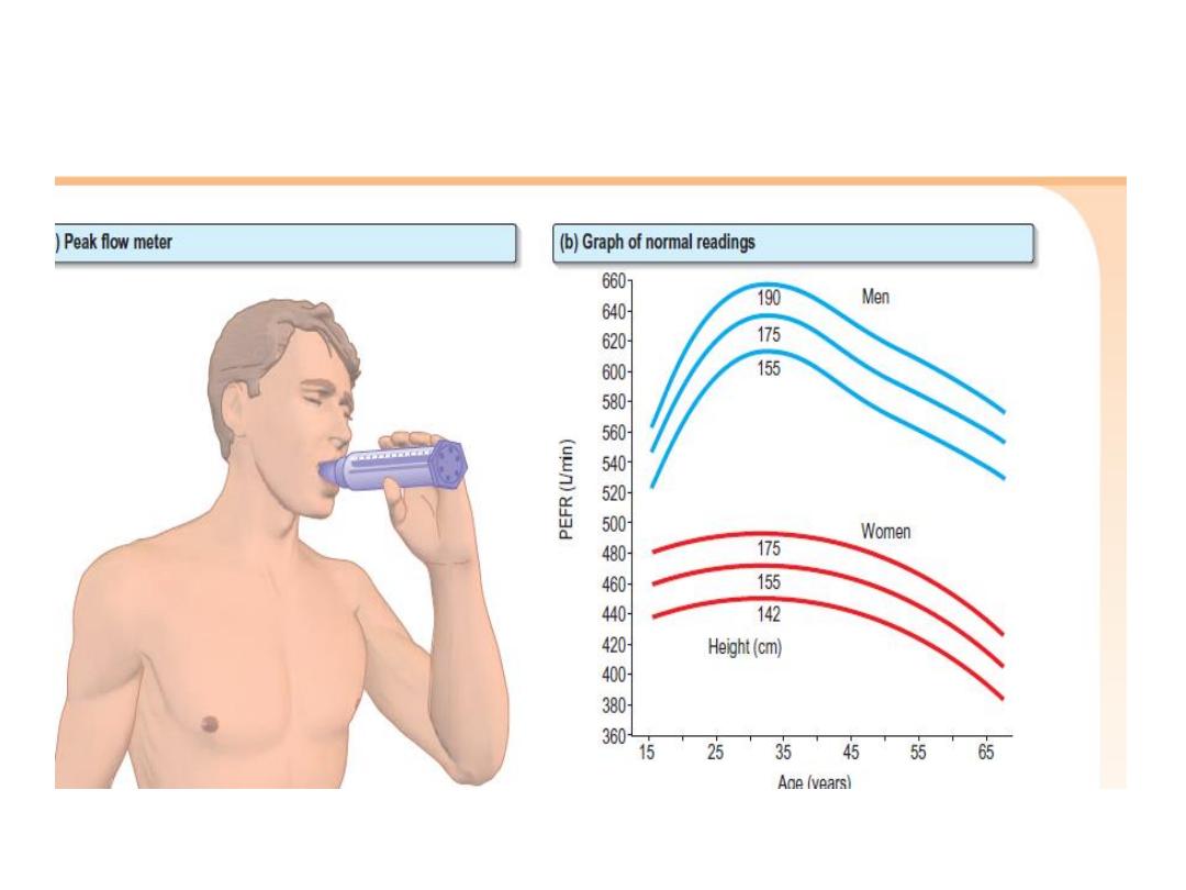
Peak flowmeter
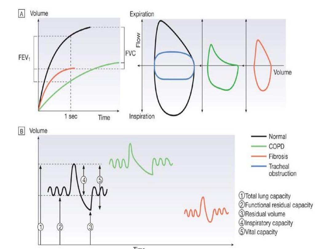

• Lung volumes
• spirometry.
• Body plethysmography
• Transfer factor

Q
• QUIZE