Dr. Suroor Mohammed
The Nervous System is formed of a number of cells, which are of 2 types:
1. Nerve cells = Neurons
2. Supporting cells = Glial cells
1.
NEURONS
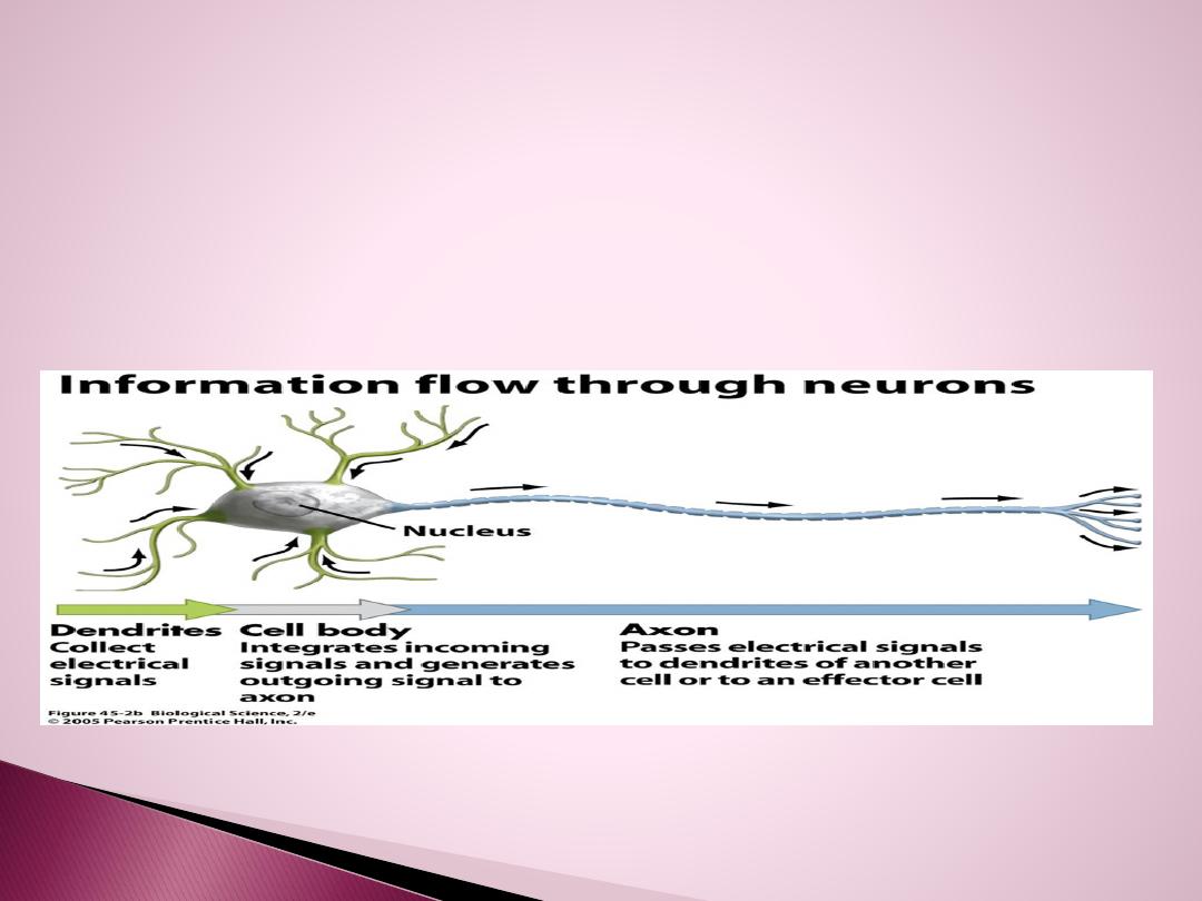
It is the basic structural unit of the NS.
It generates electrical impulses → transmitted from one part of the body
to another.
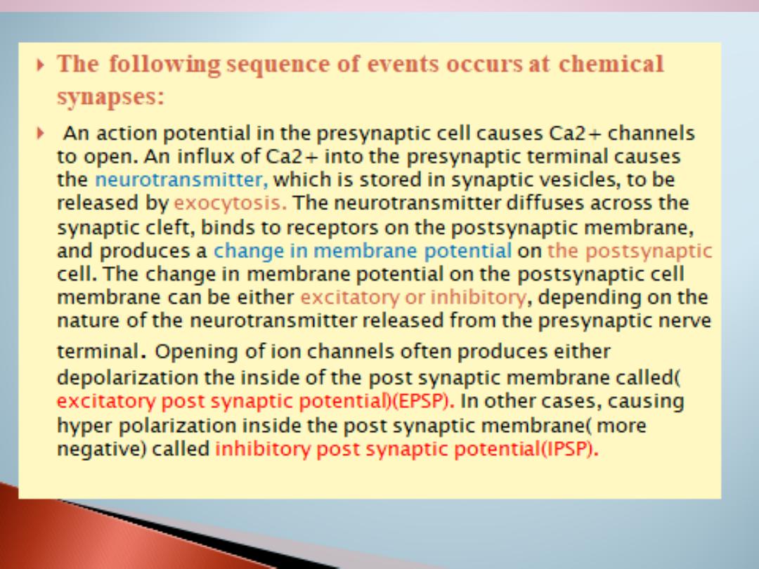
In most neurons: electrical impulses → release of chemical messengers
(= neurotransmitters) to communicate with each other.
Neurons are integrators: their output = the sum of the inputs they
receive from thousands of other neurons that end on them.
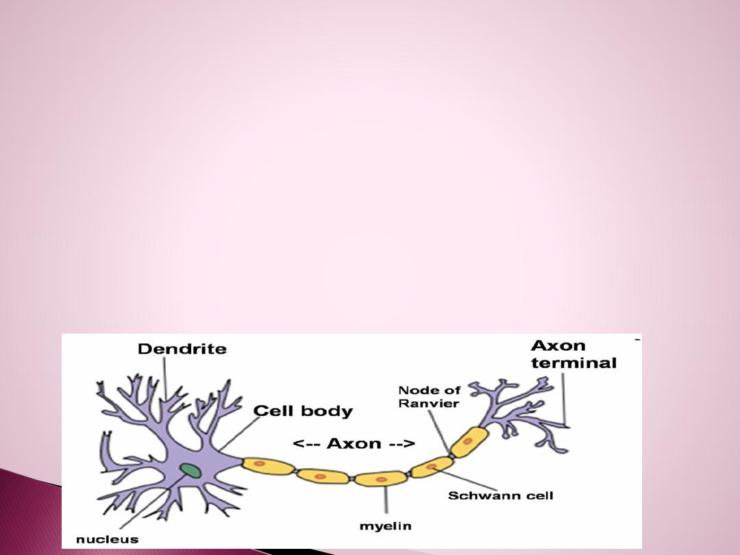
Neurons occur in a wide variety of shapes and sizes, but they share
common features. They all possess 4 parts:
1. Cell Body ( Soma):
It contains nucleus & organelles
→ provide energy & sustain metabolic activity of cells.
2. Dendrites:-
Usually 5-7 process (or more) highly branched (up to
400,000) → to increased surface area.
receive most input & Transmit impulses toward cell body only.

3. Axon
= Nerve Fiber:
- Usually single & long (few μm to 1m).
- Transmits impulses away from soma toward target
cell.
-
Axon hillock or initial segment
(= beginning of
axon + part of soma where axon joins it) is the
trigger zone where electric signals are generated in
most neurons. Signals are then propagated along
axon.
- Near its end the axon undergoes branching.
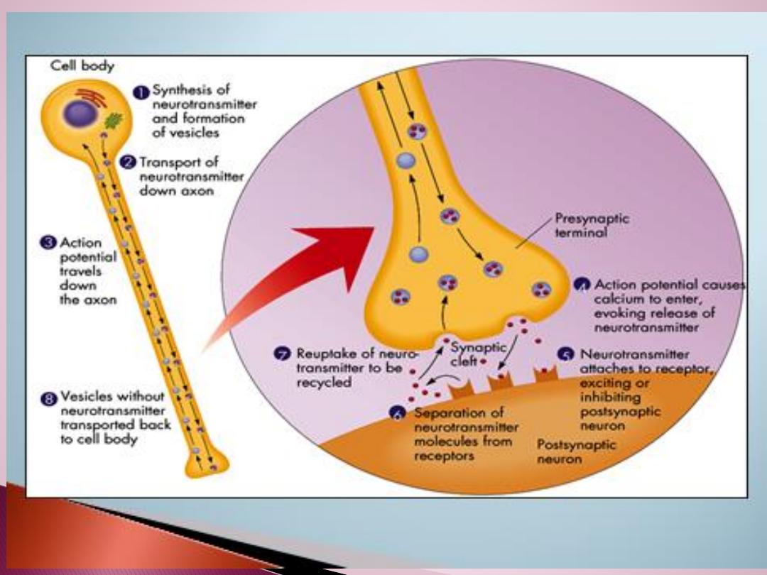
4. Axon Terminal
- Each branch of the axon ends in an axon terminal.
- Responsible for the release of neurotransmitters
(NT) from axon. NT diffuse out of the axon terminal
to next neuron or to a target cell

2.Supporting cells:
There are
sex categories
of supporting cells:
1.Schwann cells
, which form myelin sheaths
around peripheral axons.
2. Satellite cells
or ganglionic gliocytes , which
support neuron cells bodies within the ganglia
of the PNS.
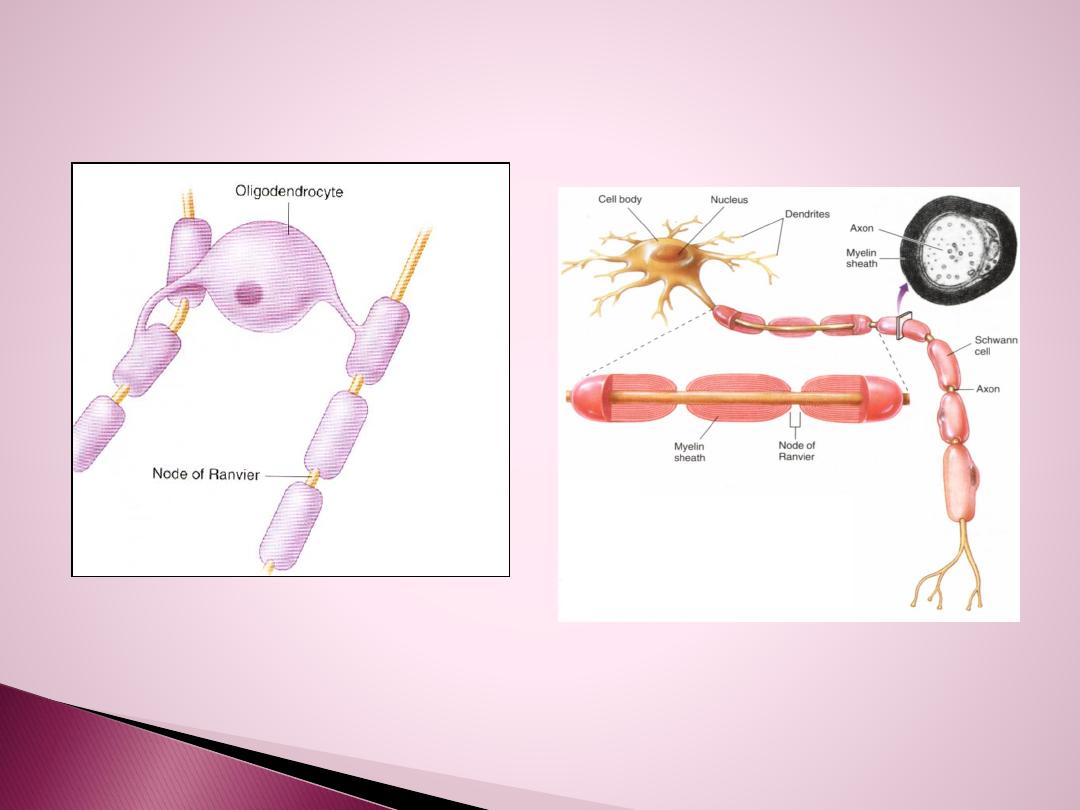
3. Oligodendrocytes,
which form myelin
sheaths around axons of CNS. Unlike Schwann
cells, they may branch to form myelin on up to
40 axons

4. Microglia,
which migrat through the CNS and
phagocytose foreign and degenerated material.
5. Astrocytes,
which help to regulate the
external environment of neurons in the CNS.
6. Ependymal cells
, which line the cavities of the
brain and the central canal of the spinal cord.
Axons of most (but not all) neurons are coated by a protective
layer = myelin sheath termed as “
myelinated
neurons”.
Myelin sheath is formed by the following cells:
1. In peripheral NS (PNS): by Schwann cells
2. In central NS (CNS): by oligodendrocytes.

Schwann Cells
- They are glia-like cells.
- During embryonic development, these cells
attach to growing axons & wrap around them →
concentric layers of plasma membrane.
- Myelin sheath of an axon is formed of many
Schwann cells that align themselves along length
of axon.
- Nucleus is located in outermost layer. Each
segment is separated from the next by a small
unmyelinated segment called node of Ranvier.
- Plasma membrane of Schwann cells is 80%
lipid → myelin sheath is mostly lipid → appears
glistening white to the naked eye.

Function of myelin sheath:
1. Myelin sheath helps to insulate axons &
prevents cross-stimulation of adjacent axons.
2. Myelin sheath allows nerve impulses to travel
with great speed down the axons, “jumping” from
one node of Ranvier to the next.
***Some nerve fibers are “unmyelinated”. Their
axons are covered by a Schwann cell, but there are
no multiple wrappings of membrane which
produces myelin. These axons conduct impulses
at a much lower rate.
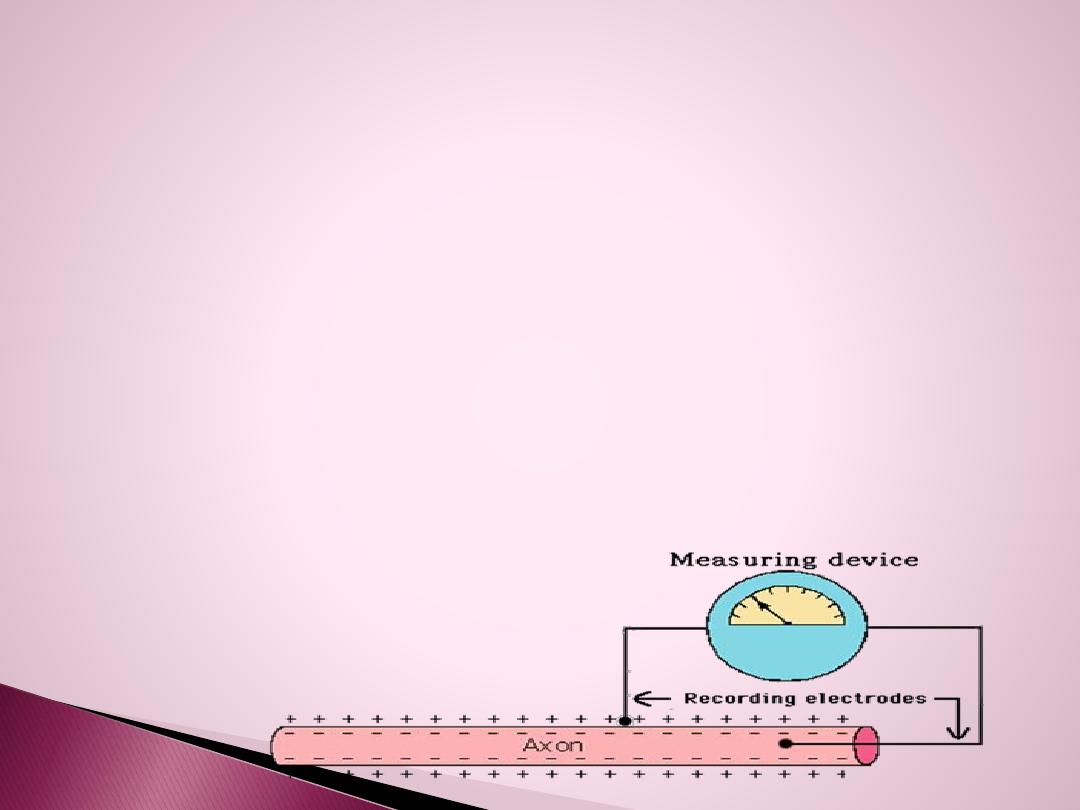
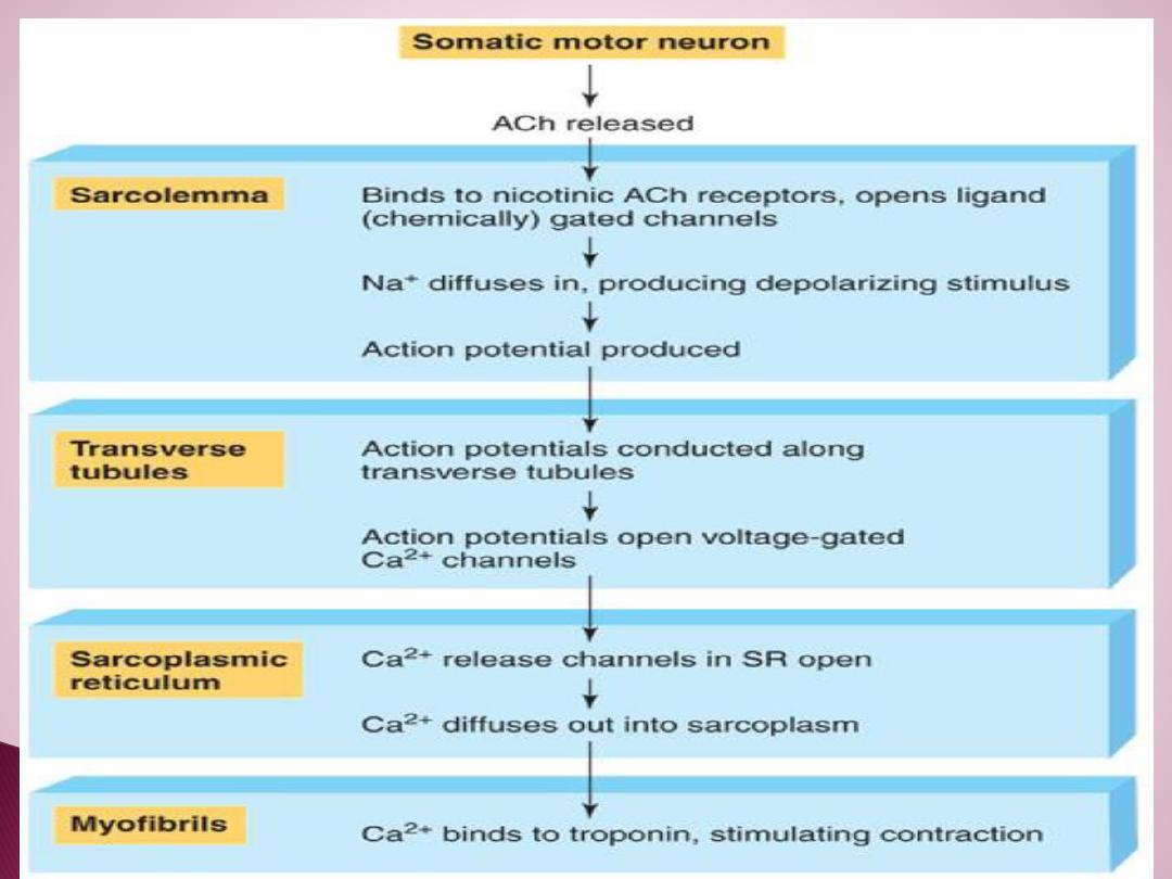
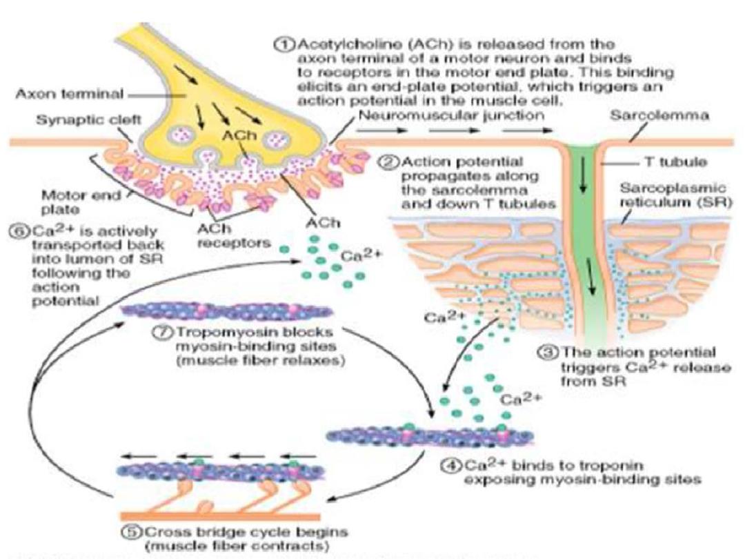
Nerve Impulse or Action Potential
Is the electrical current moving from the dendrites to cell body to axon.
It results from the movement of ions (charged particles) into and out a
neuron through the plasma membrane
Resting Membrane Potential *RMP*
The resting membrane potential is the potential difference that exists
across the membrane of excitable cells such as nerve and muscle in the
period between action potentials (i.e., at rest).
Is the difference in electrical charge on the outside and inside of the
plasma membrane in a resting neuron (not conducting a nerve impulse).
The
outside
has a
positive
charge and the
inside
has a
negative
charge.
We refer to this as a polarized membrane.
A
resting neuron is at about -70mV
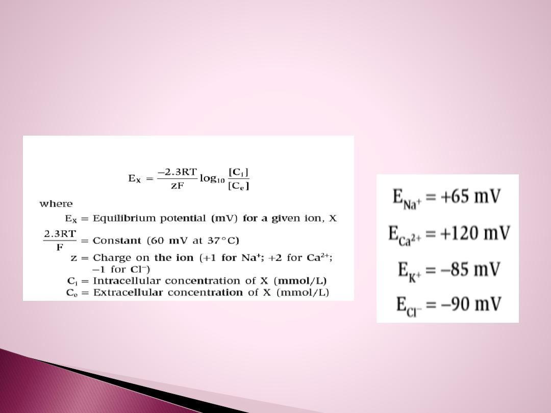
Nernst Equation
The Nernst equation is used to calculate the
equilibrium potential for an ion at a given concentration difference across a
membrane, assuming that the membrane is permeable to that ion. By
definition, the equilibrium potential is calculated for one
ion
at
a
time

At rest, The
K+
conductance or permeability is
high
and K+
channels are almost fully
open,
allowing K+ ions to diffuse
out
of the cell down the existing concentration gradient. This
diffusion creates a K+ diffusion potential, which drives the
membrane potential toward the K+ equilibrium potential. At
rest,
the Na+
conductance is
low,
and, thus, the resting
membrane potential is
far
from the Na+ equilibrium potential.
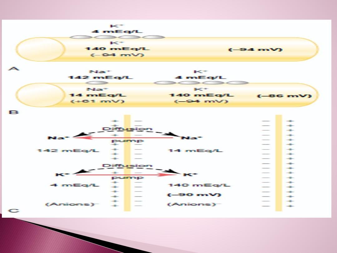
Because of the high ratio of potassium ions inside to outside, Therefore, if
potassium ions were the only factor causing the resting potential, the
resting potential inside the fiber would be equal to –94 mV.
The difference is due to :
1.There is
30 times more K+ inside the cell
than outside and about 15 times
more Na+ outside than inside.
2.There are
also large negatively charged proteins
trapped inside the cell.
(This is why it is negative inside.)
3. The action of
the Na+/K+ pumps
, that pump out 3 Na ions for every 2 K
ions that they transport into the cell.
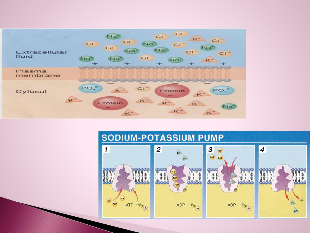
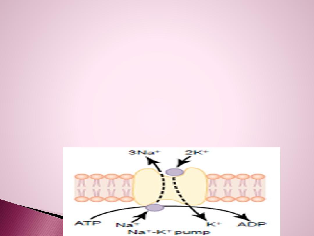
Why so much K+ inside
Special protein channels called sodium-potassium pumps moving 3 Na+ out
and bringing 2 K+ back in, when the cell is at rest.
**In a resting cell there are no open channels for Na+ to easily move back into
the cell. However, there are some K+ channels open at all time.
**Na+ causes the outside to be positive forcing more K+ into the cell. (Lots of
potassium ions inside the resting cell.
There is continuous pumping of three sodium ions to the outside for
each two potassium ions pumped to the inside of the membrane. The
fact that more sodium ions are being pumped to the outside than
potassium to the inside causes
continual loss of positive
charges from
inside the membrane; this creates an additional degree of negativity
Therefore, the net membrane potential of k+ with all these factors
operative at the same time is about –90 mV .

Alterations in the membrane potential are achieved by varying the membrane
permeability to specific ions in response to stimulations.
The physiology of neurons and muscle cells are their ability to
produce
and
conduct these changes in membrane potential, such an ability is termed
excitability or irritability.
If appropriate stimulation cause positive charges to flow into the cell. This
change is called
depolarization
(hypo polarization).
A return to the RMP is known as
repolarization
.
If stimulation cause the inside of the cell to become
more negative
than the
RMP this change is
called hyper polarization
which can be caused either by
positive charges leaving the cell or by negative charges enter the cell.
Any potential not the RMP called membrane potential.
Any stimulus can cause action potential
called threshold stimulus
.
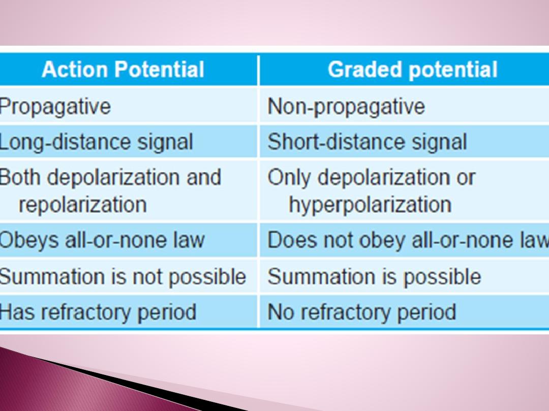
Electrotonic potential
is a local potential and cannot be propagated and
produced by sub threshold stimulus.

Action potential or ( nerve impulse)
The shape of action potential is the same in all the
nerves but it's magnitude change from one nerve to
another but it remain
uniform
shape.
When the axon membrane has been
depolarized
to a
threshold level, the
Na+gates open
and the membrane
becomes permeable to Na+, this permits Na+ to enter
the axon by diffusion which further depolarized the
membrane(make the inside less negative or more
positive).
Since the gates for the Na+channels of the axon
membrane are voltage regulated, this additional
depolarization opens more Na+channels and makes the
membrane even more permeable to Na+and more Na+
can enter the cell and induce a depolarization that opens
even more voltage– regulated Na+gates

A
positive
feedback loop is thus created, the
explosive increase in Na+permeability results in a
rapid
reversal
of the membrane potential in that
region from(– 70mv) to (+30mv). At that point in
time, the channels of Na+ close (become
inactivated).
At this time, voltage–
gated K+ channels open
and
K+ diffuse rapidly out
of the cell, and make the
inside of the cell less positive or
more negative
. This
process is called
repolarization
and represents the
completion of a negative feed back loop.
Once an action potential has been completed, the
Na+– K+ pump
will extrude the extra Na+ that has
entered the axon and recover the K+ that has
diffused out of the axon.

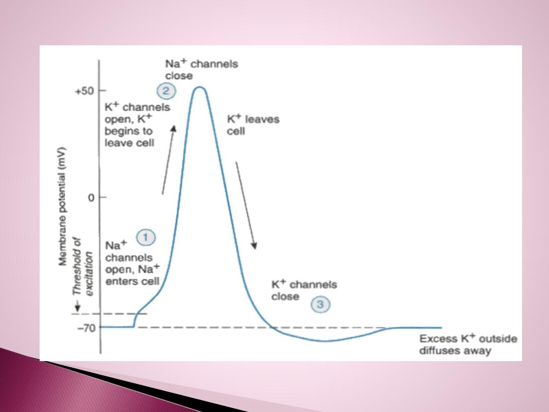
Phases of action potential
The first portion ,
local response
is due to slowly
opening of voltage gated Na+channels.
At the
firing
level (–55mv), full complete opening of
voltage gated Na+ channels, and Na+will rush very
rapidly to cell and membrane potential will reach (
+35mv). So the
depolarization
is due to opening of the
voltage gated Na+channels
.
At ( +35mv) the Na+entarce will stop because:
1. The opening of voltage gated Na+ channels are time
limited for short constant period and this limited time
cause depolarization will reach only to (+35mv) and then
stop.
2. At (+35mv) K+ channels are opened.

So
depolarization
from (–70mv to +35mv) is due
to activation of Na+ channels.
At (+35mv)
opening of K+ voltage gated
channels and K+ go outside according to
concentration gradient by diffusion. The channels
are opened completely from the first time and
repolarization
will start from (+35mv) to (–55mv),
at this point there will be in activation ( closure)
of K+ channels.
Na+ ions
concentration inside will
increase
and
this will cause stimulation to Na+–K+ pump to
exclude Na+ and carry K+ inside, till it reach to (–
70mv) again
( RMP),
so that after potential ( after
depolarization) phase due to Na+– K+ pump.

There will be loss of energy during action potential, so at
after depolarization to put the membrane potential again
equal to RMP by Na+–K+ pump is called {
recharging of
nerve},
so any stimulus at this phase the nerve will
not
response to it.
Why at( –55mv)Na+ channels will not open again ?
When Na+ channels inactivated, they need time more than
0.1msec. to return to their original conformation, and to
open Na+ channels again at (–55 mv) must apply stimulus
mor.e than the first one

Repolarization of the action potential
.
The upstroke is terminated,
and the membrane potential repolarizes to the resting level as a result of
two events.
1.The inactivation gates on the Na+ channels
respond to depolarization
by closing, but their response is slower than the opening of the activation
gates.
2.
Depolarization opens K
+
channels and increases K+ conductance
to
a
value even higher than occurs at rest.
The combined effect of closing of the Na+ channels and greater opening of
the K+ channels makes the K+ conductance much higher than the Na+
conductance. Thus, an outward K+ current results, and the membrane is
repolarized.
Hyperpolarizing afterpotential (undershoot).
For a brief period
following repolarization, the K+ conductance is higher than at rest and
the membrane potential is driven even closer to the K+ equilibrium
potential . Eventually, the K+ conductance returns to the resting level, and
the membrane potential depolarizes slightly, back to the resting
membrane potential.

All or Non law of action potential
If we apply
sub threshold
stimulus for the nerve, we get
no action
potential because it is un able to bring RMP to firing level. But if we
apply threshold stimulus, action potential will produced, and any
increase in the stimulus, there is no change in the magnitude and shape
or duration of action potential of the same nerve.
The shape, magnitude, duration and amplitude of action potential is the
same always all the same all the time and not change regardless to the
strength of stimulus to the same nerve
If a stimulus
is strong enough
to generate an action potential (reaches
threshold), the impulse is
conducted
along the entire length of the
neuron at the
same strength

Refractory periods:
Means the nerve will
not
respond to stimulus
during action
potential and it is of two types:
Absolute RP.
→located between the start of
depolarization
until one third of repolarization. The
nerve never
respond to
any stimulus
whatever it's strength, due to full, complete
activation of Na+ channels and so no extra channels are
opened, and then at (+35mv), there will be in activation of
Na+ channels and it need time to return back to it's original
condition.
Each nerve has got specific absolute RP, and this is important
to limit the number of action potential generated by the
neurons.
Relative RP.
→ This period involve from third of
repolarization
to the end of repolarization. If we apply stimulus stronger
than the original stimulus, the nerve will respond by new
action potential, because the Na+ channels will open and can
overcome the repolarization effects of the open K+ channels.

Factors effecting the conduction velocity of
nerve impulses
1)_Diameter of the axon
: which is directly proportional with the
speed of conduction.
All peripheral nerves are mixed nerves ( the nerve contain many
axons with different threshold levels and different diameter).
Maximal stimulus
:
is the stimulus when applied to nerve it will
stimulate all axons in the nerve.
Compound action potential:
Algebraic summation of all action
potentials of all the axons in the mixed nerve.
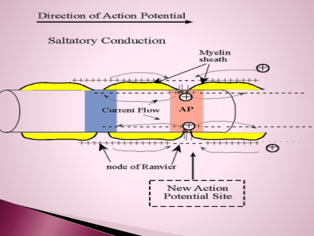
2)_ Myelin sheath
: myelinated nerve is faster than un myelinated
nerve because, myelin sheath is an insulator material, so the
depolarization and repolarization will occur between two nods of
Ranveir, the action potential in myelinated nerve will jump and
called Saltotary conduction, while in un myelinated nerve the action
potential will walk.
3). Hypoxia
( low O2 to the tissue) , it depress the conduction.
4). Local anesthesia.
5). Temperature
.

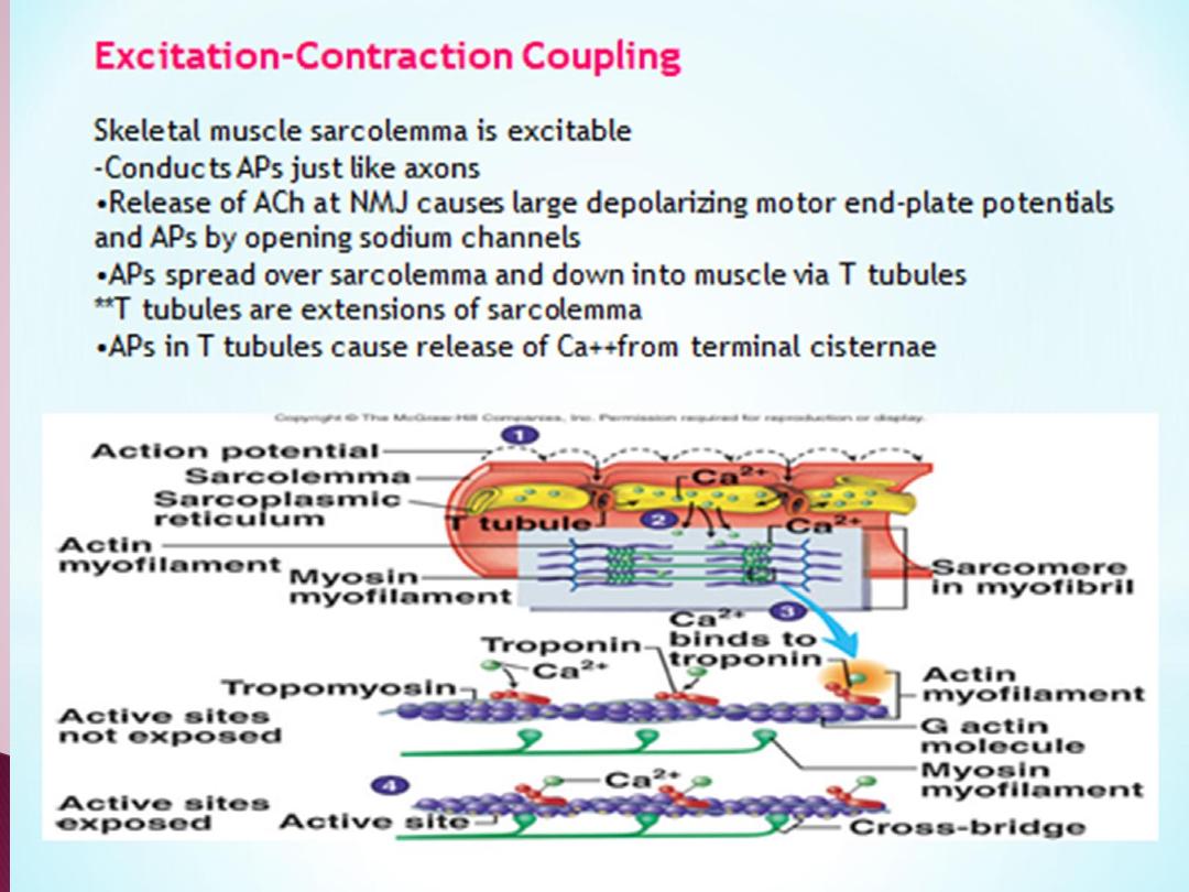
Skeletal muscles
About 40% of the body mass is skeletal muscle, and perhaps another 10%
is smooth muscle and cardiac muscle.
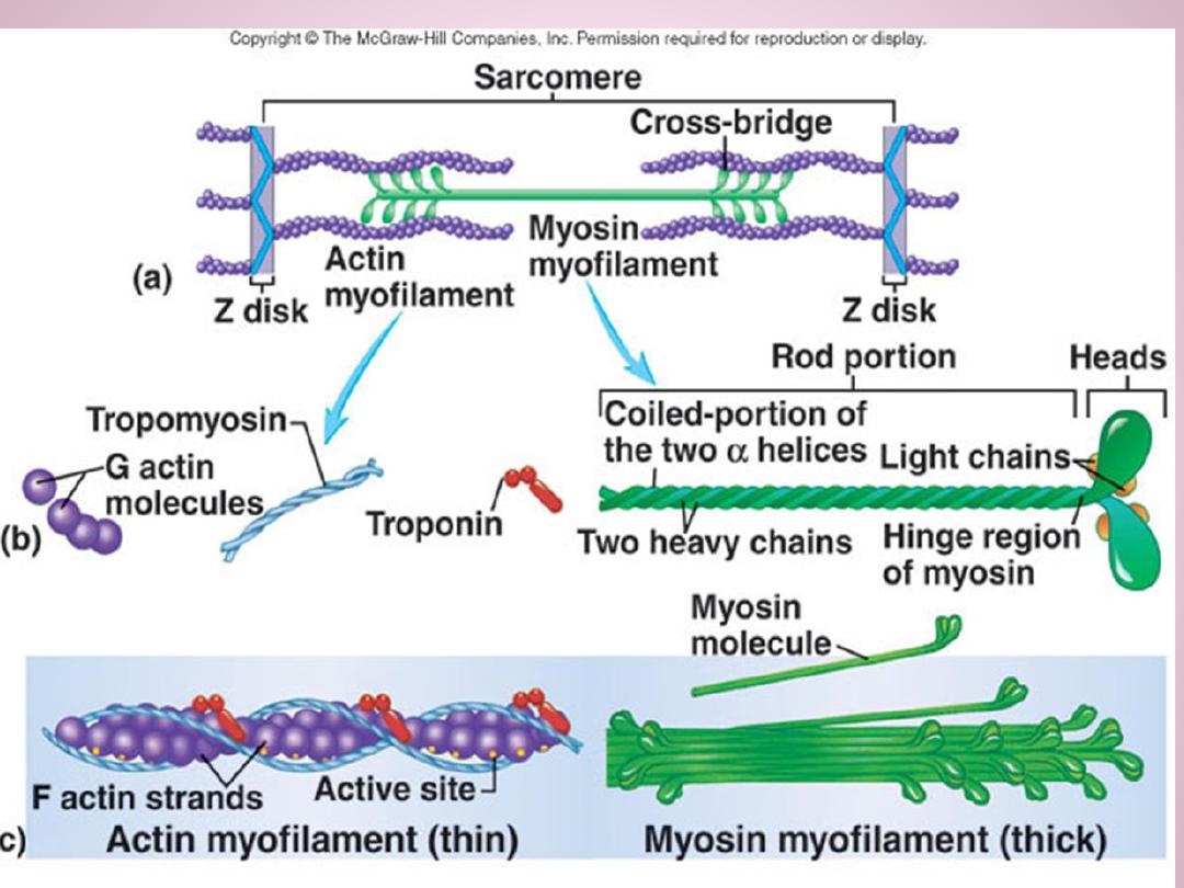
Skeletal muscle fibers
are long, cylindrical ,multinucleated cells ,with
peripheral nuclei. Each muscle fiber is composed of subunits called
myofibrils
that extend the length of the fiber. each myofibril is composed
of about 1500
myosin
thick filaments and 3000
actin t
hin filaments , that are
responsible for muscle contraction.
.
•
Cross-bridges.
The small projections from the sides of the myosin
filaments are cross-bridges. They protrude from the surfaces of the
myosin filament along its entire length except in the center. Myosin
crossbridges interact with actin filaments causing contraction.
•
Z disc.
The ends of the actin filaments are attached to Z discs .The Z
disc passes across the myofibril and from one to another, attaching and
aligning the myofibrils across the muscle fiber. The entire muscle fiber
therefore has light and dark bands, giving skeletal and cardiac muscle
astriated appearance
Sarcomere
. The portion of a myofibril that lies between two successive
Z discs is called a sarcomere..