CARDIOVASCULAR
SYSTEM

Objective:
1. What is the action potential?
2. What is differences between the membrane
properties of cardiac and skeletal muscle account for
the prolonged action potential and the plateau in
cardiac muscle?

cardiac action potential
is a brief change in voltage membrane potential
across the cell membrane of heart cell
This is
caused by the movement of charged atoms
(called ions) between the inside and outside of the
cell, through protiens called ion channels.

•
The cardiac action potential differs to action
potentials found in other types of electrically
excitable cells, such as nerves, as well as
varying within the heart also, this is due to
the presence of different ion channels in
different cells

•
Unlike the action potential in skeletel muscle
cell, the cardiac action potential is not initiated
by nervous activity. Instead, it arises from a
group of specialized cells, that have automatic
action potential generation. In healthy hearts,
these cells are found in the right atrium and are
called the sinoatrial node .

They produce roughly 60-100 action potentials
every minute. This action potential passes along
the cell membrane causing the cell to contract,
therefore the activity of the SAN results in a
resting heart rate of roughly 60-100 beats per
minute.

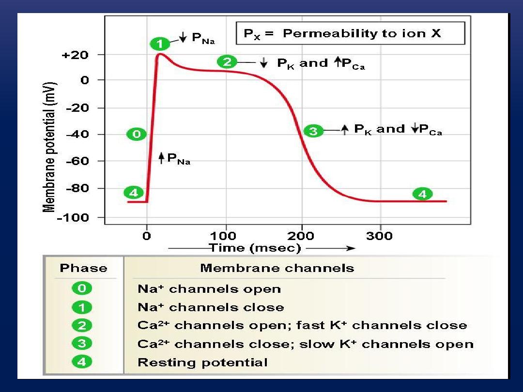
The action potential recorded in a ventricular
muscle fiber averages about 105 millivolts,
which means that the intracellular potential rises
from a very negative value, about −85 millivolts,
between beats to a slightly positive value, about
+20 millivolts, during each beat.

After
the
initial
spike,
the
membrane
remains
depolarized for about 0.2 second, exhibiting a plateau,
followed at the end of the plateau by abrupt repolariza-
tion.
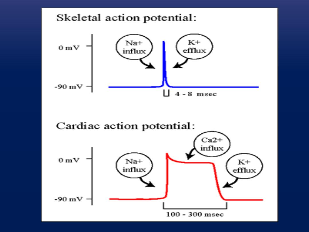
The presence of this plateau in the action potential
causes ventricular contraction to last as much as 15
times as long in cardiac muscle as in skeletal muscle.


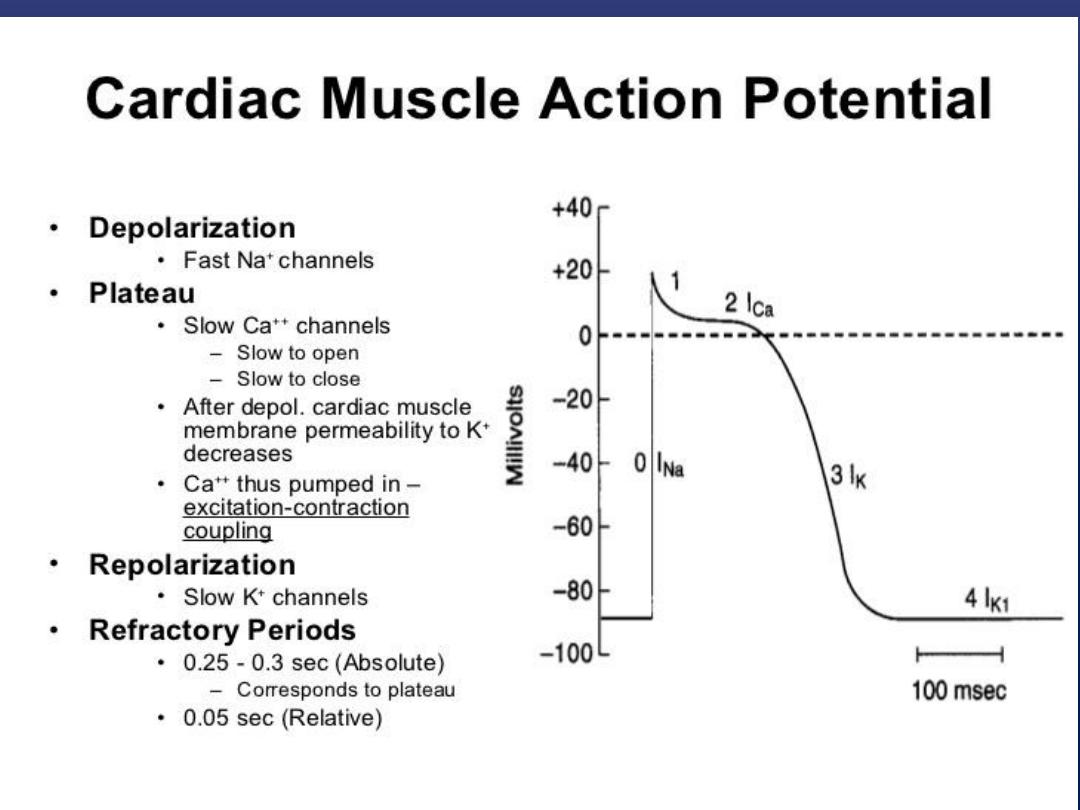
PHASES OF CARDIAC MUSCLE ACTION
POTENTIAL
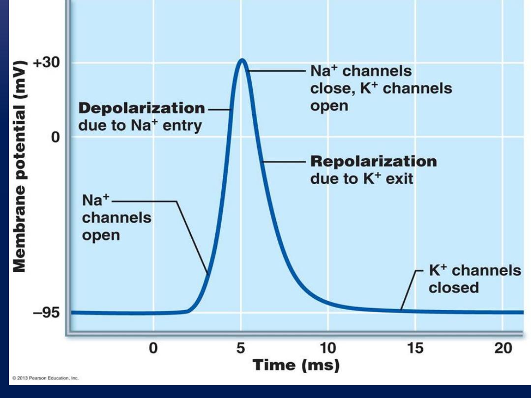
Phase 0 (depolarization), fast sodium
channels
membrane potential becomes more
positive. Voltage-gated sodium channels (fast
sodium channels) open and permit sodium to
rapidly flow into the cell and depolarize it.
The membrane potential reaches about +20
millivolts before the sodium channels close.
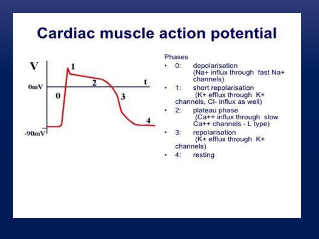

Phase 1 (initial repolarization), fast sodium
channels close
. The sodium channels close,
the cell begins to repolarize, and potassium
ions leave the cell through open potassium
channels.
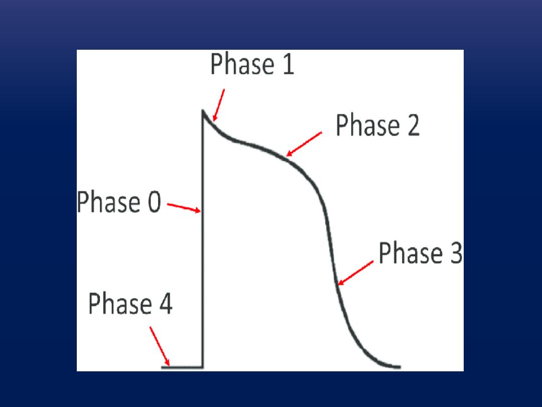

Phase 2 (plateau), calcium channels open and fast
potassium channels close.
A brief initial repolarization occurs and the action
potential then plateaus as a result of (1) increased
calcium ion permeability and (2) decreased potassium
ion permeability.

The voltage-gated calcium ion channels open
slowly during phases 1 and 0.Potassium channels
then close, and the combination of decreased
potassium ion efflux and increased calcium ion
influx causes the action potential to plateau
.

Phase 3 (rapid repolarization), calcium channels
close and slow potassium channels open.
The
closure of calcium ion channels and increased
potassium ion permeability, permitting potassium
ions to rapidly exit the cell, ends the plateau and
returns the cell membrane potential to its resting
level.
Phase 4 (resting membrane potential) averages
about −90 millivolts.

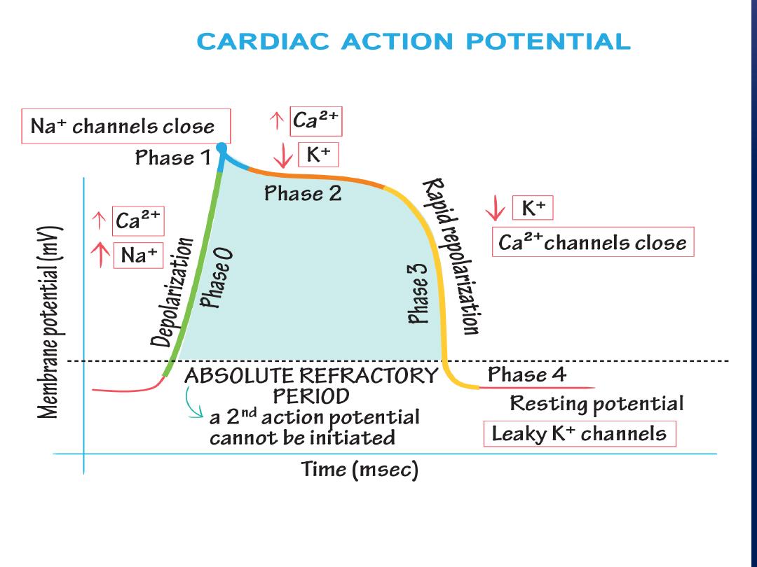
Refractory Period of Cardiac Muscle
.
Cardiac muscle, like all excitable tissue, is
refractory to restimulation during the action
potential. Therefore, the refractory period of the
heart is the interval of time during which a normal
cardiac impulse cannot re-excite an already excited
area of cardiac muscle.

The normal refractory period of the ventricle is
0.25 to 0.30 second, which is about the duration of
the prolonged plateau action potential. The
refractory period of atrial muscle is much shorter
than that for the ventricles (about 0.15 second for
the atria compared with 0.25 to 0.30 second for the
ventricles).

What is differences between the membrane
properties of cardiac and skeletal muscle account
for the prolonged action potential and the plateau
in cardiac muscle?
At least two major differences between the
membrane properties of cardiac and skeletal
muscle account for the prolonged action potential
and the plateau in cardiac muscle.

First, the action potential of skeletal muscle is caused
almost entirely by the sudden opening of large numbers
of
fast sodium channels
that allow tremendous numbers
of sodium ions to enter the skeletal muscle fiber from
the extracellular fluid. These channels are called “fast”
channels because they remain open for only a few
thousandths of a second and then abruptly close. . At
the end of this closure, repolarization occurs, and the
action potential is over within another thousandth of a
second or so.

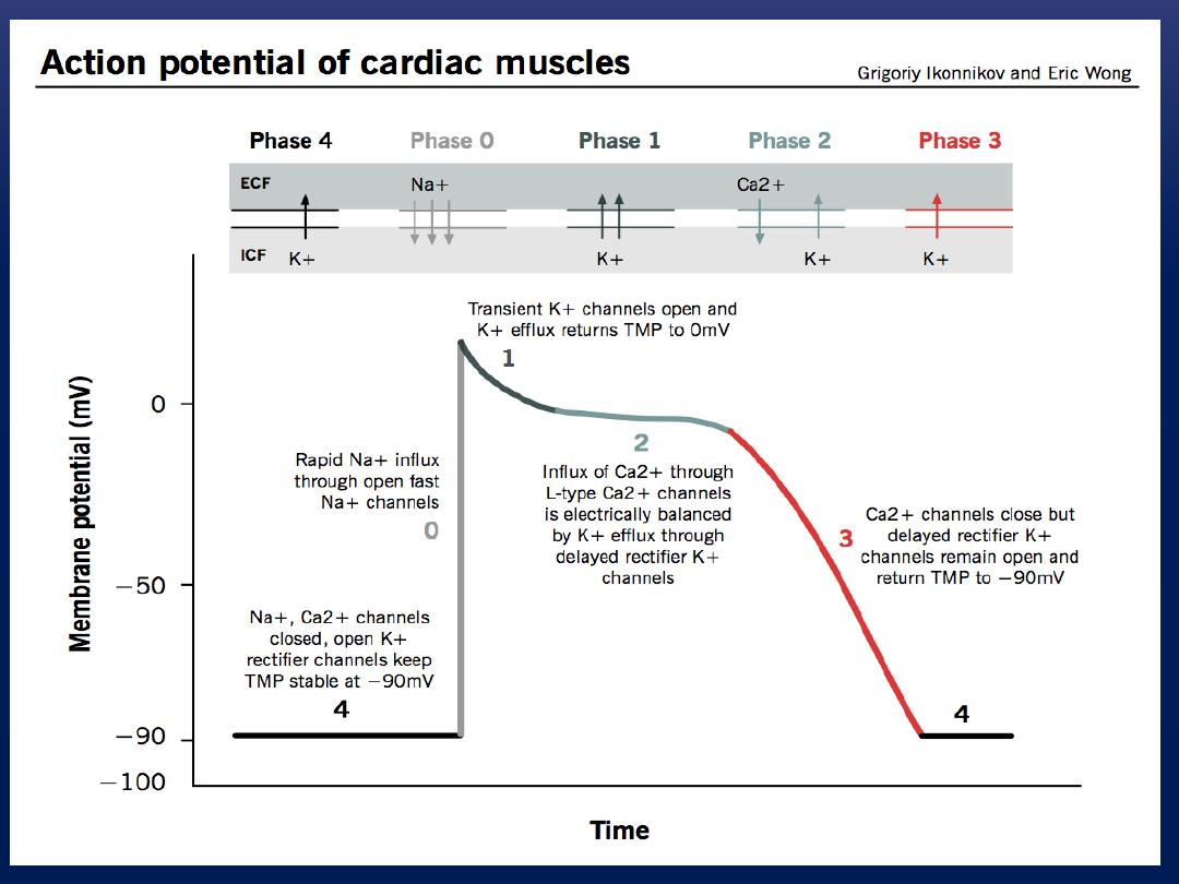
In cardiac muscle, the action potential is caused by
opening of two types of channels: (1) the same
voltage-
activated fast sodium channels
as those in skeletal
muscle and (2) another entirely different population of L-
type
calcium channels (slow calcium channels),
which
are also called calcium-sodium channels. This second
population of channels differs from the fast sodium
channels in that they are slower to open and, even more
important, remain open for several tenths of a second.

During this time, a large quantity of both calcium
and sodium ions flows through these channels to
the interior of the cardiac muscle fiber, and this
activity maintains a prolonged period of
depolarization, causing the plateau in the action
potential.

The second major functional difference between
cardiac
muscle and
skeletal
muscle that helps
account for both the prolonged action potential
and its plateau is this: Immediately after the onset
of the action potential, the permeability of the
cardiac muscle membrane for
potassium
ions
decreases about fivefold, an effect that does not
occur in skeletal muscle.

This decreased potassium permeability may
result from the excess calcium influx through
the calcium channels just noted.
Regardless of the cause, the decreased
potassium permeability greatly decreases the
outflux of positively charged potassium ions
during the action potential plateau and thereby
prevents early return of the action potential
voltage to its resting level..

When the slow calcium-sodium channels do
close at the end of 0.2 to 0.3 second and the
influx of calcium and sodium ions ceases, the
membrane permeability for potassium ions also
increases rapidly; this rapid loss of potassium
from the fiber immediately returns the membrane
potential to its resting level, thus ending the
action potential


What is the correct pathway for the heart's
conducting system?
1. SA node → AV node → Bundle
branches → Bundle of His → Purkinje
2. SA node → AV node → Bundle of
His → Bundle branches → Purkinje fibers
3. SA node → AV node → Bundle of
His → Purkinje fibers → Bundle branches
4. SA node → AV node → Purkinje
fibers → Bundle of His → Bundle
5. branches AV node → SA node → Bundle of
His → Bundle branches → Purkinje fibers

In a cardiac muscle cell, the membrane potential
increases rapidly...
1. when potassium gates open and potassium diffuses
into the cardiac muscle fiber.
2. when potassium gates open and potassium diffuses
out of the cardiac muscle fiber.
3. when the sodium gates open and sodium diffuses
into the cardiac muscle fiber
4. when the sodium gates open and sodium diffuses
out of the cardiac muscle fiber.