Histology of skin dr. Ahmed Alhuchami

Hair
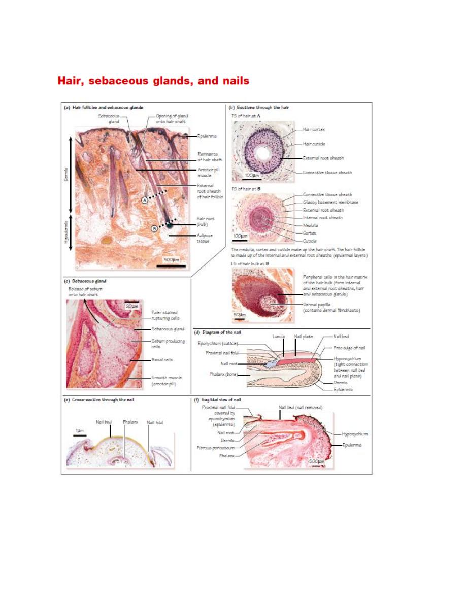
Hairs are made up of hair follicles and hair shafts .The hair shaft is made up of
columns of dead keratinized cells(hard keratin) organized into three layers :
• a central medulla , or core (not seen in fine hairs);
• a keratinized cortex ;
• a thin hard outer cuticle , which is highly keratinized.
Hair follicles are tubular invaginations of the epidermis, which develop as down
growths of the epidermis into the dermis. The hair follicle contains the following.
• An external root sheath ( ERS) , which is continuous with the epidermis. This
layer does not take part in hair formation. A glassy basement membrane
separates the ERS from the surrounding connective tissue.
• An internal root sheath ( IRS ), which lies inside the ERS. The IRS contains
keratinized cells derived from cells in the hair matrix. The type of keratin found
here is softer than that found in the hair itself. The IRS degenerates at the point
where the sebaceous gland opens onto the hair.
Hair follicle stem cells in the hair matrix , which is found in the hair bulb , are
responsible for forming hair . The stem cells proliferate, move upwards, and
gradually become keratinized to produce the hair. These stem cells also form the
ERS and IRS , and sebaceous glands.
The dermis forms a dermal papilla at the base of the hair follicle/ hair bulb, which
provides the blood supply for the hair. It is separated from the hair matrix by a
basement membrane.
Hair follicles can become inflamed, due to bacterial infections (e.g.,
Staphylococcus aureus ), resulting in a tender red spot or pustule (folliculitus).
Contraction of the arrector pili muscle , a small bundle of smooth muscle cells
associated with the hair follicle, raises the hair, and forms ‘ goose bumps ’ . This
helps to release sebum from the gland into the duct, and to release heat.

Pigmentation of hair
Hair color depends on the pigment melanin, produced by melanocytes in the hair
matrix. Differences in hair color depend on which additional forms of melanin,
pheomelanin (red or yellow) and eumelanin (brown or black), are present. The
pigment is produced by melanocytes in the hair matrix, and is then transferred to
keratinocytes, which retain this pigment as they differentiate and form hair. In old
age, melanocytes stop producing melanin, and hair turns white.
Hair growth
Hair follicles alternate between growing and resting phases. Hair is only produced
in the growing phase (this can be several years in the scalp). Hair falls out in the
resting phase. This can be permanent, resulting in baldness.
Cutting hair does not change its growth rate.
Sebaceous g lands
These glands are branched, acinar holocrine glands found next to hair follicles
.The cells rupture to secrete an oily sebum into the lumen of the hair follicle (
holocrine secretion).
The ruptured cells are continuously replaced by stem cells ( basal cells ), located
at the edges of the gland.
Nails
Nails (or nail plates ) consist of a strong plate of hard keratin, and they protect the
distal end of each digit . The nail plate is a specialized layer of stratum corneum .
It is formed by the nail bed ( nail matrix ) underneath the nail plate. Proliferating
cells in the basal layer of the nail bed move upwards continuously. As the cells
move upwards they are displaced distally and gradually transformed into hard
keratin, which lengthens and strengthens the nail plate. The tightly packed, hard,
keratinized epidermal cells in the nail plate have lost their nuclei and organelles.
Nails grow at a rate of about 0.1 – 0.2 mm per day.
The proximal end of the nail plate extends deep into the dermis to form the nail
root . The nail root is covered by the proximal nail fold . The covering epithelium
of this nail fold is called the eponychium . The outer thick corneal layer of the

eponychium extends over the dorsal layer of the nail, to form the cuticle , which
protects the base of the nail plate. If the cuticle is lost, the nail bed can become
infected. The eponychium also contributes to the formation of the superficial
layer of the nail plate.
The distal edge of the nail has a free edge . Here, the nail plate is firmly attached
to the underlying epithelium, which is known as the hyponychium ( hypo means ‘
below ’ ). This region of epithelium contains a thickened layer of stratum
corneum.
The tight connection between the nail plate and the underlying epithelium
protects the nail bed from bacterial and fungal infections. If this connection is
disrupted, then a fungal infection of the nail bed can cause onychomycosis .
Pigmentation of nails
The pink color of nails derives from the color of the underlying vascular dermis.
The nail itself is thin, hard, and relatively transparent. The white crescent at the
proximal end of the nail is called the lunula . The underlying epithelium is thicker
here, which explains the white color of the lunula. The increased epithelial
thickness means that the pink color of the dermis does not shown.