EMBRYOLOGY OF FACE AND SKULL
I. DEVELOPMENT OF FACE (weeks 4-8)
A. Formation of Face Primordia
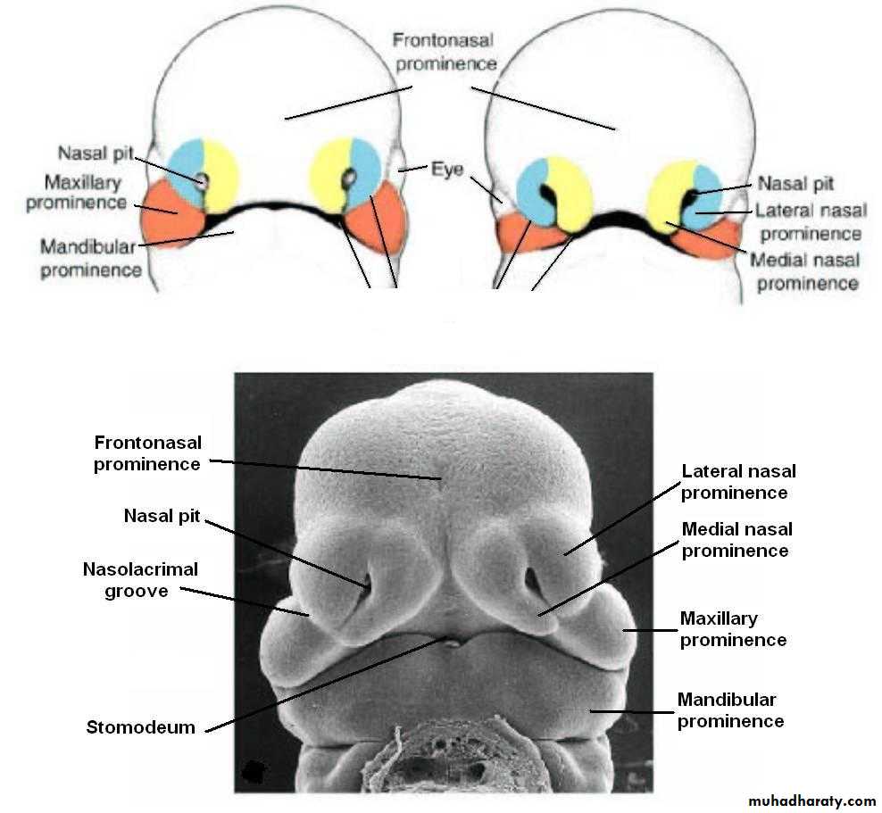
1. The face develops from the fusion of five face primordia which develop during week four and fuse during weeks five-eight. These primordia are ectodermal swellings or prominences of the cranial end of the head and some of its pharyngeal arches. They are filling with proliferating mesoderm and neural crest. They include one midline frontonasal (frontal) prominence, two (bilateral) maxillary prominences (or swellings) and two (bilateral) mandibular prominences (or swellings). The mandibular and maxillary prominences develop from pharyngeal arch I. The frontonasal prominence is not an arch derivative). These prominences encircle the stomodeum, or primitive mouth opening. This opening is originally covered by buccopharyngeal membrane, which is itself formed by fusion of the ectoderm with the underlying endoderm.
B. Development of face: fusion of face primordia: (Weeks 5-8)
1. Before any fusion takes place, the frontonasal prominence develops two sets of bilateral swellings or placodes:Nasal placodes develop close to the Stomodeum opening. They are the future site of the nasal cavities.
2. Steps in Fusion Process
a. The mandibular prominences fuse with each other in the midline.
b. Both maxillary prominences fuse with the frontonasal prominence. The nasolacrimal groove marks the line of fusion on each side.
c. The nasal placodes of the frontonasal prominence develop medial and lateral nasal processes or swellings. They encircle nasal pits which will become nasal openings.
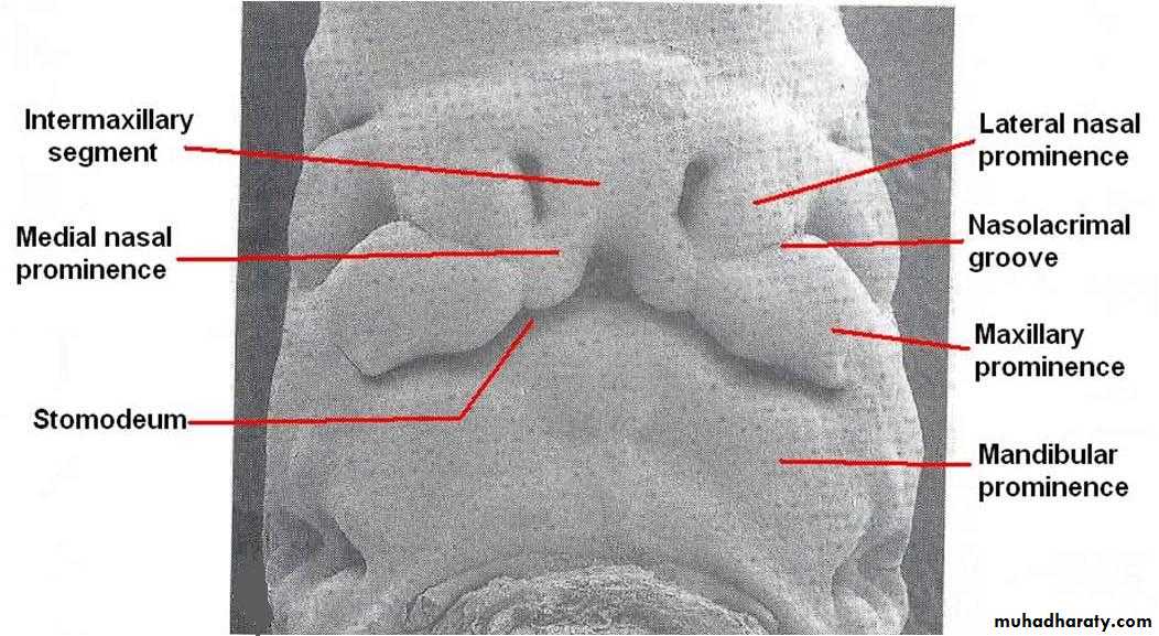
d. Fusion of medial nasal processes:
The medial nasal prominences grow towards each other and fuse in the midline. This fusion forms the intermaxillary process. This process elongates into the stomodeum opening, allowing it to fuse with the maxillary prominences to each side of it.
C. Fate of external Face Primordia:
Externally, the facial prominences form the structures of the forehead, nose, and all of the lower face and lips:
1. Frontonasal Prominence
a. Forms the forehead.b. Forms the nasal cavities and surface of the nose: The nasal pits deepen to form openings to the nasal cavities, while the nasal swellings form the dorsum and apex of the nose.
c. The medial intermaxillary segment forms the medial portion of the upper lip, called the philtrum.
2. Maxillary Prominences: Form the lateral upper lip.
3. Mandibular Prominences: Form the lower lip, and the inferior &lateral face.
4. The final form of the face results from changes in the proportion of these structures.
II. DEVELOPMENT OF THE PALATE (5-12 weeks)
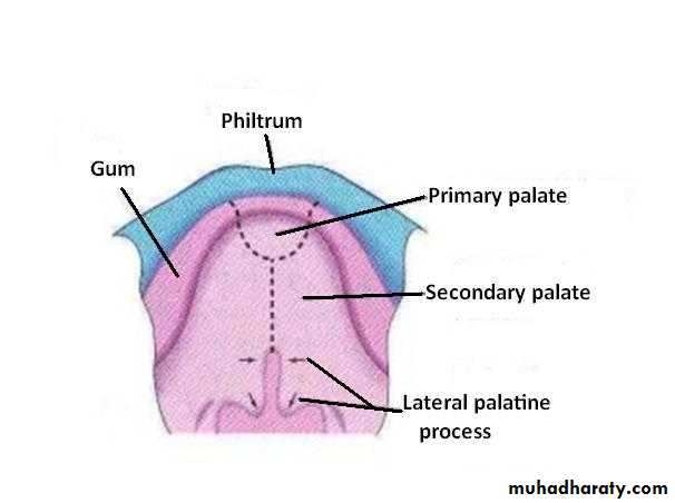
The face primordia form important internal structures of the PALATE. Development of the palate involves the formation of the primary palate, the secondary palate, and fusion of their processes. These structures develop from internal derivatives of the facial primordia. Many abnormalities can occur in these fusion processesA. Primary Palate
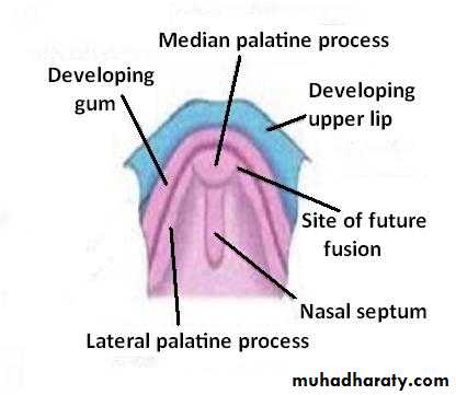
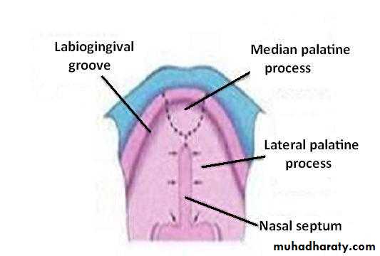
Forms from an internal swelling of the intermaxillary process, called the median palatine process.
B. Nasal cavities
1. Formed by deepening of the nasal pits. Initially they are separated from the oral cavity by the intermaxillary process, which forms the floor of the nasal cavities. Continued growth causes the nasal cavities to break through to the oral cavity at the primitive choana. At this point the floor of the nasal cavity is now formed by the Primary palate developing from the intermaxillary segment.
C. Nasal Septum
1. Forms from another internal midline swelling of the frontonasal prominence. This swelling grows into the nasal cavity towards the tongue, where it encounters the primary palate and the developing secondary palate.
D. Secondary Palate
1. Forms from two lateral palatine shelves or processes which develop as internal projections of the maxillary prominences. They grow towards the midline, where they fuse with each other, the primary palate, and the nasal septum. Fusion starts anteriorly. The fusion of the palate and nasal septum separates the nasal cavities from each other, and from the mouth.
E. Hard and Soft Palates
1. Palatine bone forms from mesoderm of the maxillary prominence (archI) in the anterior portion of the secondary palate, forming the hard palate.
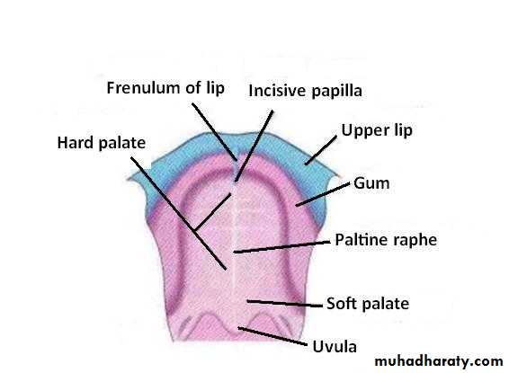
2. The posterior portion of the secondary palate forms the soft palate, which contains no bony or cartilagenous elements. Skeletal muscles in the soft palate are derived from pharyngeal arches I & IV
III. DEVELOPMENT OF THE SKULL
The skull can be divided into two regions:
Neurocranium: Bones which surround and encase the base of the brain & form support capsules for sense organs
Viscerocranium: Bones which surround the structures of the face and palate. Each has separate origins, as follows:
A. Neurocranium (brain skeleton)
1. Cartilagenous: chondrocranium, Formed by endochondral ossification of 3 pairs of cartilage models which have a dual origin: cranial 2 pairs derived from neural crest; caudal pair derived from mesoderm: sclerotomes of occipital somites (these bones can be thought of as modified vertebral elements). These bones form the base of the skull; they extend as a plate from the anterior skull to surround the foramen magnum. They also form capsules surrounding and protecting sensory organs (eyes, inner ear, and nasal cavities). These bones are: the sphenoid, occipital, ethmoid, petrous part of temporal bone.
2. Membranous: Formed directly by intramembranous ossification from mesoderm. These bone form the flat bones of the vault of the skull around the brain (calveria). They include the frontal and parietal bones.
B. Viscerocranium (face skeleton)
These bones are all formed from derivatives of pharyngeal arches I-III, which means they are derivatives of either neural crest or mesoderm.1. Cartilagenous bones
Malleus, incus, stapes (Arches I and II) Hyoid (Arches II and III)
2. Membranous bones
Maxilla, zygomatic, squamous temporal (maxillary prominence of Arch I)
Mandible (mandibular prominence of arch I)