


HERPESVIRUS DISEASES
PRIMARY HERPETIC STOMATITISOr also known as
PRIMARY HERPETIC GINGIVOSTOMATITIS
Page:235-238.
Cryo-electron microscopy reveals structure of a herpesvirus capsid:
All herpesvirus infections are more common in immunosuppression, particularly HIV infection. Infection can then be more severe, persistent or recurrent.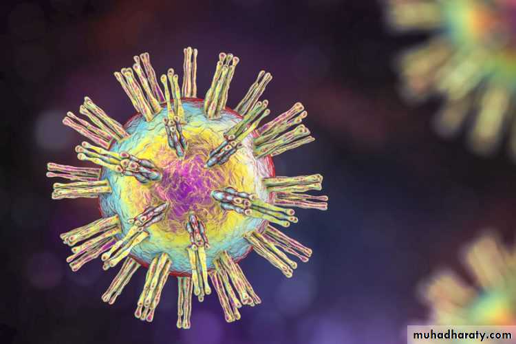
Herpes simplex virus(cryo-electron micrograph)
The herpesvirus is genetically and structurally one of the most complex viruses. It spreads within the host population efficiently, causing a range of diseases in humans, including congenital disorders and cancers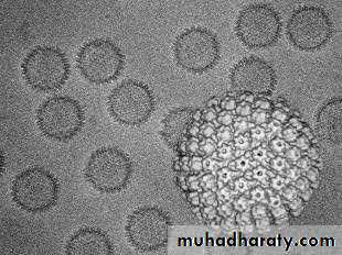
PRIMARY HERPETIC STOMATITIS
Primary infection (systemic infection in a non-immune individual) is caused by Herpes simplex virus, usually type 1.Herpes viruses have been considered almost ubiquitous(being present everywhere at once).
Free virus is transmitted by close living conditions, through saliva in early childhood.
Clinical features
The great majority of primary infections are subclinical or completely asymptomatic. Only 1% of those infected develop any symptoms, and these are often minimal. Most patients with clinical infection are children aged younger than 6 years.Clinical features
The vesicles develop on the oral mucosa approximately one week after transmission. The hard palate, gingiva and dorsum of the tongue are favoured sites . The vesicles are dome-shaped, tense and filled with clear fluid and increase from 1 mm in diameter to 2–3 mm.Symptoms depend on extent of ulceration, but the ulcers are painful and often interfere with eating.
There is a degree of fever and systemic upset with enlarged cervical lymph nodes.
This can be severe, particularly in adults.
Clinical features:
Oral lesions usually resolve within a week to 10 days, but malaise can persist so long that an adult may not recover fully for several weeks.Herpetic vesicle. The vesicle is formed by accumulation of fluid within the prickle cell layer. The virusinfected cells, identifiable by their enlarged nuclei, can be seen inthe floor of the vesicle, and a few are floating freely in the vesicle fluid
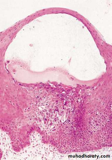
Pathology:
The DNA virus targets epithelial cells, and replication leads to cell lysis. Clusters of infected cells break down to form the vesicles in the upper epithelium . Virusdamaged epithelial cells with swollen nuclei and marginated chromatin (ballooning degeneration) are seen in the floor of the vesicle and in direct smears from early lesions.A smear from a herpetic vesicle. The distended degenerating nuclei of the epithelial cells cluster together to give the typical mulberry appearance.
Infected cells fuse with normal adjacent cells, spreading the infection and forming multinucleated cells.
The image shows a vesicle has ruptured to form anulcer and the epithelium at the margin contains enlarged, darkly staining virus-infected cells liberating free virus into the saliva.
The full thickness of the epithelium is destroyed to produce a
sharply defined ulcer associated with an inflammatory infiltrate.
Herpetic stomatitis: key features
• Usually caused by Herpes simplex virus type 1• Transmitted by close contact
• Usually affects children younger than 6 years
• Vesicles, followed by ulcers, affect any part of the oral mucosa
• Gingiva usually involved
Lymphadenopathy and fever of variable severity
• Smears from vesicles show ballooning degeneration of viral-damaged cells
• Rising titre of antibodies to HSV confirms the diagnosis
• Supportive treatment important
• Aciclovir very effective if given in first 48 hours
Latency:a state of inactivity.
Herpes simplex and zoster are neurotropic as well as epitheliotropic viruses. After the immune response develops and mucosal infection subsides, the virus can remain hidden from the immune response in the sensory nerves that supply the site of the primary infection. Virus is transported back along the nerves from the mucosa to the neurone cell bodies in the ganglia where it establishes a lifelong latent infection. During latency there are no symptoms, no virus replication occurs and the patient is not infectious.Reactivation of the latent infection depends on the host cell, not the virus.
after which it travels back down the neurones to infect the
skin or mucosa.