
NEOPLASIA
Cancer is the second leading cause of death after the cardiovascular diseases.
Even more agonizing than the associated mortality is the emotional and physical
suffering caused by neoplasms, the only hope for controlling cancer lies in learning
more about its pathogenesis, and great steps have been made in understanding the
molecular basis of cancer .
So that we will talk about :
Definition.
-
-What are the basic components
-How the tumors are designated
-what is the basic classification of the tumors
-
What are the differences between benign & malignant tumors
.
-How can you define tumor Grade & tumor Stage
-Mechanisms of invasion & metastasis
-Molecular basis of cancer
-Kinetic of tumor cell growth- factors that affect the rate of tumor growth
- How do growing tumors develop a blood supply? (Tumor angiogenesis)
-Tumor immunity -Host defense against tumor
-How the tumor cells can escape the immune system.
-Causes of cancer –Carcinogenesis
Before we discuss the features of cancer cells and the mechanisms of
carcinogenesis, it is useful to summarize the
fundamental and shared characteristics of
cancers:
• Cancer is a genetic disorder caused by DNA mutations. Most pathogenic mutations
are either induced by exposure to mutagens or occur spontaneously as part of aging.
• Genetic alterations in cancer cells are heritable, being passed to daughter cells upon
cell division.
• Mutations and epigenetic alterations impart to cancer cells a set of properties that are
referred to collectively as cancer hallmarks. These properties produce the cellular
phenotypes that give the natural history of cancers as well as their response to various
therapies.
Basic research has elucidated many of the cellular and molecular abnormalities that
give rise to cancer and govern its pernicious behavior. These insights are in turn
leading to a revolution in the diagnosis and treatment of cancer .

NOMENCLATURE:
Defcinition
Neo
plasm
=
New
growth
Abnormal mass of tissue The growth exceeds & is uncoordinated with that of
normal tissue. Persists in the same excessive manner of growth even after
cessation of the stimuli which evoked the growth. Purposeless growth , competes
with the normal cells for energy & blood supply.
All neoplasms have two basic components
1- The “transformed” neoplastic cells (the parenchyma)
2- The supportive stroma.
The latter is composed of non-transformed (non-neoplastic) elements, such as
connective tissues and blood vessels.
Basic classification of the tumors
Three categories
1- Benign tumors
2- Malignant tumors
- Primary
- Secondary ( metastatic)
3- Borderline – potentially malignant tumors
Premalignant tumors
BENIGN TUMORS
In general their names end with the suffix “oma”.
Benign mesenchymal tumors are named after their tissue of origin + “oma”
Examples
Leiomyoma
Lipoma
Chondroma,
Schwannoma, etc.
Benign epithelial tumors
are named after their tissue of origin, sometimes combined
with architecture + “oma”.
Examples
Adenomas are tumors arising from glandular tissue and usually form glandular
patterns
Cystadenomas as above but with cystic components
Papillary cystadenoma as above but with papillary (warty or finger-like projections)
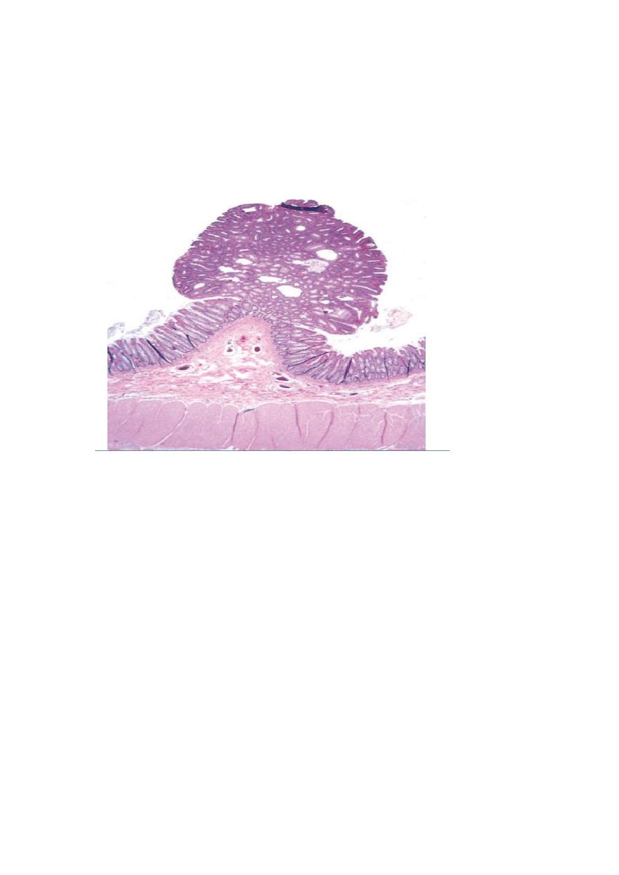
Papillomas characterized by the production of finger like projections.
MALIGNAT TUMORS
These are generally called cancers.
Their nomenclature is based on their appearance (the morphology of their
parenchymal cells) and the presumed tissue of origin.
They’re broadly divided into two categories
1-
Carcinomas
arising from epithelial cells
Examples include
- Squamous cell carcinomas,
- Adenocarcinoma,
- small cell undifferentiated carcinomas, etc.
2-
Sarcomas
arising from or differentiating towards mesenchymal tissues
Examples include
- Osteosarcomas
- Leiomyosarcomas,
- Rhabdomyosarcomas.
Some tumors have more than one parechymal cell type, these include
1.
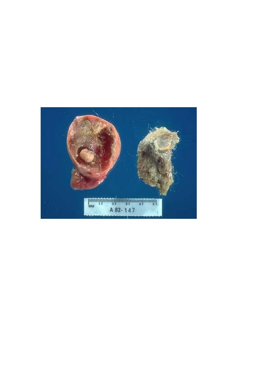
Teratomas
, which are tumors of germ cell origin, showing differentiation along all
the three germ layers (Ectoderm: like skin and its adnexae such as hair follicles and
sebaceous glands, Endoderm: like gut epithelia and Mesoderm: like bone, cartilage,
muscle, etc), thus a variety of parenchymal cell types may be seen in any one of these
neoplasms.
Examples include
- teratoma of the ovary
- teratoma of the testis
2.
Mixed tumors;
these differ from teratomas in that they are derived from one
germ cell layer, that differentiates into more than one parenchymal cell type.
Examples include
- Pleomorphic adenoma of salivary glands
- Fibroadenoma of breast
EXCEPTIONS
Exceptions to the above mentioned rules include tumors that are always malignant
such as
o
Lymphomas
(tumors of lymphoid tissue).
o
melanomas
(malignant tumors of melanocytes)
o
Seminomas and Dysgerminomas
(tumors of primitive
germ cells) .

CHARACTERISTICS OF BENIGN AND MALIGNANT TUMORS
BENIGN TUMORS
MALIGNANT TUMORS

Rate of growth
Slower
Faster
Histological features
Similar to tissue of origin.
Nuclei are normal.
Cells uniform in size and
shape.
Many differ from tissue of
origin.
Enlarged pleomorphic nuclei,
hyerchromasia, Prominent
nucleoli, Increased mitotic
activity, abnormal mitosis.
Cellular pleomorphism in size
and shape.
Clinical effects
Local pressure effects.
Hormone secretion.
Cured by adequate excision.
Local pressure and tissue
destructive effects.
Inappropriate hormone
secretion.
Not cured by local excision
because of metastasis.
Paraneoplastic syndromes.
CHARACTERISTICS OF BENIGN AND MALIGNANT NEOPLASMS
There are three fundamental features by which most benign and malignant tumors
can be distinguished:
1-
differentiation and anaplasia.
2-
local invasion .
3-
and metastasis.
DIFFERENTIATION AND ANAPLASIA
Differentiation
is the extent to which tumor cells resemble comparable normal cells
of the tissue of origin.
In most benign tumors the constituent cells closely mimic corresponding normal cells.
Malignant tumors display a range of differentiations that form the basis of tumor
grading.
Lack of differentiation (anaplasia) is the hallmark of malignant cells.
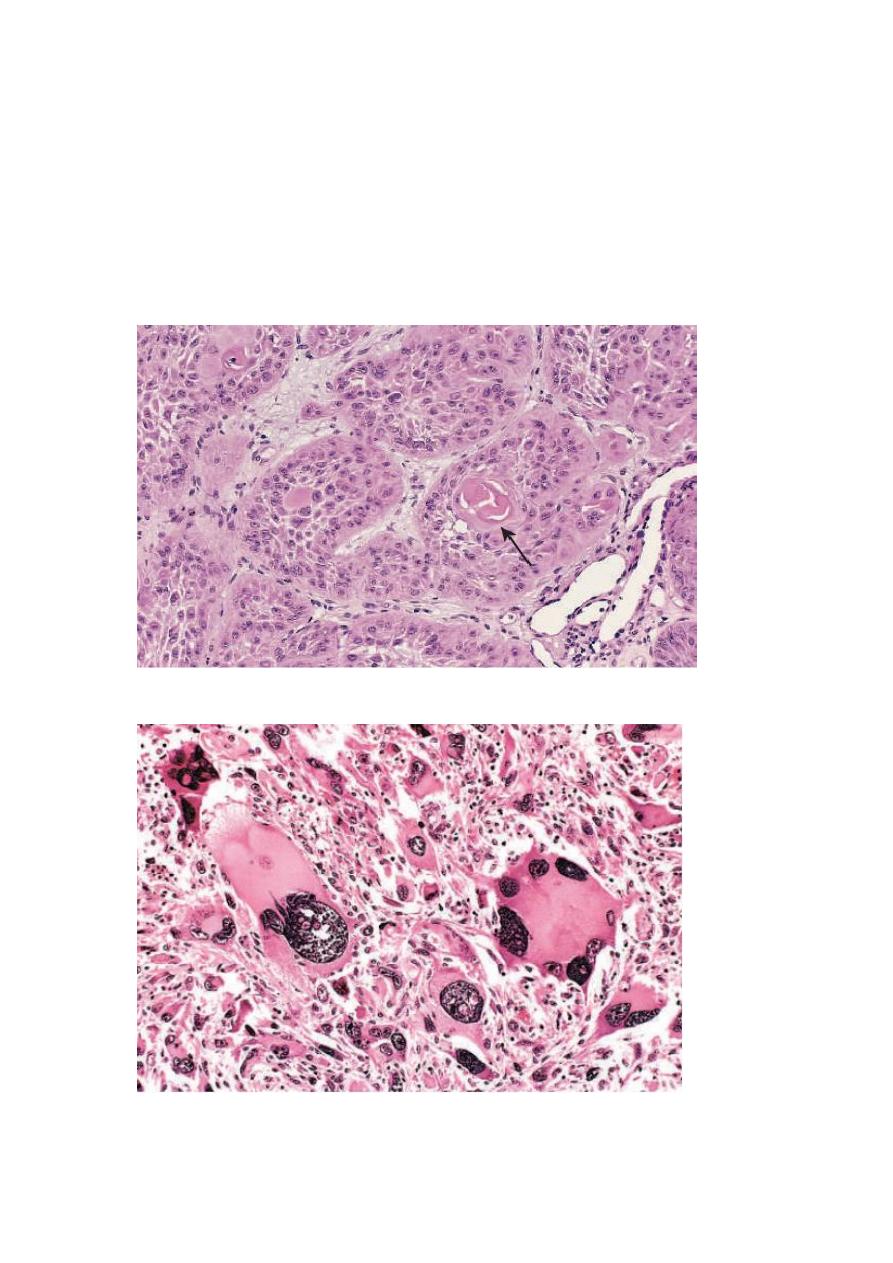
Well differentiated SCC of skin
Pleomorphic malignant tumor (rhabdomyosarcoma)
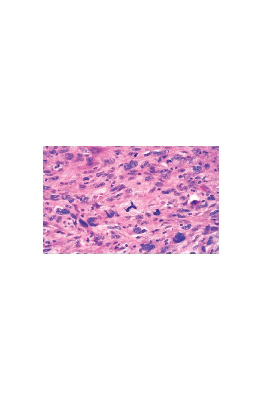
Histopathological features of anaplasia:
Cellular and nuclear pleomorphism
refers to variation in size and shape of cells
and their nuclei.
Hyperchromatism
refers to dark staining of nuclei due to abnormally increased
chromatin (nucleic acids contents), a reflection of aneuploidy.
Increased nuclear-cytoplasmic ratio (N/C)
reaching nearly 1:1 (instead of the
normal 1:4-6).
Abundant mitoses
reflecting increased proliferative activity
Abnormal mitoses,
e.g., tripolar spindles (normally mitosis is bipolar).
Tumor giant cells
containing a single giant polypoid nucleus or multiple nuclei.
Prominent nucleoli
Cytoplasmic basophilia
reflecting active protein synthesis.
Loss of orientation and disarray of tissue architecture (loss of polarity).
High-power detailed view of anaplastic tumor cells shows cellular and nuclear variation in size and
shape. The prominent cell in the center field has an abnormal tripolar spindle.
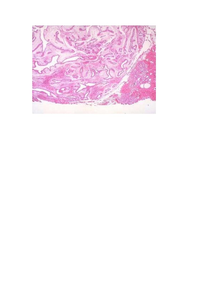
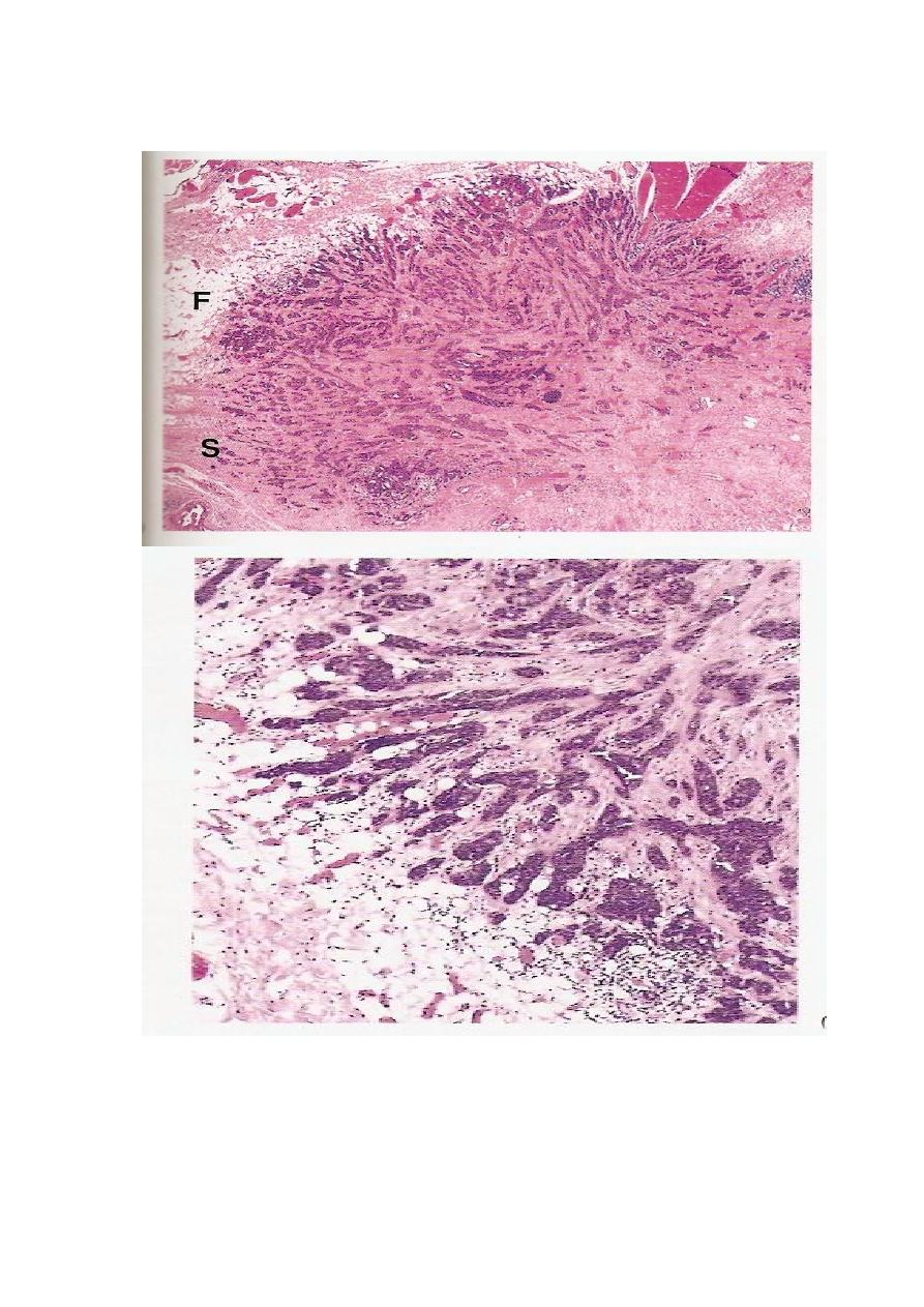
LOCAL INVASION
Most benign tumors grow as cohesive expansile masses that develop a rim of
condensed connective tissue or “capsule” at the periphery . They don’t penetrate the
capsule or the surrounding normal tissues. The line of cleavage between the capsule
and the surrounding normal tissues facilitates surgical enucleation.
Malignant tumors are invasive (infiltrative), they invade and destroy normal
surrounding tissues. They usually lack a well-defined capsule or line of cleavage,
thus, their enucleation is impossible, and their surgical removal requires removal of a
considerable margin of healthy apparently uninvolved tissue.
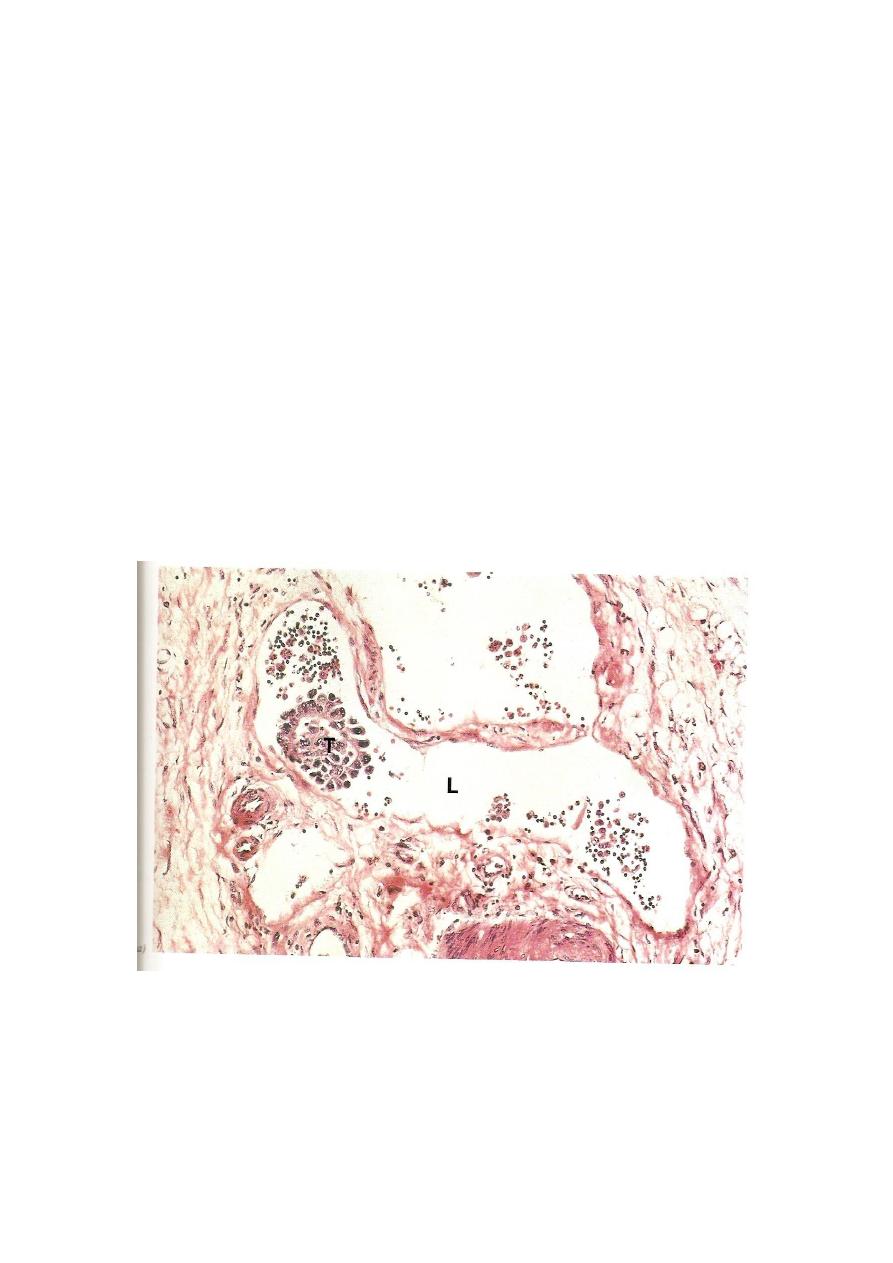
METASTASIS
This process involves invasion of blood vessels, lymphatics and body cavities by
the malignant tumor, followed by the transport and growth of secondary tumor cell
masses that are discontinuous with the primary tumor. These are called
secondaries.
Metastasis is the absolute criterion of malignancy.
Routes of tumor spread and metastasis
1.
Local spread
this occurs by invasion into the adjacent tissues.
2.
Invasion of lymphatics (lymphatic spread).
This is followed by spread of the
tumor to regional lymph nodes and ultimately to other sites in the body. It is
common in the initial spread of carcinomas. Not all enlarged lymph nodes located
at the sites of drainage of a malignancy means necessarily a metastasis. This is
because immune responses to tumor antigens can result in nodal enlargement too.
The latter is through the development of lymphoid hyperplasia.
3.
Invasion of blood vessels (hematogenous spread).
This is typical of all
sarcomas, but is also the favored route for certain carcinomas (e.g., renal cell
carcinoma). Because of their thinner walls, veins are more readily and thus more
frequently invaded than arteries. Lungs and liver are the commonest site of
hematogenous spread because they receive the systemic and portal venous blood
respectively. Other major sites are the bones and brain.
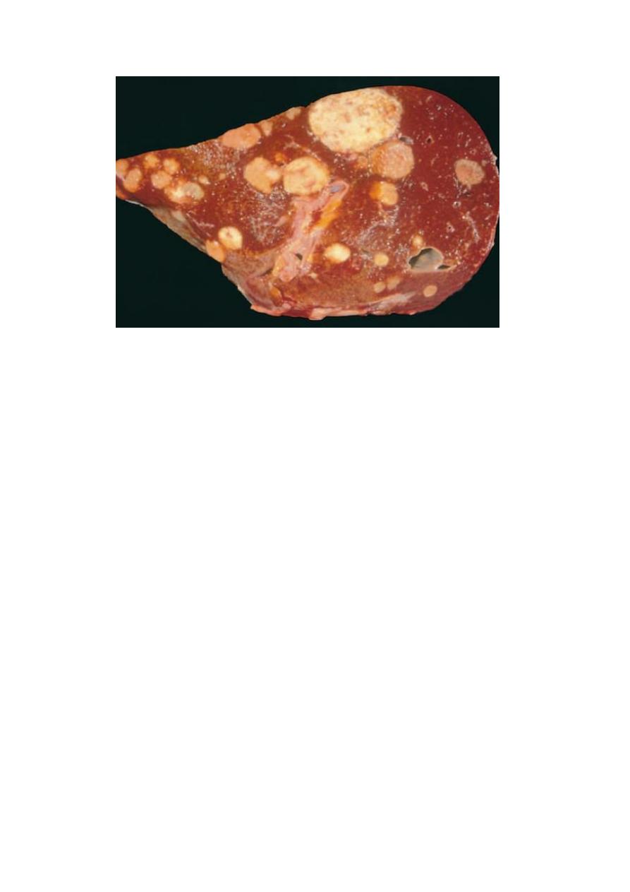
A liver studded with metastatic cancer
4.
Spread into body cavities (transcelomic spread).
This occurs by seedlings of
surfaces of peritoneal, pleural, pericardial and subarachnoid spaces. Carcinoma of
the ovary spreads transperitoneally to the surface of the liver or other abdominal
viscera (transcelomic spread).
