Orthomyxoviruses
د. انتظار علاوي جعفر / فرع الاحياء المجهريه / كليه الطب / جامعه ذي قارPhD. M.Sc. Microbiology

These viruses are classified under two families
These are:• Orthomyxoviridae, consisting of influenza viruses.
• Paramyxoviridae, consisting of parainfluenza, mumps, measles, respiratory syncytial, and Newcastle disease viruses.
The genus Orthomyxovirus includes influenza viruses, the causative agents of worldwide epidemics of influenza.
Human diseases associated with influenza virus are presented in Table 1.
Disease
Symptoms
Influenza in adults
Fever, malaise, myalgia, sore throat, and nonproductive cough
Influenza in children
Similar to that in adults but with high fever, abdominal pain, vomiting, otitis media, myositis, and croup
Complications
Primary viral pneumonia; secondary bacterial pneumonia; neurological complications and Reye’s syndrome
Orthomyxoviruses
All these viruses were grouped under myxovirus (myxa meaning mucus) due to their affinity to mucins (glycoproteins on cell surface as their receptors).Influenza Viruses
Influenza viruses belong to the family of Orthomyxoviridae and are the causative agents of influenza, a respiratory disease in humans with well-defined systemic symptoms that occurs in sporadic, epidemic, and pandemic forms. Influenza A and B viruses cause substantial morbidity and mortality in humans and a considerable financial burden worldwide, whereas influenza C viruses cause sporadic outbreaks of mild respiratory disease, mainly in children.Epidemic refers to an increase, often sudden, in the number of cases of a disease above what is normally expected in a population within a geographic area
Pandemic refers to an epidemic that has spread over several countries or continents, usually affecting a large number of people.
Properties of the Virus
MorphologyInfluenza viruses are spherical or filamentous, enveloped particles 80–120 nm in diameter.
It is composed of a characteristic segmented single-stranded RNA genome, a nucleocapsid, and an envelope (See-Fig).
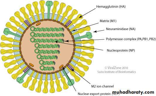
(Source: ViralZone:www.expasy.org/viralzone, SIB Swiss Institute of Bioinformatics)
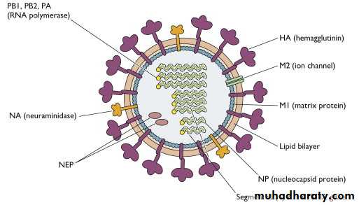
Structure of Influenza virus
The viral genome is a single-stranded antisense RNA. The genome consists of an RNA-dependent RNA polymerase, which transcribes the negative-polarity genome into mRNA.The RNA genome is segmented and consists of eight segments in Influenza A & B and seven segments in influenza C viruses. These segments code for different proteins which are NS1, NS2, NP, M1, M2, M3, HA, and NA.
The genome is present in a helically symmetric nucleocapsid surrounded by a lipid envelope. The envelope has an inner membrane protein layer and an outer lipid layer. The membrane proteins are known as matrix or M protein and are composed of two components M1 and M2.
Two types of spikes or peplomers project from the envelope:
(a ) The triangular hemagglutinin (HA) peplomers and
(b ) The mushroom-shaped neuraminidase (NA) peplomers.
18 hemagglutinin (HA) and 11 NA subtypes of influenza A viruses are found in nature.
Antigenic and genomic properties
Influenza viruses have two types of antigens:
Group-specific antigens: The ribonucleoprotein (RNP) antigen, or the “soluble” antigen, is the group-specific antigen.
Influenza viruses are divided into types A, B, and C on the basis of variation in this nucleoprotein antigen.
Type-specific antigens: The surface antigen, or “viral” antigen, or “V antigen” is composed of two virus-encoded proteins, Hemagglutinin (HA) and NA, which are the type-specific antigens.
Hemagglutinin( HA)
HA is a trimer and is composed of two polypeptides, HA1 and HA2, responsible for hemadsorption and hemagglutination. The hemagglutinin consists of 500 spikes, The triangular-shaped HA is inserted into the virus membrane by its tail end. The distal end, which contains five antigenic sites (designated as HA1–HA5), is responsible for binding of virion to host cells.Influenza viruses adsorb many avian and mammalian erythrocytes. Hemagglutinin binds with the sialic acid cell receptor, and initiates the infection in the host cell. Adsorption of erythrocytes occurs at 4°C, but at 37°C there is detachment of the red cells due to destruction of the glycoprotein receptors by the viral enzyme, neuraminidase.
The hemagglutinin agglutinates certain red blood cells, which is inhibited by the neutralizing antibodies. This forms the basis of the hemagglutination inhibition test used in the serodiagnosis of influenza.
Hemagglutinin has potency to undergo antigenic variations.
The nucleotide and amino acid sequences of the polypeptides, HA1 and HA2, undergo radical changes in antigenic shift.
In antigenic drift, only minor changes take place in the compositions of HA antigenic sites.
Neuraminidase (NA)
The NA is a glycoprotein and tetramer. It consists of 100 mushroom-shaped spikes. The NA is inserted into the virus membrane by its hydrophobic tail end. The distal end contains antigenic as well as enzymatically active sites. The NA causes hydrolysis of red cells, hence causes elution or detachment of the cells adsorbed to virion particles. The function of the neuraminidase is to cleave the neuraminic acid and to release progeny virions from the infected host cells.Aartjan J.W. te Velthuis and Ervin Fodor, 2017
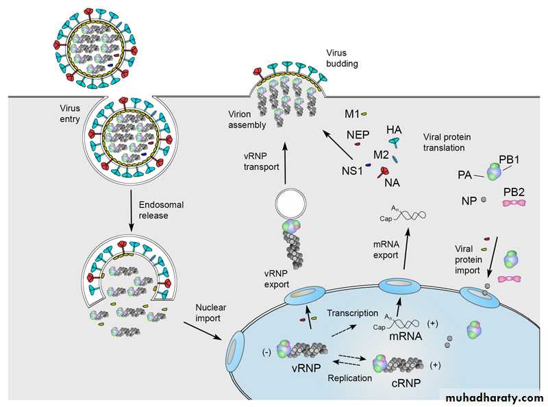
Influenza A virus replication cycle.
Influenza A virus replication cycle.Influenza A virus replication cycle.
• Viral infection initiates with the binding of a virion to cell surface receptors containing sialic acid, followed by the endocytosis of the virion.• After fusion of the viral and endosomal membranes, the viral ribonucleoproteins (vRNPs) are released into the cytoplasm and then transported into the nucleus.
• In the nucleus the viral RNA polymerase transcribes the vRNA segments into mRNAs.
• Replication …continued
• Viral mRNA is exported to the cytoplasm for translation by cellular mechanisms. The viral RNA polymerase also performs replication of vRNA by copying it into complementary RNA (cRNA), which serves as a template for the production of more vRNA. Newly synthesised viral polymerase and nucleoprotein are imported into the nucleus and bind to cRNA and vRNA to assemble vRNPs and cRNPs, respectively.• Following nuclear export, progeny vRNPs are transported across the cytoplasm on recycling to the cell membrane, where assembly of progeny virions takes place.
• Mature virions are released by budding.
Antigenic variations
Antigenic variation is a unique feature of influenza virus.
The surface antigens HA and NA show variations and are primarily responsible for antigenic variations exhibited by influenza viruses. The internal RNP antigen and M protein are stable, hence do not contribute to the antigenic variations. Antigenic variations are of two types: antigenic shift and antigenic drift.
Antigenic shift
Antigenic shift: The abrupt, drastic, discontinuous change. This occurs due to major antigenic changes in HA or NA antigens, and is caused by replacement of the gene for HA by one coding for a completely different amino acid sequence. The antigenic shift is characterized by alteration of virtually all the antigenic sites of the HA. Antigenic shift has been demonstrated in type A influenza virus only.Antigenic shift variants appear less frequently, about every 10 or 11 years. It is demonstrated that pandemic strains are the recombinant strains, originated from some animal or bird reservoir, either spreading to humans directly by
host range mutation or as a result of a recombination between human and nonhuman strains. The completely novel antigens that appear during antigenic shift are acquired by genetic reassortment.
The donor of the new antigens is probably an animal influenza virus. Type A viruses have been identified in pigs, horses, and birds, and animal influenza viruses possessing antigens closely related to those of human viruses.
Antigenic drift
Is gradual, sequential, regular antigenic change in influenza virus. This occurs due to minor antigenic changes in the HA or NA occurring at frequent intervals. This is caused even by a single mutation affecting HA glycoprotein. The antigenic drift is characterized by changes in certain epitopes in the HA, while others are being conserved. Antigenic drift variants occur very frequently, virtually every year. This is responsible for emergence of the strains that cause yearly influenza epidemics.
Antigenic drift
Antigenic shift• Gradual, sequential, and regular variation at periodic intervals
Abrupt, drastic, and
discontinuous variation in antigenic structureResults in a different strain
Related to predecessor strainResults in a new strain
Not related to predecessor strain
Antigenic drift is due to
mutation and selection
Antigenic shift is due to gene
reassortment
Responsible for epidemics
of influenza
Responsible for epidemics as well as pandemics of influenza
Table: Differences between antigenic shift and antigenic drift
Key Points
Influenza A virus shows maximum antigenic variations.Influenza B virus does not undergo antigenic shift because influenza B virus is the only human virus for which there is no animal source of new RNA segments. However, influenza B virus undergoes antigenic drift.
Antigenic variation never occurs in type C influenza virus beacuse its lack of NA.
Influenza epidemics and pandemics
Influenza epidemics are of two types. Yearly epidemics are caused by both type A and type B viruses. The rare, severe influenza pandemics are always caused by type A virus. Antigenic shift and antigenic drift are the two different mechanisms responsible for producing the strains that cause these two types of epidemics.Gene reassortment
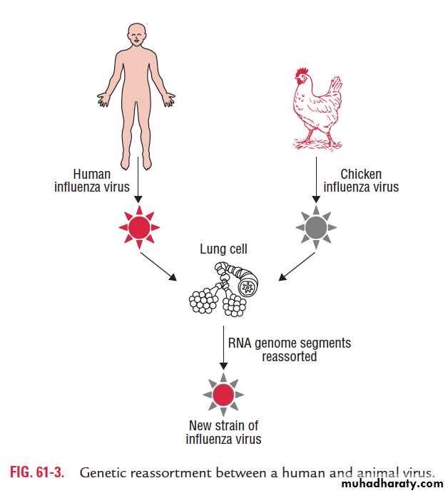
Because the influenza virus genome is segmented, genetic reassortment can occur when a host cell is infected simultaneously with viruses of two different parent strains. This process of genetic reassortment accounts for the periodic appearance of the novel types of influenza A strains that cause influenza pandemics.Influenza viruses of animals, such as aquatic birds, chickens, swine, and horses show high host specificity. These animal viruses are the source of the RNA segments that encode the antigenic shift variants that cause epidemics among humans.
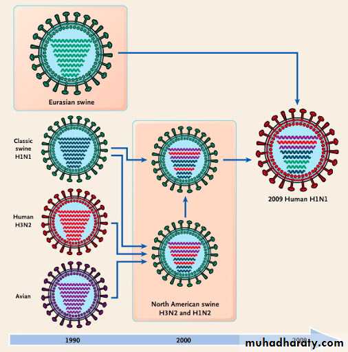
For example, if a person is infected simultaneously by an avian and human influenza strains, then it is possible that a genetic reassortment could occur in infected cells in humans. The reassortment could lead to emergence of a new human influenza A virus, the progeny of which will contain a mixture of genome segments from the two strains (e.g., a new variant of human influenza A virus bearing the avian virus HA).
Designation of influenza viruses
Influenza virus type A can be classified into subtypes based on the variations in their surface antigens. The WHO proposed a new system of classification in 1971 and was later modified, which takes into account the nature of both the surface antigens. According to this, the complete designation of a strain will include the(a ) type, (b ) place of origin, (c ) serial number, and (d ) year of isolation followed by (e ) antigenic subtypes of the HA
and NA in parentheses.
For example: influenza A/Singapore/1/57 (H2N2) indicates that influenza was first originated from Singapore and was isolated for the first time in the year 1957. The HA and NA antigens are H2 and N2 as shown in the parentheses.
Pathogenesis and Immunity
Influenza virus is transmitted from person to person primarily in droplets released by sneezing and coughing.Inhaled influenza viruses reach lower respiratory tract, tracheobronchial tree, the primary site of the disease. They attach to sialic acid receptors on epithelial cells by HA present on the viral envelope. Relatively few viruses are needed to infect lower respiratory tract than the upper respiratory tract. Neuraminidase of the viral envelope may act on the N -acetyl neuraminic acid residues in mucus to produce liquefaction.
Infection of mucosal cells results in cellular destruction and desquamation of the superficial mucosa. The resulting edema and mononuclear cell infiltration of the involved areas are accompanied by symptoms including nonproductive cough, sore throat, and nasal discharge. Although the cough may be striking, the most prominent symptoms of influenza are systemic: fever, muscle aches, and general prostration. In an uncomplicated case, virus can be recovered from respiratory secretions for 3–8 days.
Clinical Syndrome
Incubation period is short (1–3 days). The classic influenza syndrome is a febrile illness of sudden onset, characterized by tracheitis and marked myalgias. Headache, chills, fever, malaise, myalgias, anorexia, and sore throat appear suddenly. The body temperature rapidly rises to (38.3–40.0°C) and respiratory symptoms ensue. Nonproductive cough is characteristic. Sneezing, rhinorrhea, and nasal obstruction are common.
Patients may also report photophobia, nausea, vomiting, diarrhea, and abdominal pain. They appear acutely ill and are usually coughing. Minimal to moderate nasal obstruction, nasal discharge, and pharyngeal inflammation may be present.
Clinical Syndrome
Complications
1-Secondary bacterial infections: Life-threatening influenza is often caused by secondary bacterial infections with staphylococci, pneumococci, and Haemophilus influenzae. Pneumonia may develop as a complication and may be fatal, particularly in• Elderly persons above 60 years with underlying chronic disease.
• In people chronic cardiorespiratory disease, renal disease, etc.)
• Pregnant women.
Complications
2-Reye’s syndrome is a noted complication of influenza B infection. The condition is seen most commonly in young children and is associated with degenerative changes in the brain, liver, and kidney.3- Central nervous system complications: Guillain–Barre syndrome characterized by encephalomyelitis and polyneuritis is a rare complication of influenza virus infection
Reservoir, source, and transmission of infection
Infected humans are the main reservoir of infections for influenza A virus. Respiratory secretions of infected persons are the important source of infection. The virus is excreted in respiratory secretions immediately before the onset of illness
and for 3–4 days thereafter. Wild aquatic birds are known
reservoirs of influenza A. They secrete the viruses in their feces, which contaminates ponds and lakes. The virus is spread from person-to-person primarily by air-borne respiratory droplets released during the acts of sneezing and coughing.
Influenza B virus only causes epidemics. Infection is from
humans-to-humans. No animal reservoir hosts are known.
Laboratory Diagnosis
During an epidemic of influenza, the clinical diagnosis can be made, but definitive diagnosis depends on the laboratory methods.Specimens:
Nasal or throat washings or sputum for viral
antigen and viral RNA.
Throat gargles for isolation of viruses.
Serum for viral antibodies.
Direct antigen detection
1- Is made by demonstrating viral antigens directly on cells obtained from the nasopharynx. Immunofluorescence (IF) or enzyme-linked immunosorbent assay (ELIZA) using specific monoclonal antibodies are used to detect viral antigen.The results of the rapid tests are useful to start treatment with the NA inhibitors within 48 hours of the onset of symptoms.
Immunofluorescent staining of some Influenza Ag
Cao et al, 2014.

Eliza assay
2-Reverse Transcription-Polymerase Chain Reaction (RT-PCR) assay can identify the presence of influenza viral RNA in respiratory specimens with very high sensitivity and specificity.
3- Isolation of the virus:
Throat gargles are the specimen of choice. The specimen is collected in saline broth or a buffered salt solution and is sent immediately to the laboratory, or if delayed is stored at 4°C.
The virus is isolated from the specimen by inoculation into embryonated eggs or into certain cell cultures.