1
بسم هللا الرحمن الرحيم
Lecture 5 Neurophysioloy Dr. Noor
2
nd
stage 2020
…………………………………………………………………..
Spinal cord
Objective
1.
What is the physiologic anatomy of spinal cord?
2.
Organization of spinal cord for motor function?
Anatomy
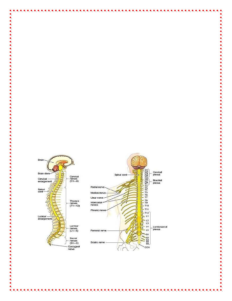
The spinal cord is a long, thin, tubular bundle of nervous
tissue and support cells and is the most important structure
between the body and the brain. The spinal cord extends from the
foramen magnum where it is continuous with the medulla to the
level of the first or second lumbar vertebrae. It is a vital link
between the brain and the body, and from the body to the brain.
The spinal cord is 40 to 50 cm long and 1 cm to 1.5 cm in
diameter. Two consecutive rows of nerve roots emerge on each of
its sides. These nerve roots join distally to form 31 pairs of spinal
nerves.
The spinal cord is a cylindrical structure of nervous tissue
composed of white and gray matter, is uniformly organized and is
divided into four regions: cervical (C), thoracic (T), lumbar (L) and
sacral (S), each of which is comprised of several segments. The
spinal nerve contains motor and sensory nerve fibers to and from
2
all parts of the body. Each spinal cord segment innervates a
dermatome.
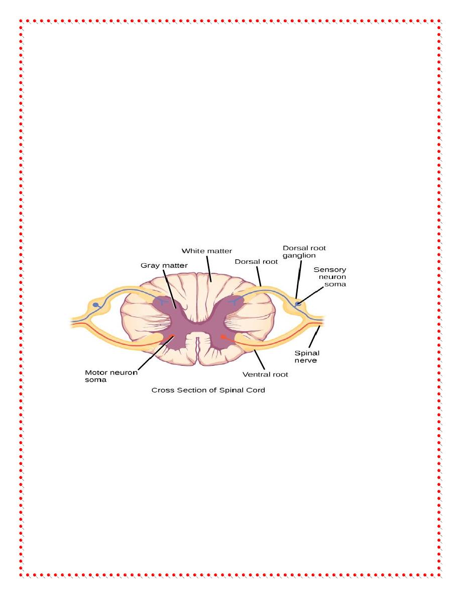
Internal Structure of the Spinal Cord
A transverse section of the adult spinal cord shows white matter in
the periphery, gray matter inside, and a tiny central canal filled
with CSF at its center. Surrounding the canal is a single layer of
cells, the ependymal layer. Surrounding the ependymal layer is the
gray matter – a region containing cell bodies – shaped like the
letter “H” or a “butterfly”.
The two “wings” of the butterfly are connected across the midline
by the dorsal gray commissure and below the white commissure .
3
Gray matter
The spinal cord plays a key role in integration of multiple
peripheral and central inputs via the system of neurons in the gray
matter. On cross-section, the gray matter in the spinal cord
includes the dorsal, ventral, and an intermediolateral horns or
columns.
White matter
The white matter includes ascending and descending tracts that are
composed of axons. The white matter also has glial cells.
Functions
The spinal cord works a bit like a telephone switchboard operator,
helping the brain communicate with different parts of the body,
and vice versa. Its three major roles are:
To relay messages from the brain to different parts of the
body (usually a muscle) in order to perform an action

4
To pass along messages from sensory receptors (found all
over the body) to the brain
To coordinate reflexes (quick responses to outside stimuli)
that don't go through the brain and are managed by the spinal
cord alone
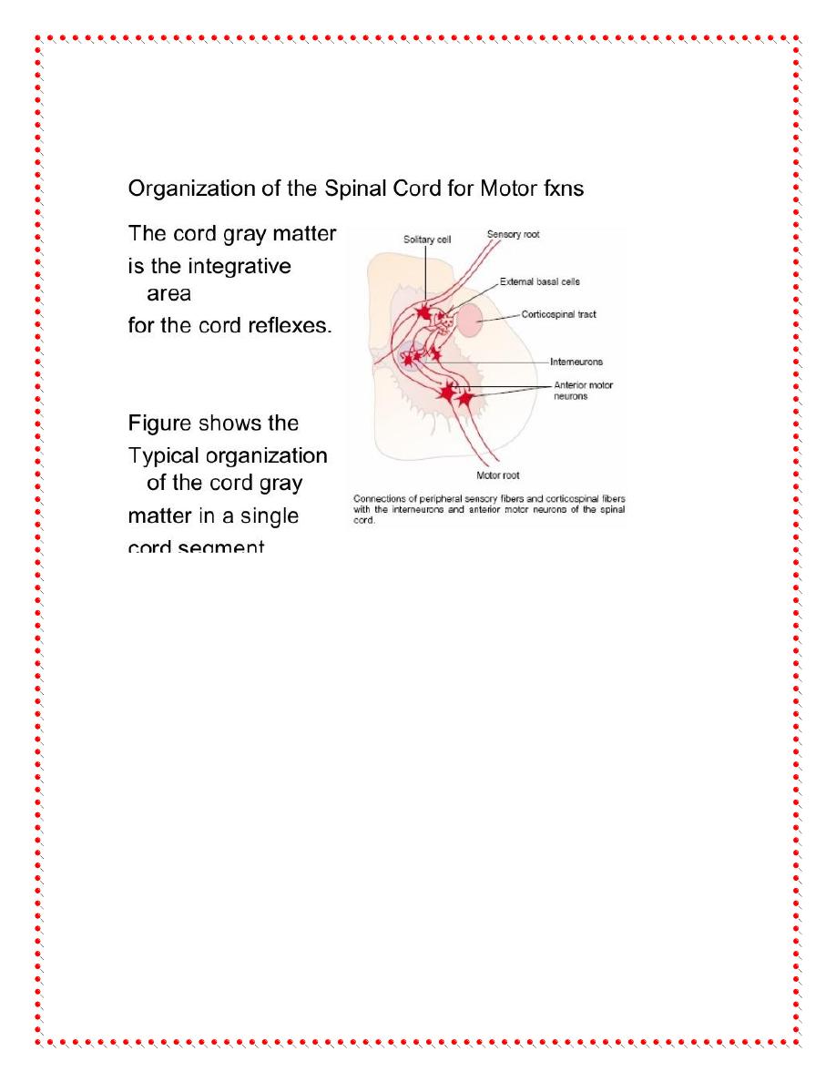
ORGANIZATION OF THE SPINAL CORD FOR MOTOR
FUNCTIONS
The cord gray matter is the integrative area for the cord reflexes.
Sensory signals enter the cord almost entirely through the sensory
roots, also known as the posterior or dorsal roots.
After entering the cord, every sensory signal travels to two
separate destinations: (1)One branch of the sensory nerve
terminates almost immediately in the gray matter of the cord and
elicits local segmental cord reflexes and other local effects, and
(2)another branch transmits signals to higher levels of the nervous
system—that is, to higher levels in the cord itself, to the brain
stem, or even to the cerebral cortex.
Each segment of the spinal cord (at the level of each spinal nerve)
has several million neurons in its gray matter, Aside from the
5
sensory relay neurons, the other neurons are of two types: (1)
anterior motor neurons and (2) interneurons.
Anterior Motor Neurons.
Located in each segment of the anterior horns of the cord gray
matter are several thousand neurons that are 50 to 100 percent
larger than most of the others and are called anterior motor
neurons . They give rise to the nerve fibers that leave the cord by
way of the anterior roots and directly innervate the skeletal muscle
fibers. The neurons are of two types, alpha motor neurons and
gamma motor neurons.

6
1.
Alpha Motor Neurons.
The alpha motor neurons give rise to large type A alpha (Aα)
motor nerve fibers, averaging 14 micrometers in diameter; these
fibers branch many times after they enter the muscle and innervate
the large skeletal muscle fibers. Stimulation of a single alpha nerve
fiber excites anywhere from three to several hundred skeletal
muscle fibers, which are collectively called the motor unit.
2.
Gamma Motor Neurons
.
Along with the alpha motor neurons, which excite contraction of
the skeletal muscle fibers, about one half as many much smaller
gamma motor neurons are located in the spinal cord anterior horns.
These gamma motor neurons transmit impulses through much
smaller type A gamma (Aγ) motor nerve fibers, averaging 5
micrometers in diameter, which go to small, special skeletal
muscle fibers called intrafusal fibers.

7
Interneurons
These fibers constitute the middle of the muscle spindle, which
helps control basic muscle “tone,”
Interneurons. Interneurons are present in all areas of the cord gray
matter—in the dorsal horns, the anterior horns, and the
intermediate areas between them.
These cells are about 30 times as numerous as the anterior motor
neurons. They are small and highly excitable, often exhibiting
spontaneous activity and capable of firing as rapidly as 1500 times
per second. They have many interconnections with one another,
and many of them also synapse directly with the anterior motor
neurons.
The interconnections among the interneurons and anterior motor
neurons are responsible for most of the integrative functions of the
spinal cord
Only a few incoming sensory signals from the spinal nerves or
signals from the brain terminate directly on the anterior motor
neurons. Instead, almost all these signals are transmitted first
through interneurons, where they are appropriately processed.
8
Thus, the corticospinal tract from the brain is shown to terminate
almost entirely on spinal interneurons, where the signals from this
tract are combined with signals from other spinal tracts or spinal
nerves before finally converging on the anterior motor neurons to
control muscle function.
Thank you
References : Guyton and Hall textbook of medical physiology,
thirteen edition