
•
It is a field of study
concerned with
joints .
Arthrology

•
Articulation between boney
surfaces wich allow free or
limited movement
The joint is the
•
A
•
B
Classification of joints
according to :
•
classification
]
Joints
Classification of Joints
Fibrous Joints
Cartilaginous Joints
Synovial Joints
Free to share, print, make copies and changes. Get yours at www.boundless.com
•
Structural Classification of Joints
•
Functional Classification of Joints
Classification of Joints
Joints > Classification of Joints
Free to share, print, make copies and changes. Get yours at www.boundless.com
•
Fibrous Joints
•
1-Sutures
•
2-Syndesmoses
•
3-Gomphoses
Fibrous Joints
Joints > Fibrous Joints
Free to share, print, make copies and changes. Get yours at www.boundless.com
•
Cartilaginous Joints: Synchodroses
•
Cartilaginous Joints: Symphyses
Cartilaginous Joints
Joints > Cartilaginous Joints
Free to share, print, make copies and changes. Get yours at www.boundless.com
•
Structure of Synovial Joints
•
Nerve and Blood Supply
•
Bursae and Tendon Sheaths
•
Stability and Range of Motion at Synovial Joints
•
Synovial Joint Movements
•
Types of Synovial Joints
Synovial Joints
Joints > Synovial Joints
Free to share, print, make copies and changes. Get yours at www.boundless.com

Free to share, print, make copies and changes. Get yours at www.boundless.com
Appendix

Key terms
• abduction
The movement that separates a limb or other part from the axis, or middle line, of the body.
• acromioclavicular joint
A joint at the top of the shoulder that is the junction between the acromion (a bony process on the
scapula) and the clavicle.
• adduction
The action by which the parts of the body are drawn toward its axis.
• anastomosis
A cross-connection between two blood vessels.
• annulus fibrosus
Fibrous ring of intervertebral disk.
• apoptosis
A type of "cell suicide" called programmed cell deaththat occurs in multicellular organisms.
• articular cartilage
A tough, elastic, fibrous connective tissue found in various parts of the body such as the joints, outer ear, and
larynx. A major constituent of the embryonic and young vertebrate skeleton, converted largely to bone with maturation.
• articulation
A joint or the collection of joints at which something is articulated, or hinged, for bending.
• ball-and-socket joint
A joint in which the ball-shaped surface of one rounded bone fits into the cup-like depression of another
bone.
• cartilaginous joints
Joints connected by fibrocartilage or hyaline cartilage. They allow more movement than fibrousjoints but less
than synovialjoints.
• circumduction
A conical movement of a body part consisting of a combination of flexion, extension, adduction, and abduction.
• condyle
A smooth prominence on a bone where it forms a joint with another bone.
Free to share, print, make copies and changes. Get yours at www.boundless.com
Joints

• connective tissue
A type of tissue found in animals that functions in binding other tissue systems (such as muscle to skin) or
organs. It consists of the cells, fibers, and a ground substance or extracellular matrix.
• convection
The movement of groups of molecules within fluids such as liquids or gases.
• diarthrosis
A joint that can move freely in various planes.
• diastasis
A separation between two parts of a bone, without fracture.
• dorsiflexion
The movement which decreases the angle between the dorsum (superior surface) of the foot and the leg, so that
the toes are brought closer to the shin.
• epiphyseal plate
The epiphyseal plate is a hyaline cartilage plate where growth occurs in children and adolescents, located in
the metaphysis at each end of a long bone .
• eversion
The condition of being turned outward.
• fibrous joints
Fixed or immobile joints that are connected by dense, tough connective tissue thatis rich in collagen fibers.
• flexion
The act of bending a joint. The counteraction of extension.
• fontanelle
An anatomical feature of the infant human skull comprising the soft membranous gaps.
• gomphoses
A joint that binds the teeth to bony sockets (dental alveoli) in the maxillary bone and mandible.
• gomphoses
A joint that binds the teeth to bony sockets (dental alveoli) in the maxillary bone and mandible.
Free to share, print, make copies and changes. Get yours at www.boundless.com
Joints

• gomphosis joints
Joints of very limited mobility. These are found at the articulationbetween teethand the sockets of maxilla or
mandible (dental-alveolar joint).
• intervertebral disc
A cartilaginous joint that allows slight movement of the vertebrae by lying between adjacent vertebrae in the
spine. It also acts as a ligament to hold the vertebrae together.
• manubrium
The broad upper part of the sternum.
• nucleus pulposus
Inner gel-like center of the vertebral disc.
• osteomyelitis
An infection of the bone and bone marrow characterized by inflammation.
• periosteum
A membrane that covers the outer surface of all bones.
• plantarflexion
The movement that increases the approximate 90 degree angle between the front part of the foot and the shin.
• prime mover
A muscle that acts directly to bring about a desired movement.
• pronation
The action of rotating the forearm so that the palm of the hand is turned down or back.
• retinacula
A band around tendonsthat holds them in place for stabilization.
• sciatica
Pain that travels down the leg from the lower back region.
• scurvy
A disease resulting from a lack of vitamin C.
Free to share, print, make copies and changes. Get yours at www.boundless.com
Joints

• Sharpey's fibres
A matrix of connective tissue consisting of bundles of strong collagenous fibers connecting periosteum to
bone.
• supination
The action of rotating the forearm so that the palm of the hand is turned up or forward
• suture
In anatomy, a suture is a fairly rigid joint between two or more hard elements such as the bony plates of the skull.
• suture
A type of fibrous joint which only occurs in the skull (cranium).
• symphysis
The cartilaginous material that adjoins and facilitates the junction of such bones, with or without synovia.
• symphysis
The cartilaginous material that adjoins and facilitates the junction of such bones, with or without synovia.
• synarthrosis
A type of joint in which two bones are connected rigidly by fibrous tissue.
• synchondrosis
A slightly moveable articulation between bones joined by hyaline cartilage.
• syndesmoses
Slightly movable articulations where the contiguous bony surfaces are united by an interosseous ligament, as in
the inferior tibiofibular articulation.
• synovial fluid
A viscous fluid found in the cavities of synovialjoints that reduces friction between the articular cartilage during
movement.
• synovial fluid
A viscous, non-Newtonian fluid found in the cavities of synovial joints. With its yolk-like consistency, its principal
role is to reduce friction between the articular cartilage of synovial joints during movement.
• synovial fluid
A viscous, non-Newtonian fluid found in the cavities of synovial joints. With its yolk-like consistency, its principal
role is to reduce friction between the articular cartilage of synovial joints during movement.
Free to share, print, make copies and changes. Get yours at www.boundless.com
Joints

• synovial joint
Also known as a diarthrosis, the most common and most movable type of joint in the body of a mammal.
• Synovial joint
The most common and most movable type of joint in the body of a mammal.
• synovial membrane
A thin membrane of joints comprised of smooth connective tissue and that secretes synovial fluid.
• tendon
A tough band of inelastic fibrous tissue that connects a muscle with its bony attachment.
Free to share, print, make copies and changes. Get yours at www.boundless.com
Joints
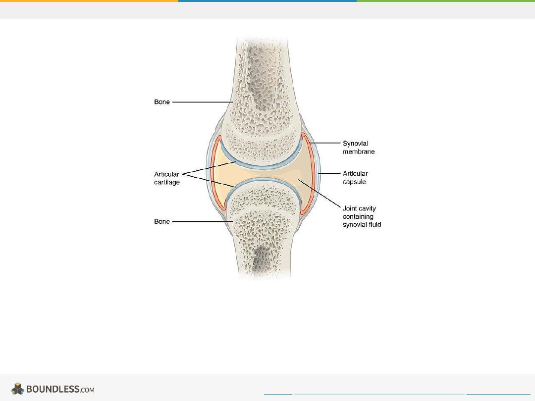
Synovial Joint
This diagram of a synovial joint delineates the articular cartilage, articular capsule, bone, synovial membrane, and joint cavity containing synovial fluid.
Free to share, print, make copies and changes. Get yours at www.boundless.com
Wikipedia Commons. "Synovial Joints."
https://commons.wikimedia.org/wiki/File:907_Synovial_Joints.jpg
Joints

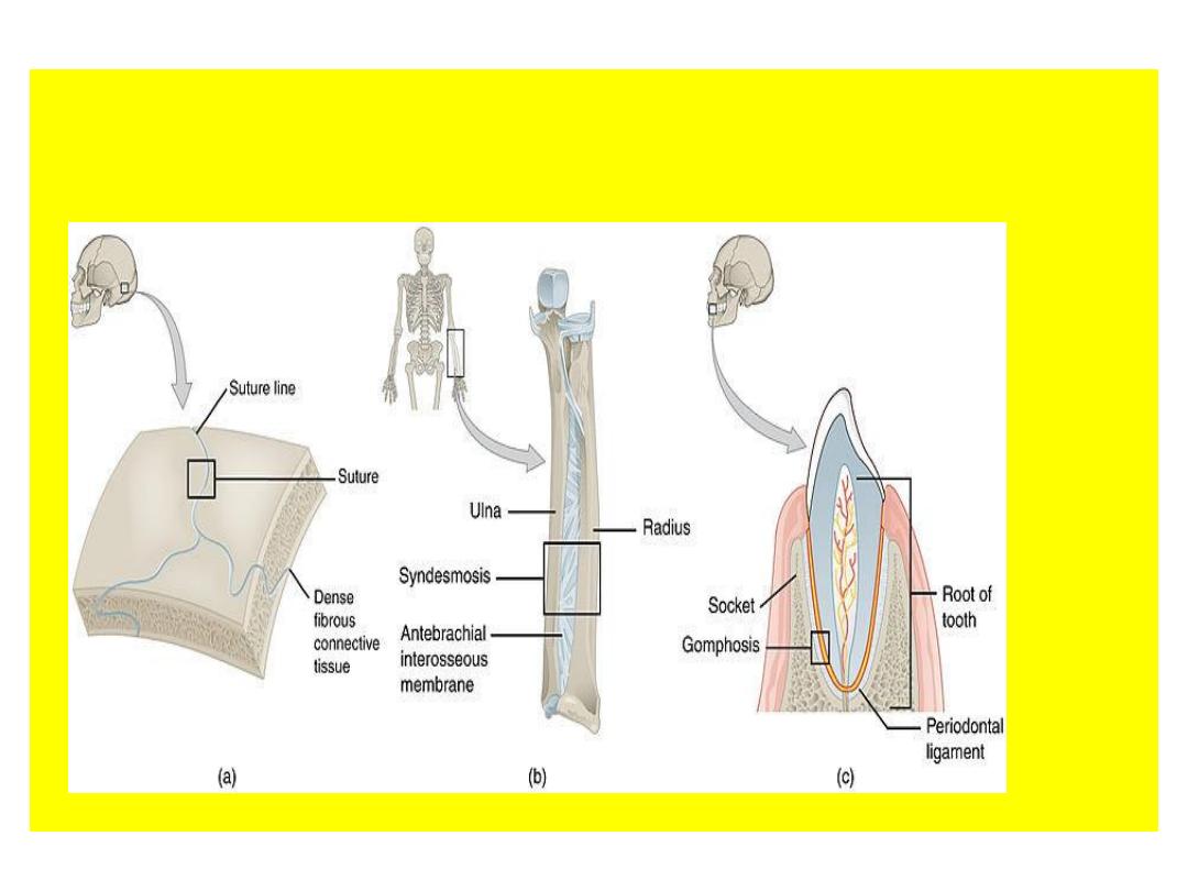
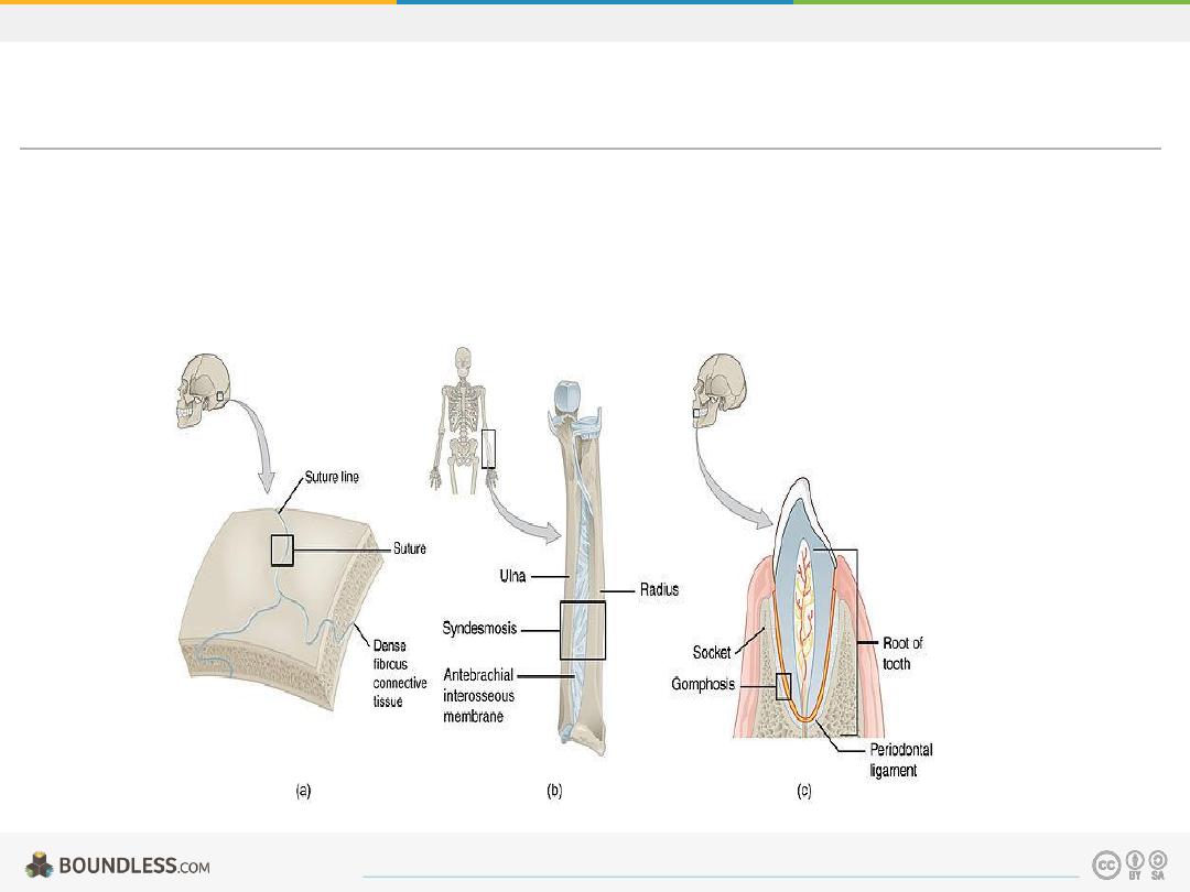
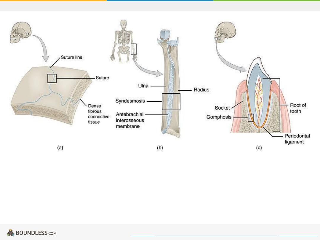
fibrous.jpg
Image demonstrating the three types of fibrous joints. (a) Sutures (b) Syndesmosis (c) Gomphosis.
Free to share, print, make copies and changes. Get yours at www.boundless.com
Wikipedia Commons. "Fibrous Joints."
https://commons.wikimedia.org/wiki/File:904_Fibrous_Joints.jpg
Joints
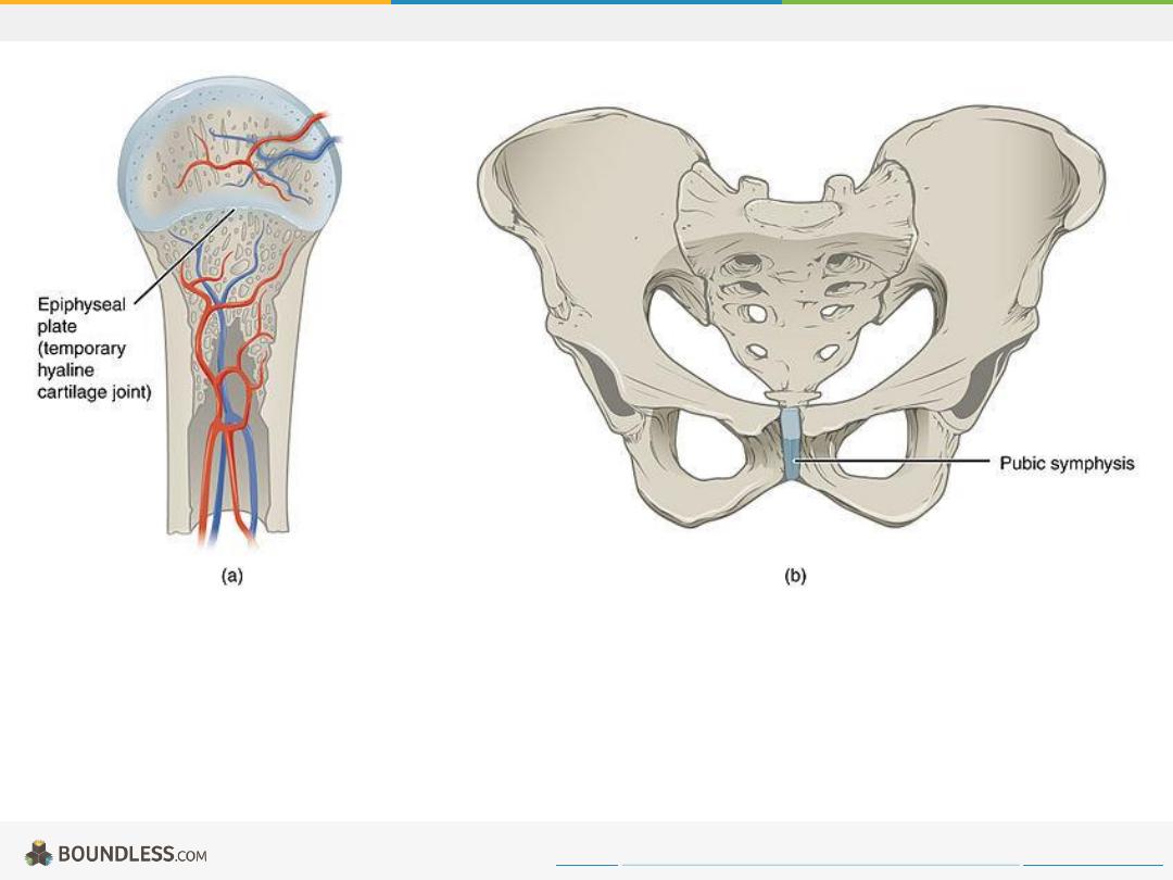
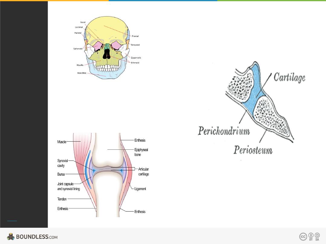
Cartilaginous Joints
Image demonstrates a synchondrosis joint with epiphyseal plate (temporary hyaline cartilage joint) indicated (a) and a symphysis joint (b).
Free to share, print, make copies and changes. Get yours at www.boundless.com
Wikipedia Commons. "Cartiliginous Joints."
https://commons.wikimedia.org/wiki/File:906_Cartiliginous_Joints.jpg
Joints
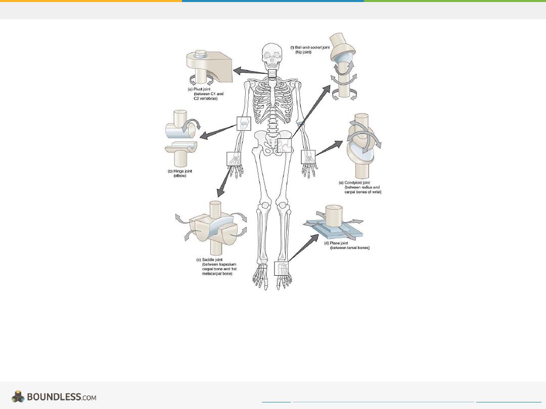
Types of Synovial Joints.jpg
Image of a skeleton and skematics of the different classes of synovial joints.
Free to share, print, make copies and changes. Get yours at www.boundless.com
Wikipedia. "Types of Synovial Joints."
https://en.wikipedia.org/wiki/File:909_Types_of_Synovial_Joints.jpg
Joints
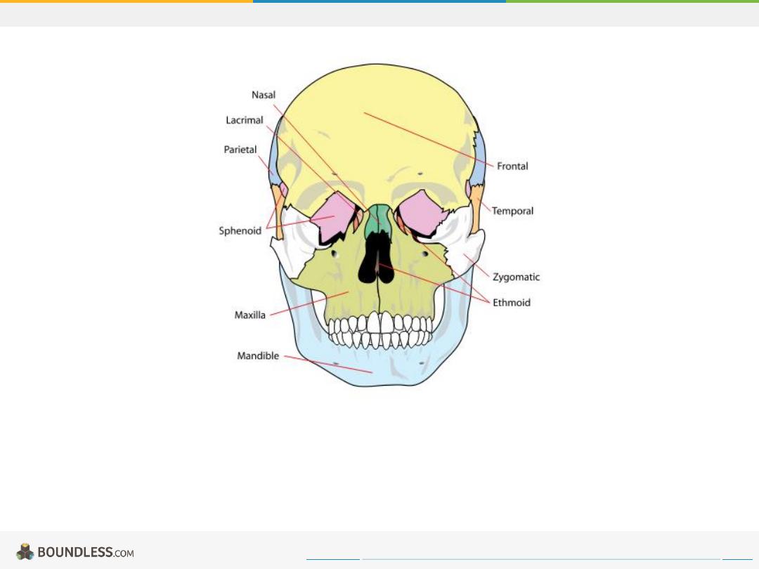
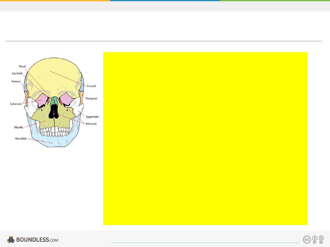
Fibrous Joints
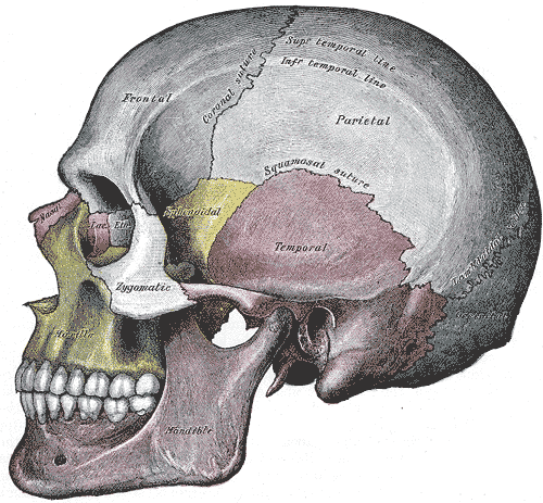
The adult skull is normally made up of 22 bones. Except for the mandible, all are joined together by sutures, semi-rigid articulations formed by bony
ossification. The presence of Sharpey's fibers permit a little flexibility.
Free to share, print, make copies and changes. Get yours at www.boundless.com
Wikimedia. "Skull bones diagram."
.svg)
http://upload.wikimedia.org/wikipedia/commons/1/1a/Human_skull_front_simplified_(bones).svg
Joints
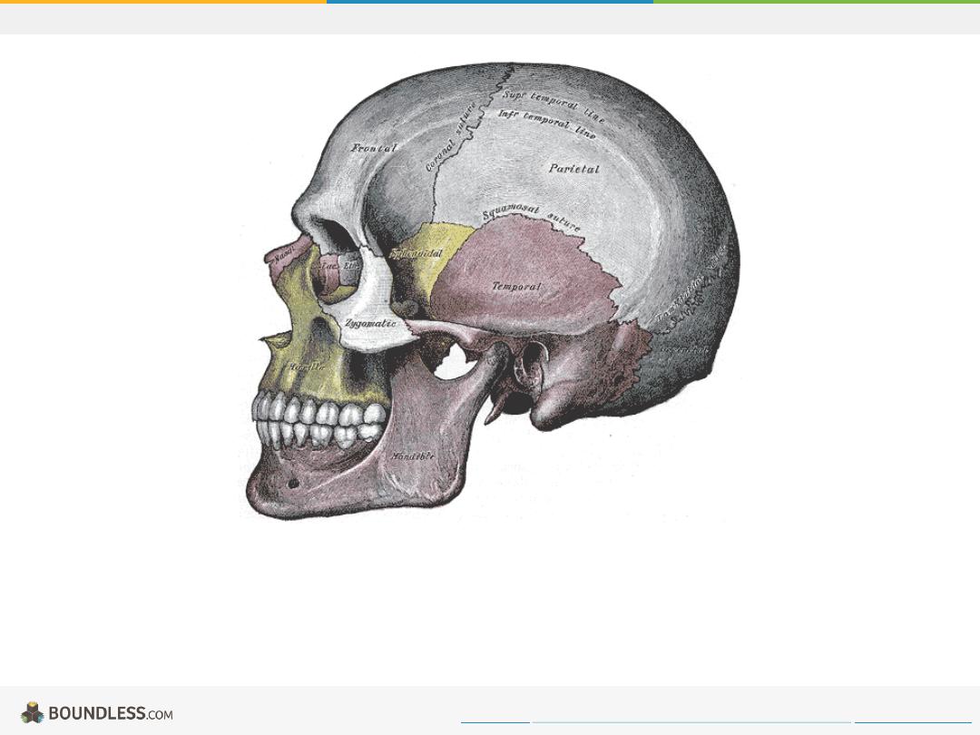
Cranial Sutures
Lateral view of skull showing the location of some of the cranial sutures.
Free to share, print, make copies and changes. Get yours at www.boundless.com
Wikimedia. "Cranial Sutures."
http://upload.wikimedia.org/wikipedia/commons/2/2c/Gray188.png
Joints
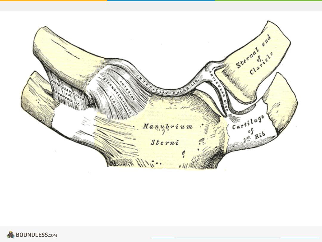
Saddle Joint
Sternoclavicular articulation. Anterior view.
Free to share, print, make copies and changes. Get yours at www.boundless.com
Wikimedia. "Sternoclavical Articulation."
http://upload.wikimedia.org/wikipedia/commons/3/3f/Gray325.png
Joints
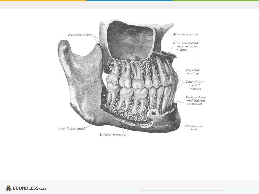
Gomphoses
This image illustrates the gomphoses joints of teeth within the jaw.
Free to share, print, make copies and changes. Get yours at www.boundless.com
http://upload.wikimedia.org/wikipedia/commons/6/6c/Gray1003.png
Joints
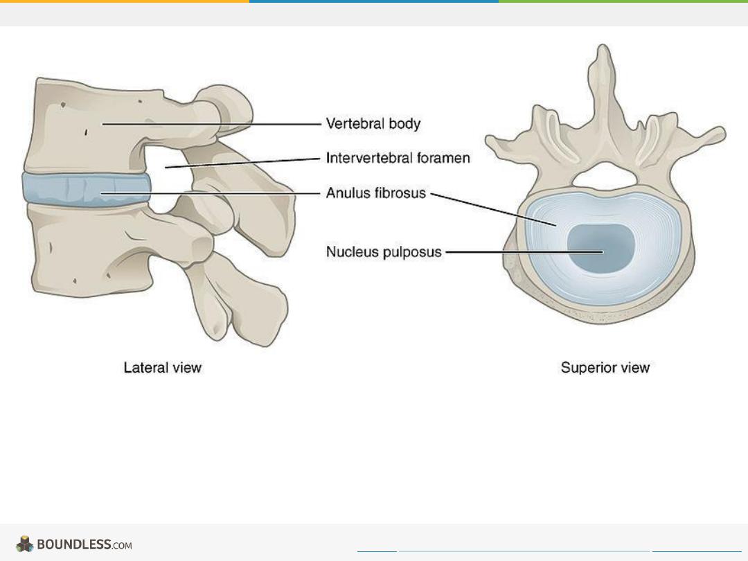
Diagram of Invertebral Disc
The lateral and superior view of an invertebral disc, including the vertebral body, intervertebral foramen, anulus fibrosis, and nucleus pulposus.
Free to share, print, make copies and changes. Get yours at www.boundless.com
Wikipedia Commons. "intervertebral disc."
https://commons.wikimedia.org/wiki/File:716_Intervertebral_Disk.jpg
Joints
Synovial Joint
An illustration of the structure of a synovial joint.
Free to share, print, make copies and changes. Get yours at www.boundless.com
http://en.wikipedia.org/wiki/File:Joint.png
Joints
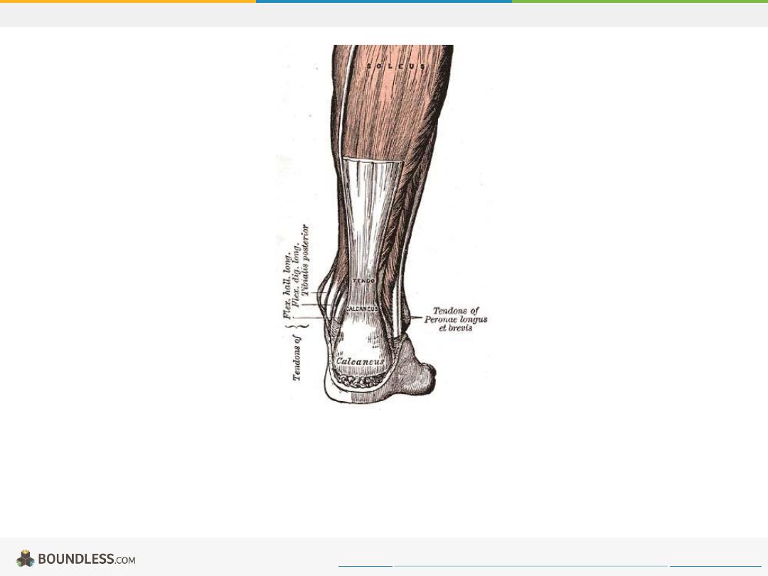
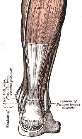
Achilles Tendon
The Achilles tendon, also called the calcaneus, provides stability and limits the range of motion at the ankle joint. It's depicted in this diagram in relation
to the tendo calcaneus.
Free to share, print, make copies and changes. Get yours at www.boundless.com
Wikimedia.
http://upload.wikimedia.org/wikipedia/commons/3/3c/Achilles-tendon.jpg
Joints
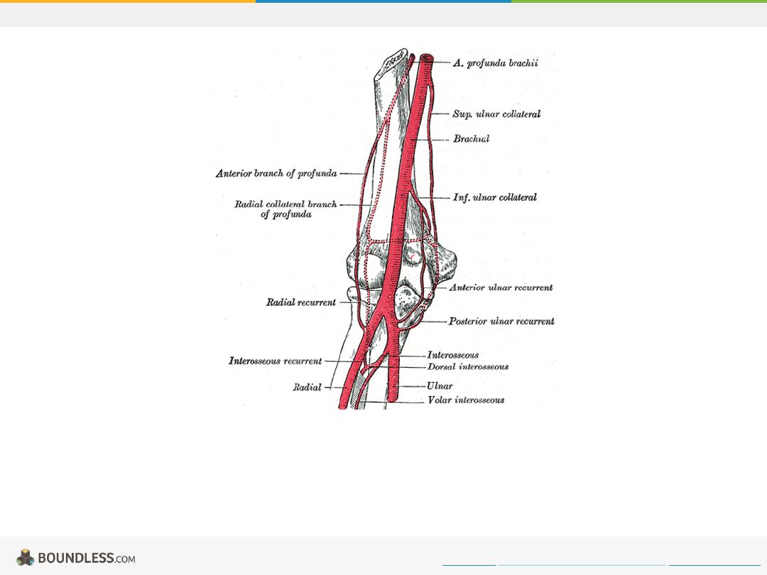
Elbow Joint
Diagram of the anastomosis around the elbow joint.
Free to share, print, make copies and changes. Get yours at www.boundless.com
Wikipedia. "Gray526."
http://en.wikipedia.org/wiki/File:Gray526.png
Joints
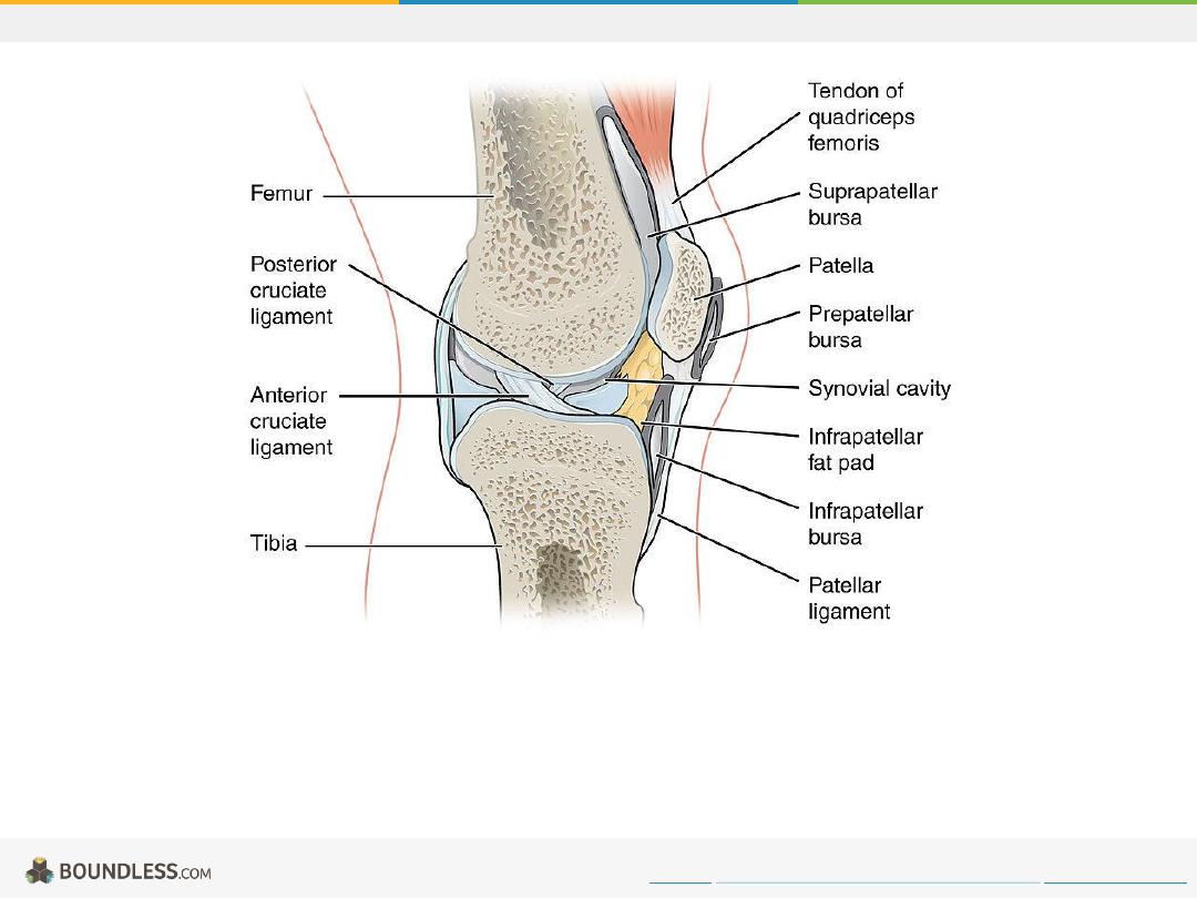
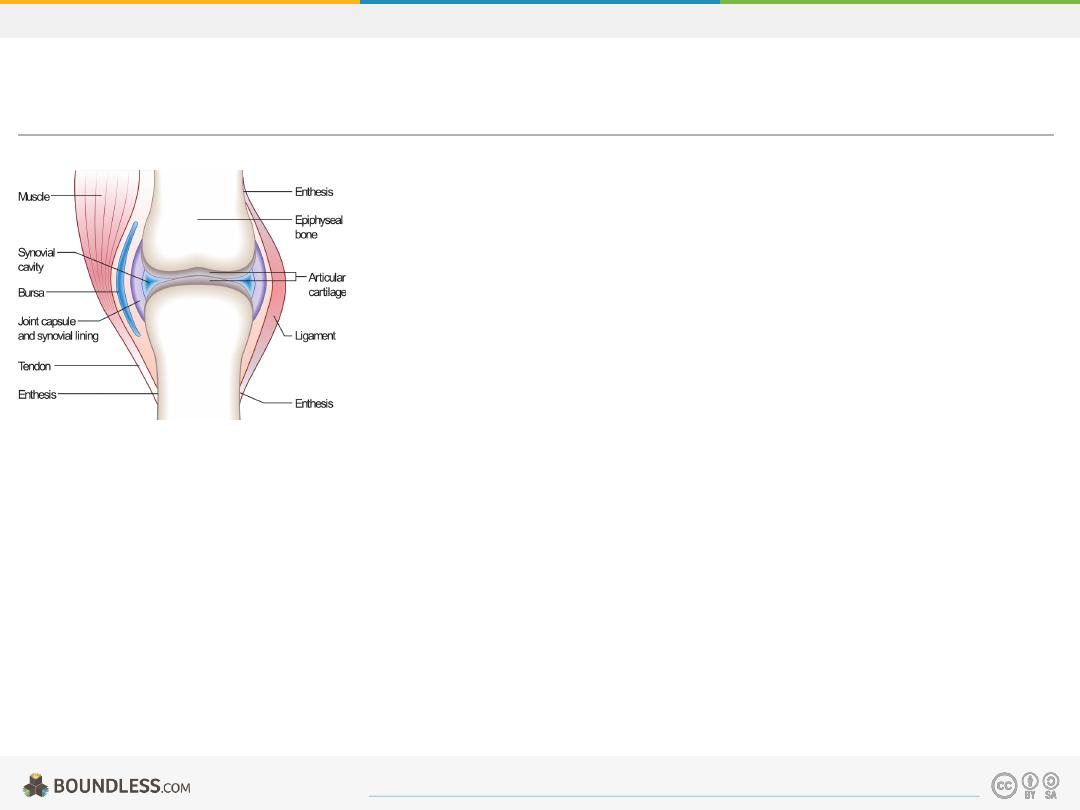
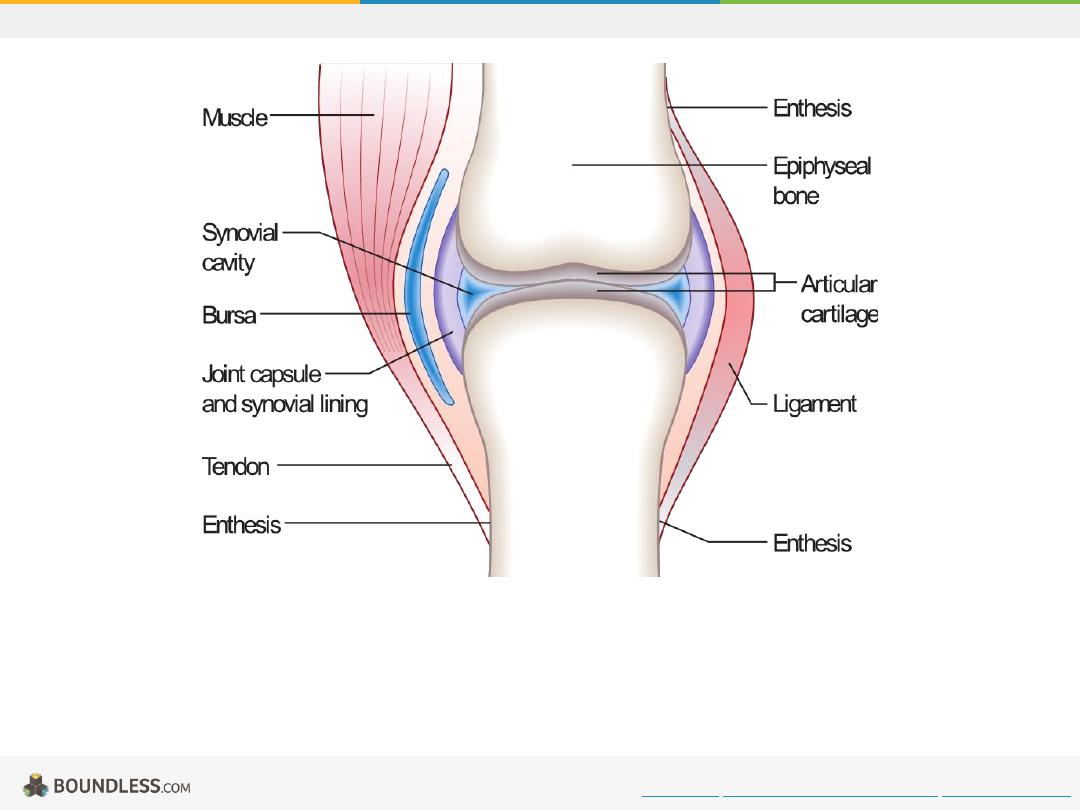
Bursa.jpg
Drawing of the knee joint showing bursae, ligaments, and tendons.
Free to share, print, make copies and changes. Get yours at www.boundless.com
Wikipedia. "Bursa.jpg."
https://commons.wikimedia.org/wiki/File:908_Bursa.jpg
Joints
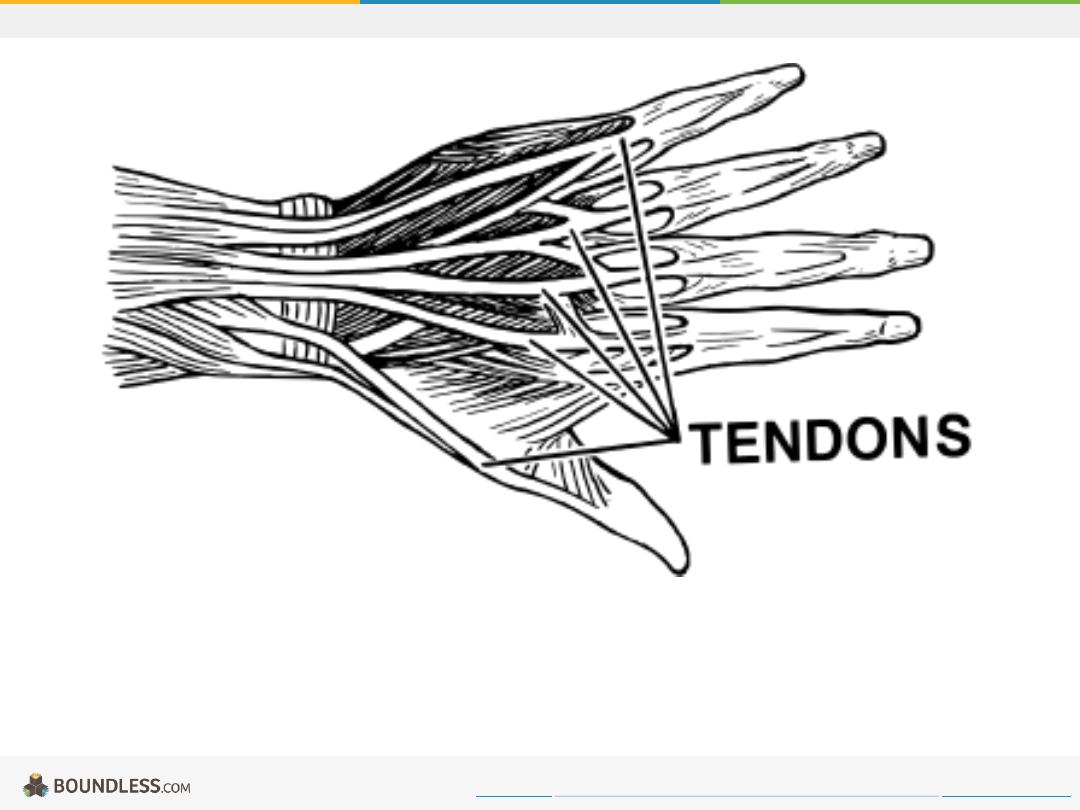
Tendons
Illustration of the location of tendons in the hand
Free to share, print, make copies and changes. Get yours at www.boundless.com
Wikimedia.
http://upload.wikimedia.org/wikipedia/commons/a/a1/Tendon_(PSF).svg
.svg)
Joints
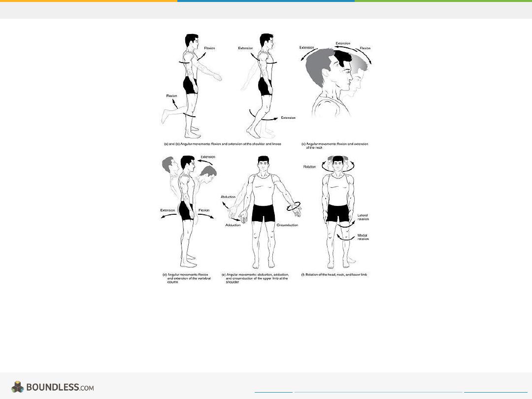
Body_Movements_I.jpg
Image demonstrating the various joint movements.
Free to share, print, make copies and changes. Get yours at www.boundless.com
https://commons.wikimedia.org/wiki/File:Body_Movements_I.jpg
Joints
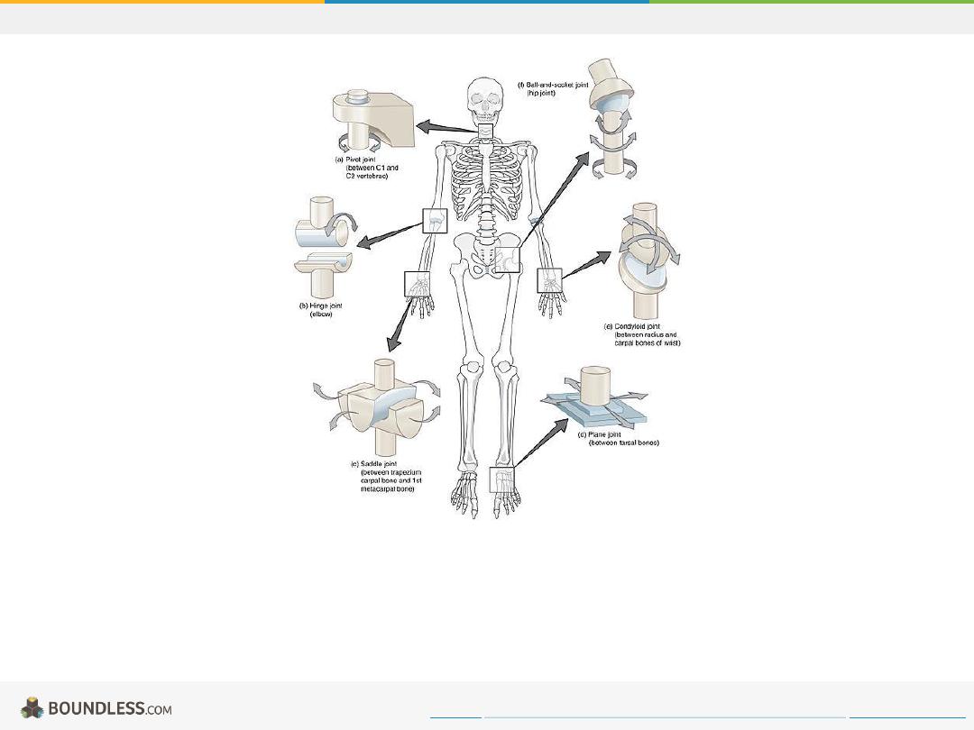
Six Types of Synovial Joints
Image demonstrating the six different types of synovial joints.
Free to share, print, make copies and changes. Get yours at www.boundless.com
Wikipedia Commons. "Synovial_Joints.jpg."
https://commons.wikimedia.org/wiki/File:909_Types_of_Synovial_Joints.jpg
Joints
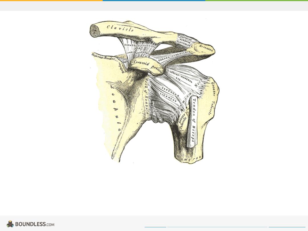
Plane Joint
The left shoulder and acromioclavicular joints, and the proper ligaments of the scapula.
Free to share, print, make copies and changes. Get yours at www.boundless.com
http://upload.wikimedia.org/wikipedia/commons/3/3b/Gray326.png
Joints
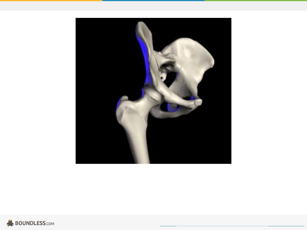
Ball and Socket Joint
Hip joint: the ball of the femur head fits in the socket of the acetabulum of the pelvis.
Free to share, print, make copies and changes. Get yours at www.boundless.com
Wikimedia.
http://upload.wikimedia.org/wikipedia/commons/9/9f/Hip.jpg
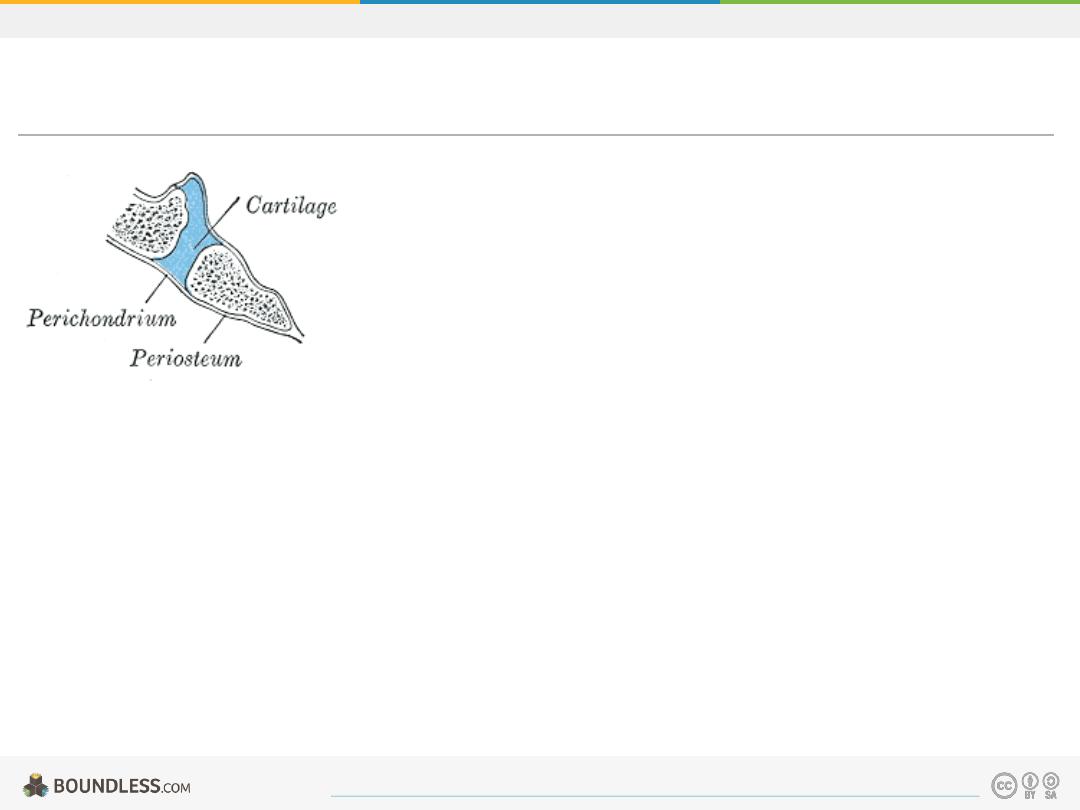
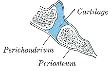
Joints
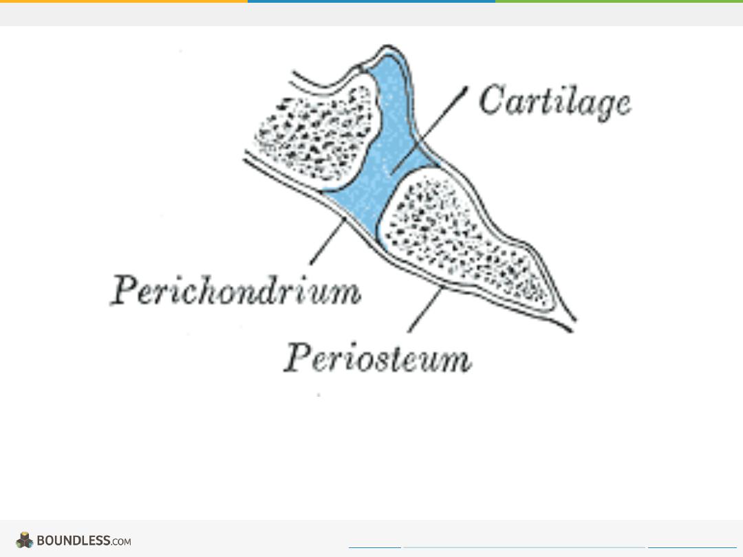
Synchondroses
Section through occipitosphenoid synchondrosis of an infant, including the cartilage, perichrondrium, and periosteum.
Free to share, print, make copies and changes. Get yours at www.boundless.com
http://upload.wikimedia.org/wikipedia/commons/7/76/Gray297.png
Joints
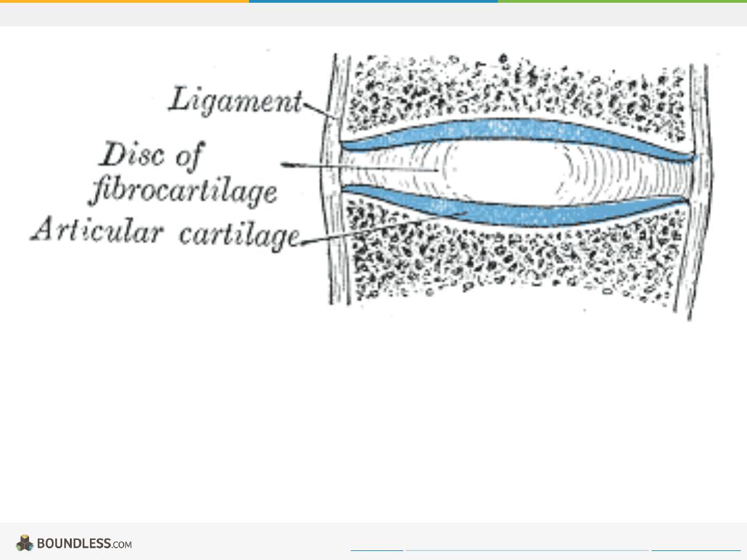
Symphyses
Diagrammatic section of a symphysis including the ligament, disc of fibrocartilage, and articular cartilage.
Free to share, print, make copies and changes. Get yours at www.boundless.com
http://upload.wikimedia.org/wikipedia/commons/b/b1/Gray298.png
Joints
Fibrous Joints
Image of fibrous joints with the tibiofibular syndesmosis demonstration in figure (b).
Free to share, print, make copies and changes. Get yours at www.boundless.com
https://en.wikipedia.org/wiki/Fibrous_joint#/media/File:904_Fibrous_Joints.jpg
Joints
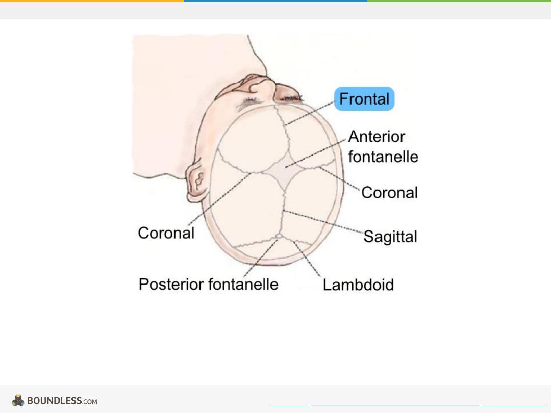
Frontal_suture_top_view.png
Drawing of human baby skull seen from the top. Cranial sutures are depicted with the frontal suture highlighted in blue.
Free to share, print, make copies and changes. Get yours at www.boundless.com
Wikipedia. "Frontal suture top view.png."
https://en.wikipedia.org/wiki/File:Frontal_suture_top_view.png
Joints