Diagnostic medical imaging
IntroductionRadiology is an older term, used when x-rays were the only testing modality using radiation available in medicine. Today, Diagnostic Imaging Service uses not only traditional x-rays but also ultrasound, computed tomography (CT) scans, and magnetic resonance imaging (MRI) in the diagnostic evaluation of patients.
Diagnostic imaging
refers to a variety of non-invasive methods for identifying and monitoring diseases or injuries via the generation of images representing internal anatomic structures and organs of the patient's body.Medical imaging
is the technique and process of creating visual representations of the interior of a body for clinical analysis and medical intervention, as well as visual representation of
the function of some organs or tissues.
Types of scan
• X-ray.• Fluoroscopy.
• Ultrasound.
• Bone densitometry.
• CT. A sensitive diagnostic tool used to image many diseases and injuries.
• MRI. A powerful tool that uses strong magnetic fields to produce images
• PET/CT.
Why is Diagnostic medical imaging important
Diagnostic medical imaging allows patients to limit or avoid invasive medical procedures and return to their lives faster, reducing the amount of time missing work and in hospitals with compounding medical bills
Imaging procedures
They are medical tests that allow doctors to see inside the body in order to diagnose, treat, and monitor health conditions. Doctors often use medical imaging procedures to determine the best treatment options for patients.The radiologist will look for areas of white, high-density tissue and note its size, shape, and edges. A lump or tumor will show up as a focused white area on a mammogram. Tumors can be cancerous or benign. ... The radiologist will check their shape and pattern, as they can sometimes be a sign of cancer.
.
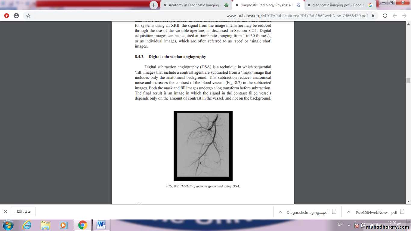
For almost half a century following the discovery of x-rays by Roentgen in 1895, radiologic imaging was mainly based on plain and contrast-enhanced radiography. Those images were created by exposing film to an x-ray beam attenuated after penetrating the body. The production of x-rays and radiographic images is described in the next chapter. In the recent half century, diagnostic radiology has undergone dramatic changes and developments. Conventional angiography, nuclear medicine, ultrasonography, and computed tomography (CT) were developed between 1950 and 1970.
Magnetic resonance (MR) imaging, interventional radiology, and positron emission tomography (PET) were developed later. Conventional radiology, including contrast-enhanced radiography and CT, uses ionizing radiation created from x-ray equipment. Nuclear medicine uses ionizing radiation that is emitted from injected or ingested radioactive pharmaceuticals in various parts of the body. Ultrasonography and MR imaging modalities use sound waves and magnetism, respectively, rather than ionizing radiation.
X- RAYS
Conventional radiography refers to plain radiographs that are generated when x-ray film is exposed to ionizing radiation and developed by photochemical process. During development, the metallic silver on the x-ray film is precipitated, rendering the latent image black. The amount of blackening on the film is proportional to the amount of x-ray radiation exposure. Plain radiography relies on natural and physical contrast based on the density of material through which the x-ray radiation must pass. Thus, gas, fat, soft tissue, and bone produce black, gray-black, gray, and white radiographic images, respectively, on film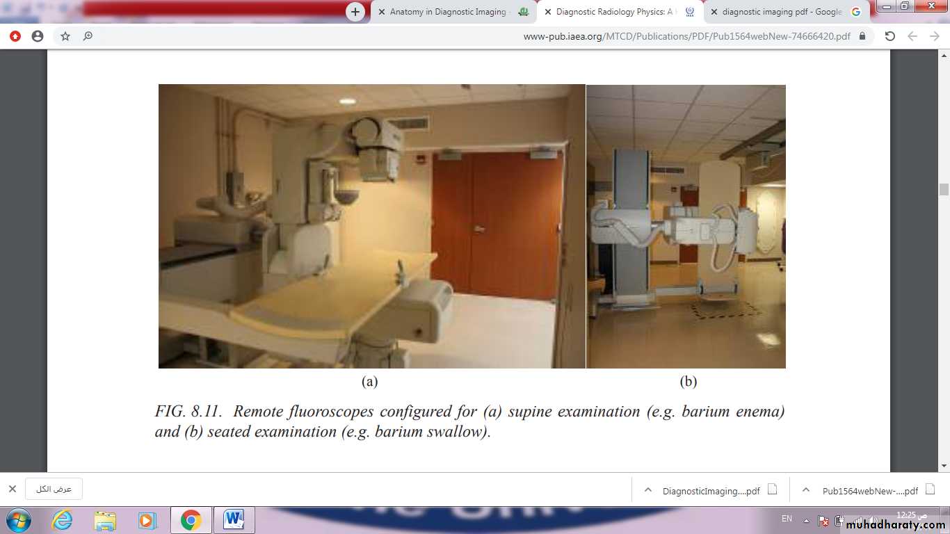
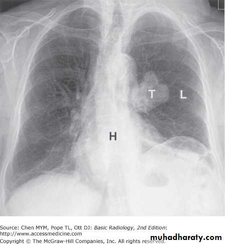
Although other image modalities such as CT, ultrasonography, and MR imaging are being used with increasing frequency to replace plain radiographs, conventional radiography remains a major modality in the evaluation of chest, breast, bone, and abdominal diseases
Dental Radiography
The tooth is a low attenuation static object that, when radiographed directly, places very limited demands on X ray generation. The image receptor is placed inside the mouth and irradiated externally. This universal low cost technique is known as an intraoral examination, with bitewing being the most common examination. When radiographs of the entire set of teeth are required, both the image receptor and the X ray source are external to the patient and the X ray beam is transmitted through the head, demanding significant X ray generation power and complex motion control for the X ray tube and image receptor. This procedure, known as an orthopantomograph (OPG)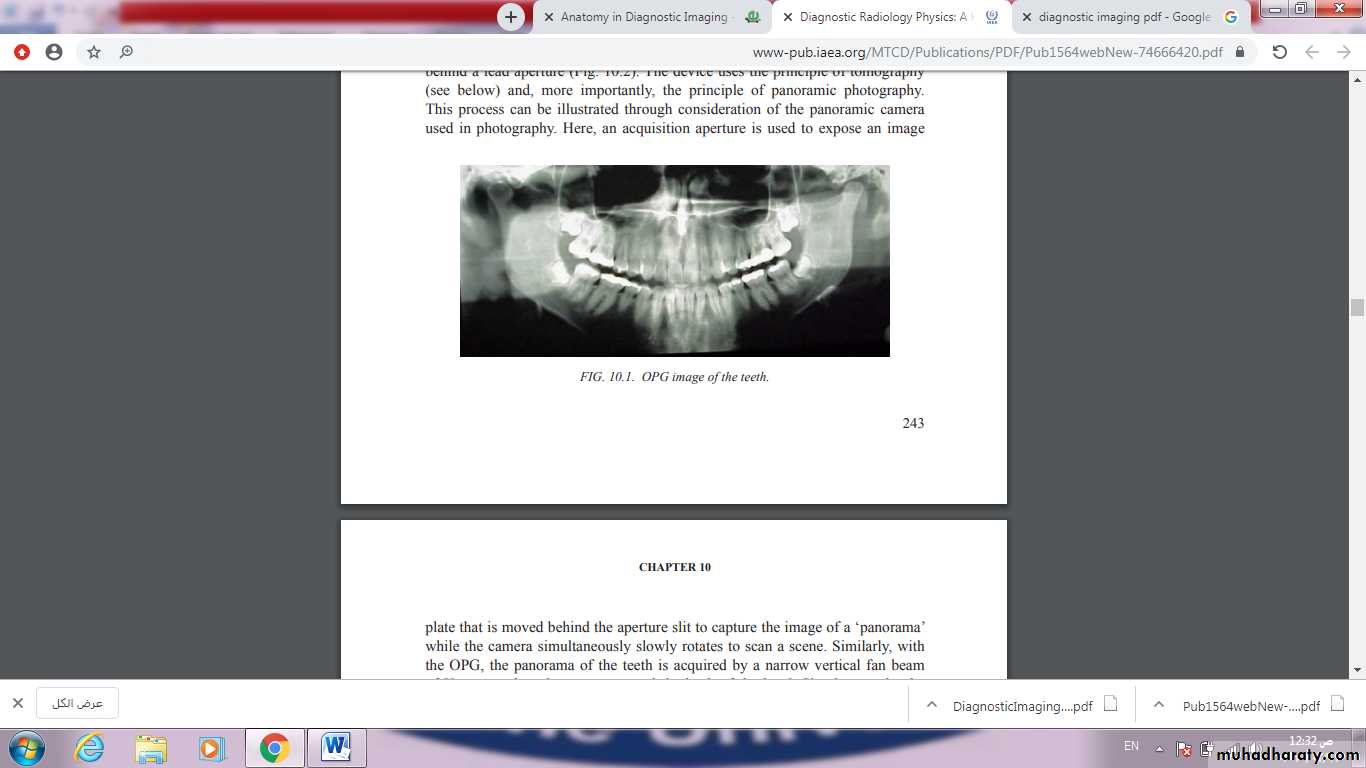
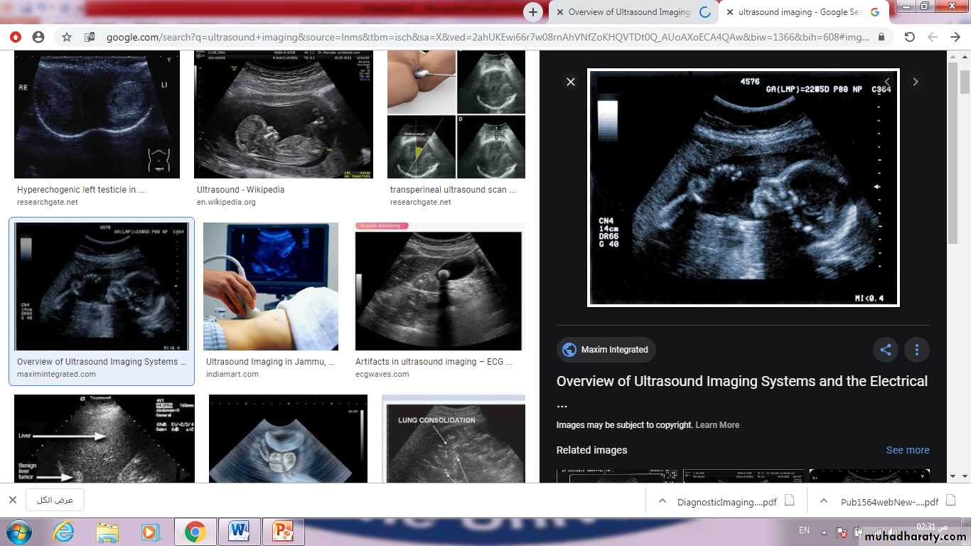
Ultrasound
Ultrasound is the most commonly used diagnostic imaging modality, accounting for approximately 25% of all imaging examinations performed worldwide at the beginning of the 21st century. The success of ultrasound may be attributed to a number of attractive characteristics, including the relatively low cost and portability of an ultrasound scanner, the non-ionizing nature of ultrasound waves, the ability to produce real time images of blood flow and moving structures such as the beating heart, and the intrinsic contrast among soft tissue structures that is achieved without the need for an injected contrast agent.The latter characteristic enables ultrasound to be used for a wide range of medical applications, which historically have primarily included cardiac and vascular imaging, imaging of the abdominal organs and, most famously, in utero imaging of the developing fetus. Ongoing technological improvements continue to expand the use of ultrasound for many applications, including cancer imaging, musculoskeletal imaging, ophthalmology and others.
Advanced diagnostic imaging procedures include computed tomography (CT), magnetic resonance imaging (MRI), and positron emission tomography/computed tomography (PET/CT) imaging devices.
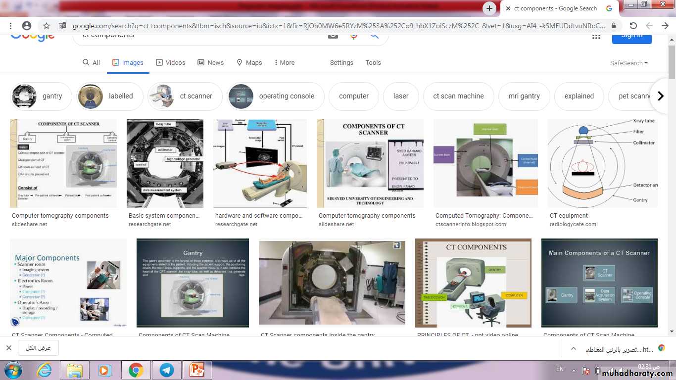
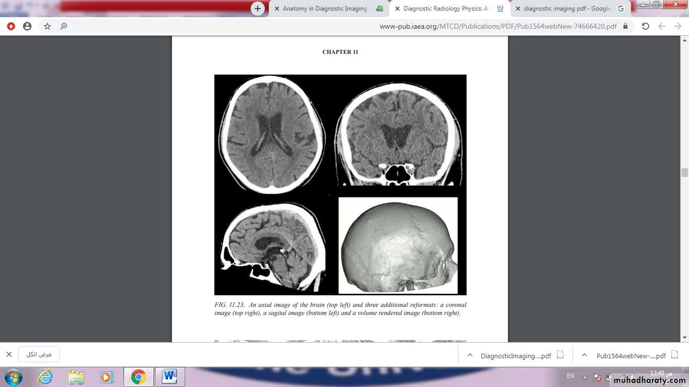
Computed Tomography
CT
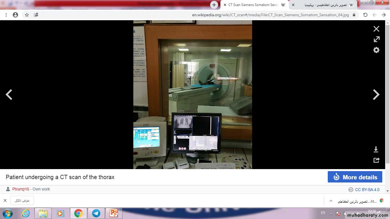
A computerized tomography (CT) scan combines a series of X-ray images taken from different angles around your body and uses computer processing to create cross-sectional images (slices) of the bones, blood vessels and soft tissues inside your body. CT scan images provide more-detailed information than plain X-rays do.
A CT scan or computed tomography scan makes use of computer-processed combinations of many X-ray measurements taken from different angles to produce cross-sectional images of specific areas of a scanned object, allowing the user to see inside the object without cutting.
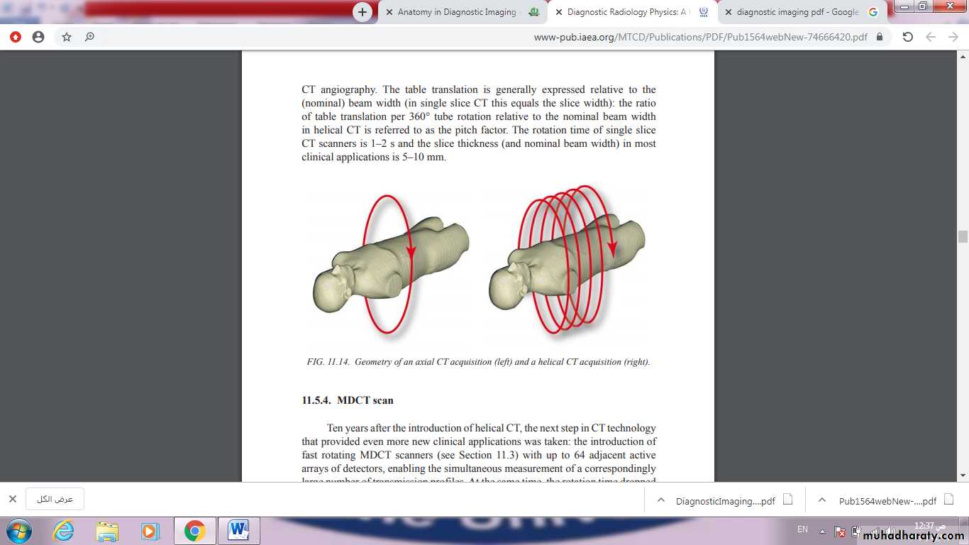
The essential physical characteristics of CT detectors are a good detection efficiency and a fast response with little afterglow. Currently, solid state detectors1 are used, as they have a detection efficiency close to 100% compared with high pressure, xenon filled ionization chambers that were used previously and that had a detection efficiency of about 70%. Solid state detectors are generally scintillators, meaning that the X rays interacting with the detector generate light. This light is converted to an electrical signal, by photodiodes that are attached to the back of the scintillator, which should have good transparency to ensure optimal detection. Typically, an antiscatter grid is mounted at the front of the detector, which consists of small strips of highly attenuating material (e.g. tungsten)
A computed tomography angiogram (CT angiogram) is a test that uses X-rays to provide detailed pictures of the heart and the blood vessels that go to the heart, lung, brain, kidneys, head, neck, legs, and arms. A CT angiogram can show blocked or narrowed areas of a blood vessels Computed tomography angiography is a computed tomography technique used to visualize arterial and venous vessels throughout the body. Using contrast injected into the blood vessels, images are created to look for blockages, aneurysms, dissections, and stenosis.
Magnetic Resonance Imaging
MRI1973, when Paul Lauterbur developed a method for spatially encoding the NMR signal by utilizing linear magnetic field gradients. About the same time, Peter Mansfield had also discovered a means of determining the spatial structure of solids by introducing a linear gradient across the object. The idea of applying magnetic field gradients to induce spatially varying resonance frequencies to resolve the spatial distribution of magnetization was a major milestone and the beginning of magnetic resonance imaging (MRI). For their work, Lauterbur and Mansfield were awarded the Nobel Prize for medicine in 2003.
Since its discovery, MRI has quickly become one of the most important medical imaging devices available to physicians today. Unlike other imaging modalities, such as X ray and computed tomography, MRI does not involve ionizing radiation. MRI also offers superior soft tissue contrast that is not possible with other imaging modalities. Furthermore, in MRI, the desired level of image contrast among different tissues can often be precisely controlled with simple adjustments to the acquisition timing parameters. MRI has become an invaluable tool for the assessment of many types of disease.
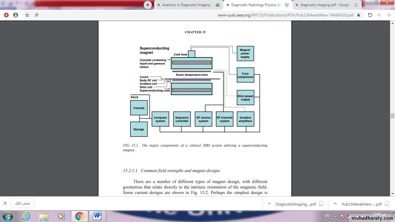
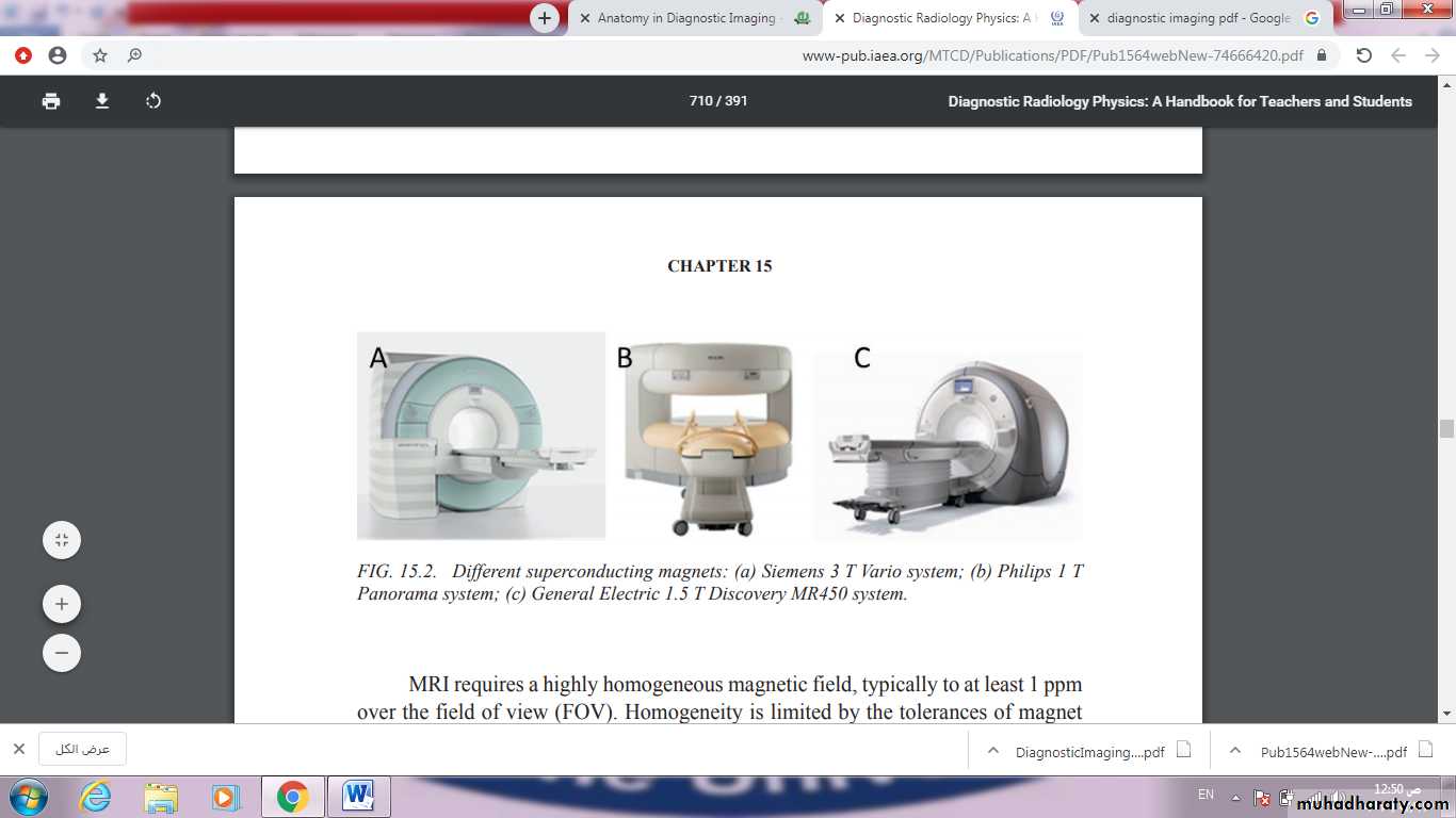
MRI systems comprise a number of major hardware components, under the control of digital systems that provide instructions, monitor system performance and acquire and process the signals that are used to create images or spectroscopic signals reporting on a wide range of tissue states. These systems are coordinated by one or more computer workstations or PCs that provide the interface to the MR operator, enabling acquisitions to be planned and executed, and images to be calculated, displayed and stored, often providing sets of measurement and analysis software addressing particular clinical questions. The next figure shows the major components of an MRI system, and these are described in more detail below.
Positron Emission Tomography/
Computed Tomography(PET/CT)
Positron emission tomography (PET) uses small amounts of radioactive materials called radiotracers or radiopharmaceuticals, a special camera and a computer to evaluate organ and tissue functions. By identifying changes at the cellular level, PET may detect
the early onset of disease before other imaging tests can.
A positron emission tomography (PET) scan is an imaging test that helps reveal how your tissues and organs are functioning. A PET scan uses a radioactive drug (tracer) to show this activity. This scan can sometimes detect disease before it shows up on other imaging tests.
A PET-CT scan combines a CT scan and a PET scan. It gives detailed information about cancer. ... The PET scan uses a mildly radioactive drug to show up areas of your body where cells are more active than normal.
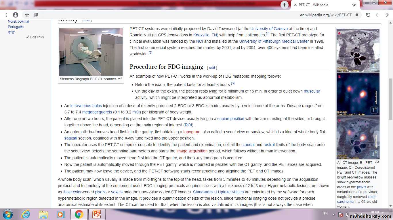
is a nuclear medicine technique which combines, in a single gantry, a positron emission tomography (PET) scanner and an x-ray computed tomography (CT) scanner, to acquire sequential images from both devices in the same session, which are combined into a single superposed (co-registered) image. Thus, functional imaging obtained by PET, which depicts the spatial distribution of metabolic or biochemical activity in the body can be more precisely aligned or correlated with anatomic imaging obtained by CT scanning. Two- and three-dimensional image reconstruction may be rendered as a function of a common software and control system.
PET-CT has revolutionized medical diagnosis in many fields, by adding precision of anatomic localization to functional imaging