Dental Radiology
Dental Radiologyد. شهرزاد سامي
د. شهرزاد سامي
Radiographic Interpretation of Mixed Lesion (part 2)
Focal Cemento-osseous Dysplasia
This usually solitary fibro-cemento-osseous lesion occupies a portion of the spectrum between the periapical and florid cemento-osseous dysplasias.
• Shape: Round, monolocular.
• Outline: —Well defined but irregular
— Not corticated.
Radiodensity: — Early stage — radiolucent
— Intermediate stage — radiolucent with patchy opacity within the radiolucency
— Late stage — densely radiopaque but often surrounded by a thin radiolucent line.
Cemento-ossifying Fibroma
Cemento-ossifying fibromas are rare fibro-osseous benign neoplasms that affect the jaws.Shape: — Round
— Monolocular.
Outline: — Smooth, well defined
— When opaque usually surrounded by a thin encapsulating radiolucent line
— Usually corticated and circumscribed.
Radiodensity: — Early stage — radiolucent
— Intermediate stage — radiolucent with gradually increasing internal radiopaque calcified patches
— Late stage — radiopaque zones coalesce to form a densely radiopaque mass with or without a radiolucent periphery.
Ameloblastic Fibro-odontoma
These rare, monolocular or multilocular odontogenic tumours resemble closely ameloblastic fibromas and also affect children. However, they usually contain enamel or dentine, either as multiple, small opacities or as a solid mass.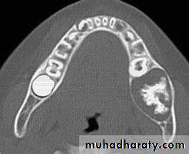
Cherubism
This rare disease of the jaws is inherited, but many cases appear spontaneously. Radiologically the lesions resemble closely other giant cell-containing lesions.• Age: Children, 2-6 years old.
Shape: — Multilocular
— Bilateral lesions typically symmetrical.
Outline: — Smooth, Well defined, Well corticated
Radiodensity: Radiolucent with internal radiopaque septa producing a multilocular appearance.
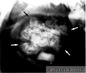
Osteosarcoma
Rare, rapidly destructive malignant tumor of bone. The cause of osteosarcoma is unknown, but genetic mutation and viral causes have been suggested. It is also known to occur in association with Paget’s disease and fibrous dysplasia after therapeutic irradiation.
Early features:
Non-specific, poorly defined radiolucent area around one or more teeth.
Widening of the periodontal ligament space.
Later features:
• Osteolytic lesion:— Monolocular, ragged area of radiolucency — Poorly defined, moth-eaten outline.
— So-called spiking resorption and/or loosening of associated teeth.
• Osteosclerotic and mixed lesions:
— Poorly defined radiolucent area
— Variable internal radiopacity with obliteration of the normal trabecular pattern
— Perforation and expansion of the cortical margins by stretching the periosteum, producing the classical, but rare sun ray or sunburst appearance
— Spiking resorption and/or loosening of associated teeth
— Distortion of the alveolar ridge.
Osteioradionecrosis
Its inflammatory condition of the bone that occur after the bone exposed to the therapeutic dose of radiation usually given to the malignancy of head and neck region.Osteoradionecrosis is characterized by the presence of exposed bone for at least 3 months occurring at any time after the delivery of radiation therapy. This exposed bone may completely sequestrate.
The radiologic identification of osteoradionecrosis relies on the identification of dead bone in the form of sequestra.
In contrast to osteomyelitis, there is no periosteal bone reaction in most cases. The presence of a pathologic fracture is suggestive of osteoradionecrosis.