
Dr. Mazin
X ray of urology
This a
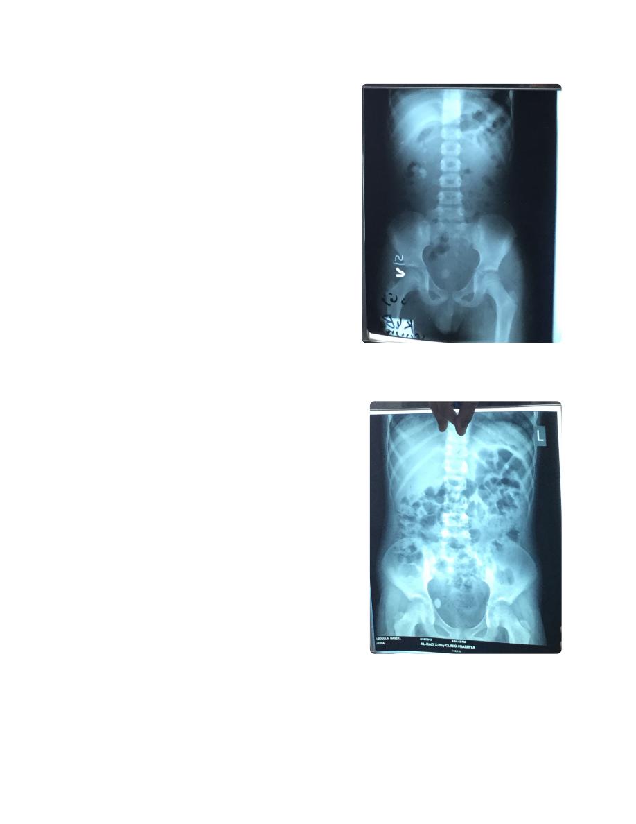
KUB film (Kidney-ureter-bladder)
•
Pubis symphysis, and xiphoid of sternum
◦
If these two not shown, it is
plain
‣
abdominal film
KUB is taken while the patient is standing.
•
Named: name of patient
•
Labeled for known direction
•
Dated
•
Showing (any findings)
•
Plain X ray means without contrast
•
Look for any
radio opaque opacity
•
Osteoporosis shows black area in bone
•
Radioopaqe opacity means white spot
•
Diffrential diagnosis
•
Foreign body in clothes
◦
Scars in skin
◦
Calcified lymph node
◦
Calcified hematoma
◦
Calcified tumor
◦
Stones
◦
◦
Double J catheter, a stent
•
J shape end for staying in position , form both
◦
ends
Indications
◦
Prophylaxis
‣
Prevent obstruction after or within surgey
•
For gynecological purpose to avoided
•
uretric injury During hysterectomy "Water
under bridge)
Treatment
‣
Bypass stone or obstruciton
•
Uretric trauma
•
Uretric surgery
•
Common side effect
◦
Irritative voiding system
‣
Hematurea
‣
Slipping and migration inside the body
‣
Don't say stone, but say most likely stone
•
Don't the the opacity in ureter or bladder, but say it in
•
pelvic region, then say most likely stone in the ureter
for example
Radio opaque in right kidney misleading with gall
•
bladder (gall bladder stones mostly radio lucent,
confirmed by lateral x ray (if anterior to mid axillary line
most likely gall bladder, if posterior to it most likely
renal) or ultra sound )
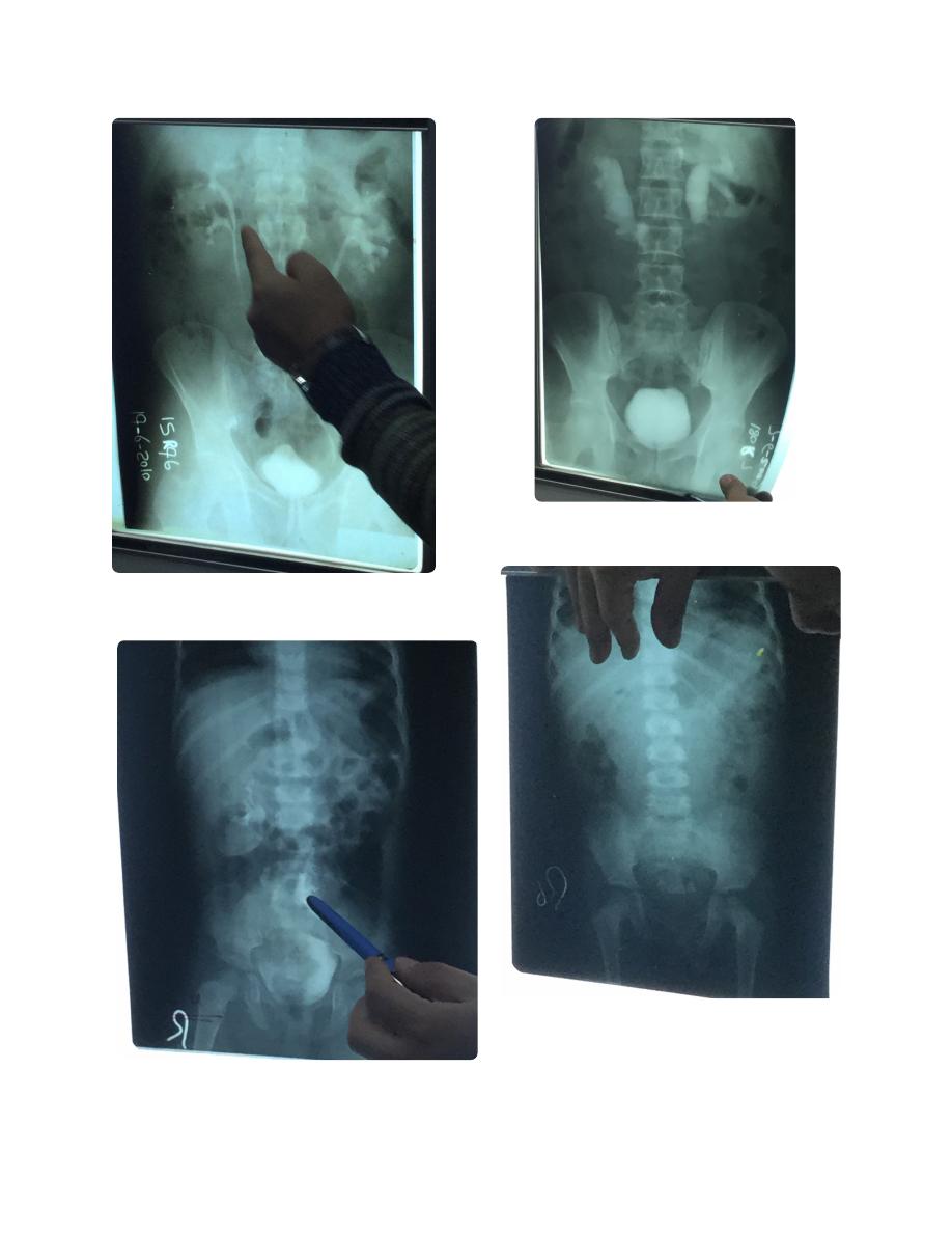
KUB film named, labeled, dated.
Showing radio opaque opacity in
pelvic lesion, most likely a vesicle
stone (secondary to upper system)
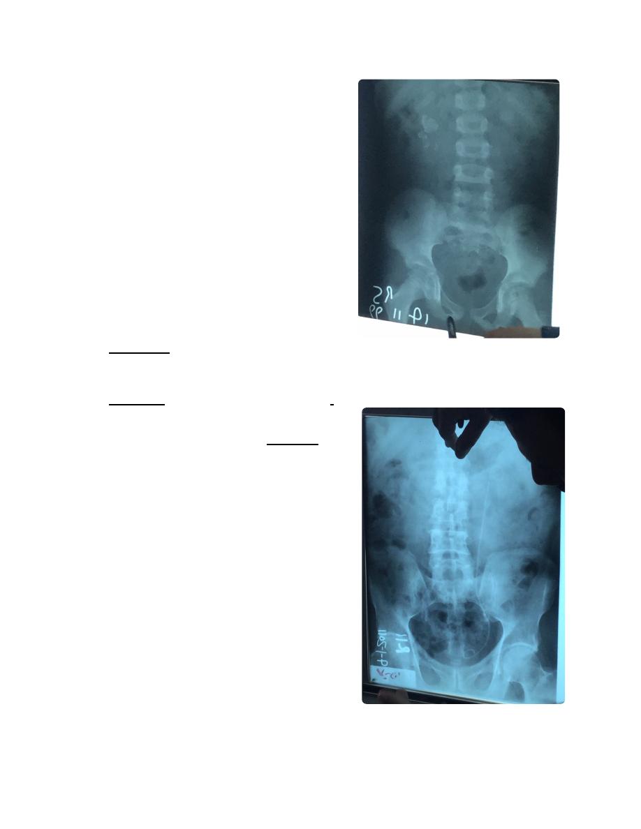
KUB, named, dated, labeled (label
should be on right not left). Showing
radio opaque opacity in pelvic
lesion, most likely uretric stone
(because lateral position, if vesical it
should be with gravity)
IVU film (intra venous urography, contrast excreted
•
exclusively by kidney) ةنولم ةعشأ
Always say I need KUB film
‣
Indications
◦
Recurrent hematurea
‣
Trauma
‣
Tumor
‣
Congenital anomaly
‣
Gynecological
‣
Recurrent UTI
‣
At night fasting, no eat in morning, take rectul
◦
suppository to reduce gasses, ask for allergy to
penciline and asthma and pregnancy
(contraindication)
Also contraindicated in DM patients who taking
◦
metformin (cause lactic acidosis), should stop it
2 days before and after, and use insulin during
this period.
Always
must be KUB before IVU
◦
First image in one minture of injection called
◦
nephrogram.
Delay means impairment in kidney, like
‣
renal artery stenosis
Second image in fifteen minutes called
◦
pyelogram (calyx and pelvis and upper ureter)
Dilated uretric means hydrouretronephrosis
‣
due to stone for example.
Third image in 30-60 min called cystogram
◦
Nephronstomy catheter (
Nephrostogram)
•
Indications
◦
Diagnostic
‣
To know level of obstruction
•
Taking biopsy
•
Therapeutic
‣
Destruction of renal stone
•
Bypass obstruction
•
Evacuation of pu
•
Pelvic kidney , 1/400000-600000
•
Liable for stasis, stones, TB, and carcinoma,
◦
Problematic in pregnancy
◦
Most common cause of bilateral hydronephrosis in
•
children is posterior valve obstruction (congenital)
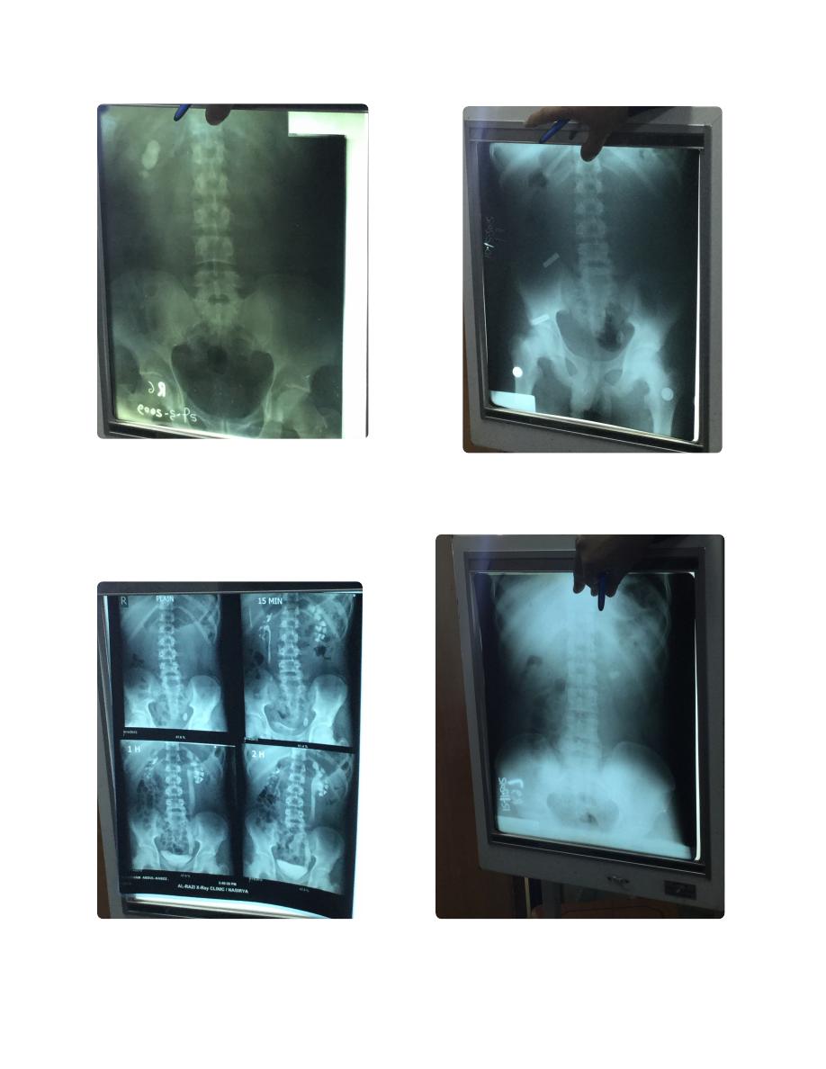
KUB film, not named, labeled, dated.
Showing multiple radio opaque
opacity in right renal area, most likely
multiple renal stones.
KUB ... etc
Showing Double J tube
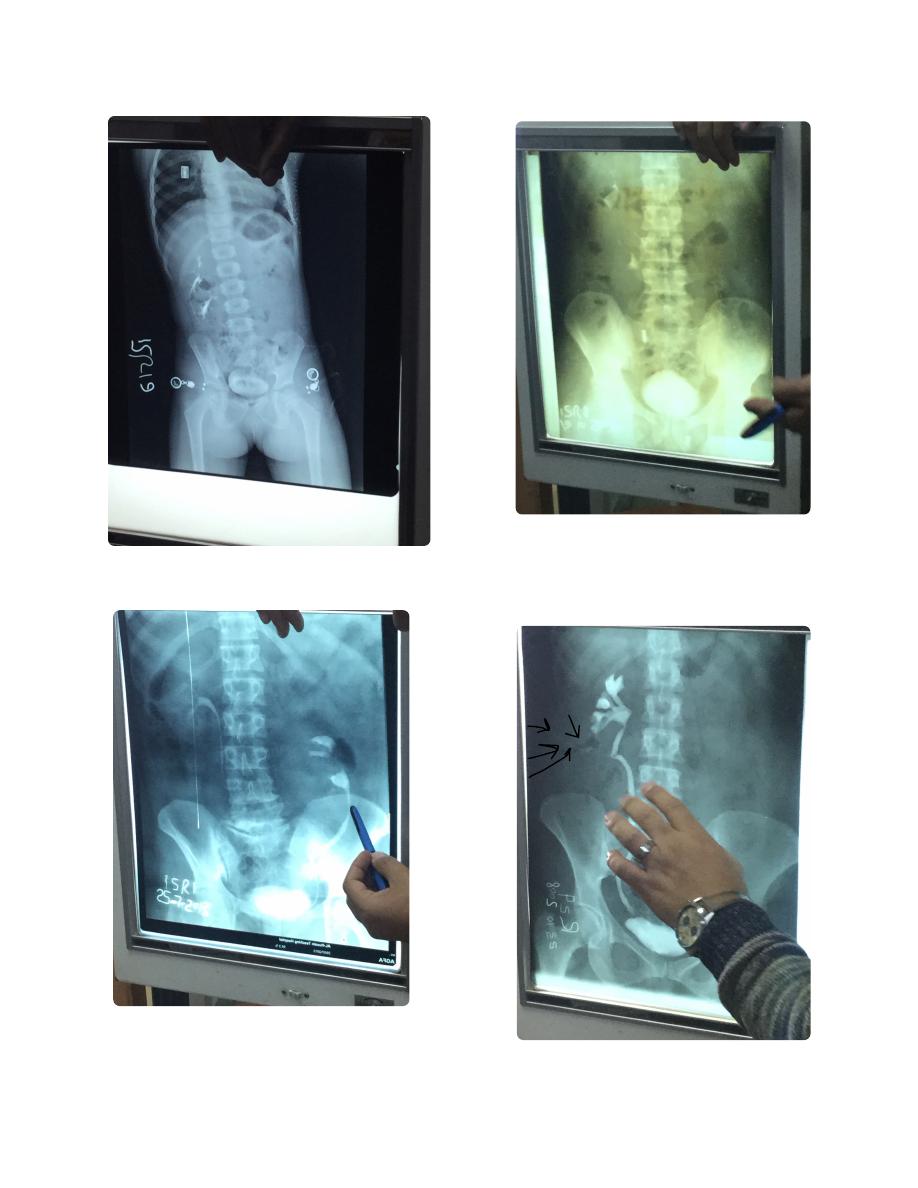
KUB ... etc. Showing radioopaque
opacity in right renal area.
(misleading with gall bladder (gall
bladder stones mostly radio lucent,
confirmed by lateral x ray (if anterior to
mid axillary line most likely gall bladder,
if posterior to it most likely renal) or
ultra sound ))
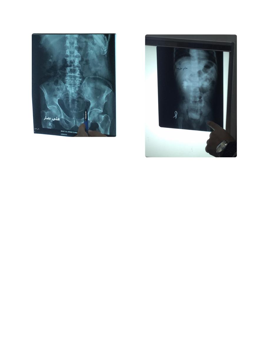
IVU, I need KUB
It is named, labeled, dated.
6KUB ... etc
Showing radio opaque opacity in
pelvic region, most likely uretric
stone.
8Plain abdominal X ray! (Symphysis pubis
not shown)
IVU , I need KUB ...etc
Ectopia
Nephrostogram (not IVU)
IVU...
Cross-renal ectopia, congenital
anomaly (But ureter does not cross
because of different origin)
IVU ...
Malrotated kidney زعالنه
IVU...
Hydronephrosis
IVU... etc
Pelvic kidney
KUB film not named not dated not labeled.
Multiple vesicular stone. (This presents as
unanimity to urinate while sitting, but able
when stand and move around)
(This is a child, epi-phiseal plates un united)
IVU ... etc
Bivid pelvis of right kidney
Ectopic note from Dr. Alaa
Stoma closed 8-12 weeks
Not earlier due to inflammation
Not after due to distal part atrophy
Complication of double J tube
(perforation)
IVU, I need a KUB
Named, not dated, labeled.
Child (un united epi phiseal plates) with
bilateral hydronephrosis, most common
cause is posterior valve obstruction
(congenital)
Scanned by CamScanner
Scanned by CamScanner
Scanned by CamScanner
Scanned by CamScanner
Scanned by CamScanner
Scanned by CamScanner
Scanned by CamScanner