
Anemia
Anaemias
•
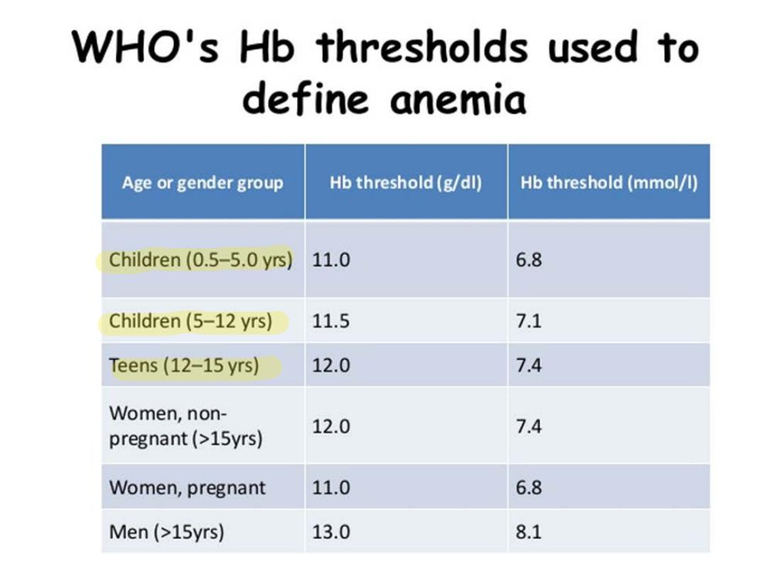
Is defined as a reduction of RBC volume or Hb
concentration below the range of values occurring in
healthy persons ( reduction in the amount of circulating
Hb resulting in decreasing in O2 carrying capacity of
blood ) .
•
Few clinical disturbances occur until the Hb level fall
bellow 7—8 gm/dl , where below this level , pallor is
evident in mucous membranes
Physiological adjustment to anemia include:--
•
1- increase cardiac output & O2 extraction
•
2- a shunting of blood towards vital organ & tissues.
•
3-2,3 DPG increases within the RBC leading O2
dissociation curve shifted to right resulting more
release of O2 to tissues .
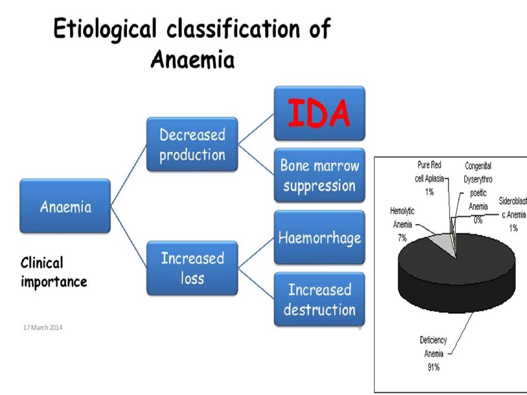

Approach patients with anemia :-
Anemia ---Hb &indices---Retic count &morphology ---:--
1- inadequate response (RPI ) < 2% :-
a- hypochromic microcytic
b- normochromic normocytic anemia .
c- macrocytic .
2-adequate response : ( RPI ) > 2% :-
1-Hbpathy
2-Hbss ,SC , S-B thal.
3-enzymopathy as G6PD def. or pyruvate def.
4-membranopathy as cong. Sperocytosis or elliptocytosis
5- extrinsic factors like DIC & HUS ,, Burns ,, wilson dis. ,, Vit
E def.
6-immune H.A as in auto immune , iso immune , drug
induced
•
Inadequate response :--
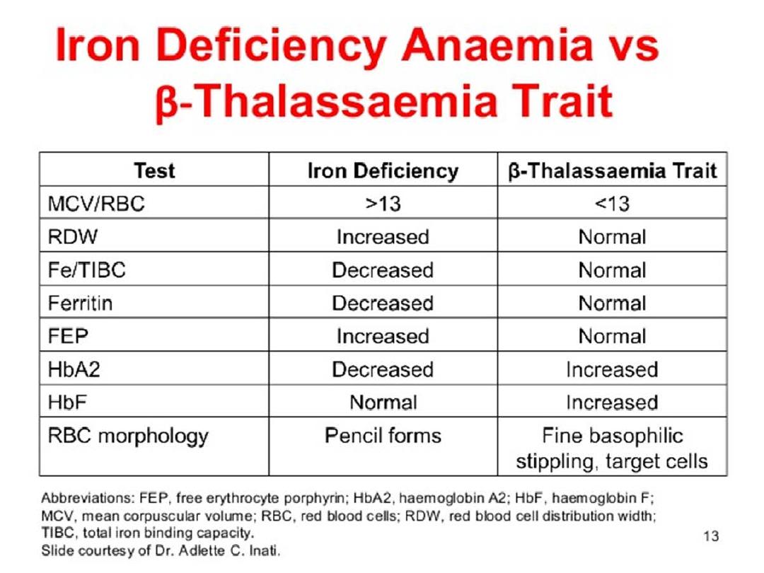
1- Hypochromic microcytic anemia as IDA , thalass.
Trait , chronic inflammatory dis. , copper and lead
poisoning , sideroblastic anemia .
2- normochromic normocytic anemia as chronic
inflammotory dis. , recent blood loss , malignancy ,
marrow failure , chronic renal failure & transiant
erythroplastopenia of childhood .
3- macrocytic anemia as in folate , Vit 12 def. ,
marrow failure , hypothyrodism , down syndrome
•
Anemia with reticulocytosis (normal retic count is 1%). :--
1- ongoing hemolysis
2- bleeding
•
Anemia with increase T.S.B hemolysis doing perpheral blood
smear
1-spherocyte as in hereditory spherocytosis , wilson dis. , auto
immune H.A .
2-sickle cell ( SCA, S. thalass. )
3- target cell ( Hb sc )
4-bite cell / blister cell ( G6PD )
5- Micro angiopathy H.A (D.I.C., H.U.S )
6- Nucleated RBC ( B-thalass. )
•
Note : reticulocyte production index( RPI) : measured by :--
observed PCV
------------------- X retic count
normal PCV

•
Anemia with low or normal retic count:---do red
blood cell size :-
1-microcytosis ( IDA, thalass.trait, chronic infla. Dis.,
copper& lead poising , sideroblastic )
2-Normochromic normocytic :--as in chronic infla. Dis.
, recent blood loss , malignancy , marrow infiltration,
chronic renal failure , TEC ) .
3- macrocytic as in vit. B12 , & folate def. ,
hypothyrodism , marrow failure , down syndrome
,aplastic anemia ( cong.or acquired )
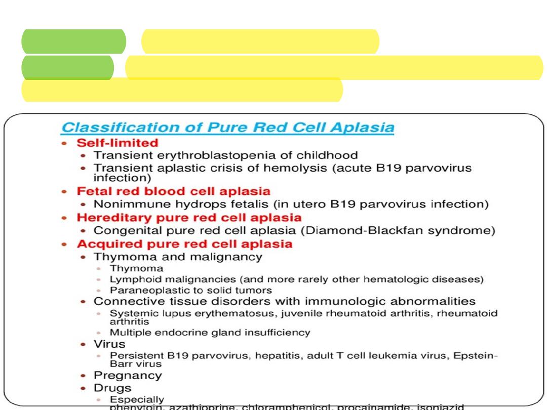
Anemia of inadequate production :--
•
1- congenital :-- hereditary pure red cell aplasia .
•
2- acquired :--TEC, assoc. with chr. H.A by parvo virus , immune def. &
others like SLE, DRUG like chloromphenicol )
Hereditary Pure red cell aplasia ( Blackfan
Diamond syndrome)
•
Is rare condition , is sporadic , although dominant
or recessive in 15% ,.& mutation is seen in 25%
Usually become symptomatic in early infancy as
profound anemia by 2-6 month of age ( 90% occur
in first year of life )
•
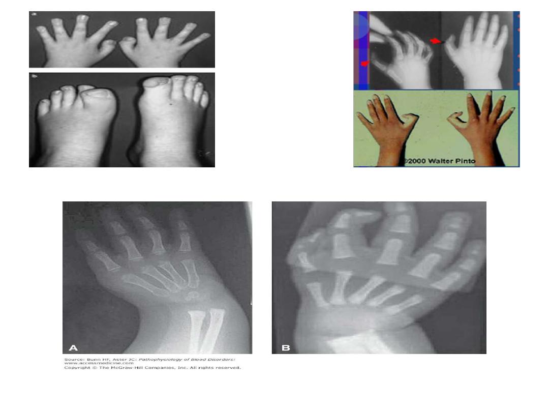
50% of affected children have cong. Anomalies like
short stature in 30%, defect in upper extremeties
and thumb defect .
•
cranio facial abnormalities (, cleft palate ,
macroglossia and cong. Malformtion) in 35-45%
,cardiac and urogenital defect .

•
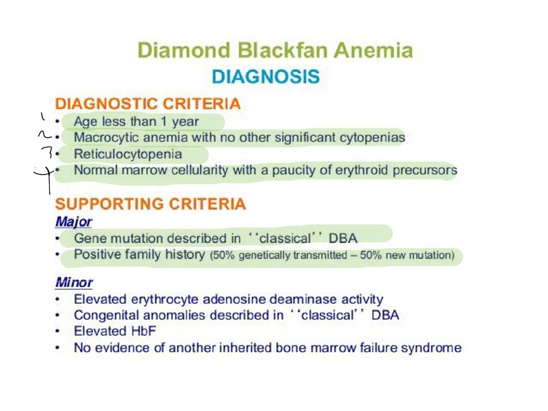
Lab. Finding :---
1- macrocytosis with no hypersegmented neutrophil .
2- very low retic count .
3-EAD(erythrocyte adenosine deaminase is increased
in most pts
4- RBC precursor is markedly reduced .
5- slight elevation of Hb F
•
DD :---
1- other causes of low retic count
2- TEC

Thumb defect ( 6 fingers)
Treatment :--
1- 80% of patients respond initially to steroid of unknown
mechanism ( 2mg\kg of prednisolone ).
•
Hb reach to normal within 4-6 wks ( increasing RBC precursor
within 1-3 wk of therapy ) .
•
Steriod is not recommended before 6 month of age even
some hematologest not given before one year of age .
•
20% of patients not respond to steroid (needs blood
transfusion )
Prognosis :--
•
50% are long term responder And 50% who respond initially
become resist later & 20% has spontaneous remission .
•
(40% is steriod dependant , 40% transfusion dependant &
20% no need treatment Although it is considered as
premalignant syndrome with acute leukemia and
mylodysplastic syndrome . .

Transiant Erythroplastopenia of Childhood:-
•
Is more common acquired red cell aplasia occurring in
children( more common than Black fan Diamond syndrome ),
IS characterized by :---
1- occur in previous healthy children between 6 month– 3 years
( mostly above one year of age ) & only 10% after 3 year of age .
2- resulting from transiant immunological suppression of
erythropoiesis & often follow viral infection .
3- very low retic count
4- normal MCV and HbF
5- virtually all children are recovered within 1—2 month
•
Blood transfusion may be necessary , while cortico steriod
with no benefit .
•
Note ;-- any child with presumed TEC who required more
than one unit of blood transfusion , you should evaluated for
another diagnosis .
Physiological Anemia of infancy
•
Is progressive decline of Hb within first few wks of
life & persists for 6-8wk , reaching in full term infant
9-11 gm\ dl at 8-12wk of age , while in preterm
infant reaching 7-9gm\dl by 3-6 wk of age .
•
Several factors are operative:--
1- suppression of erythropoeitin production with
increasing O2 delivery to tissue.
2- shortened survival of RBC of premature infant 40—
60 days instead of 120 days .
3- late hypo-regenerative anemia with absence of
reticulocyte.
4- dietary factors like folic acid ( vit.E def. & therapy
have no role in this type of anemia )

•
Treatment
1-Generally required no therapy other than ensuring
that diet of the infant contains essential nutrients for
normal haematopoiesis especially folic acid & iron
2-anemia in VLBW --- treated by erythropoitin & iron .

Megaloblastic anemia:-
•
Caused by :--
1- folate def.
2- vit. B12 def.
3-inborn error of metabolism ( blackfan diamond
syndrome)
•
Folic acid def.:---
•
Folic acid absorbed through out small intestine and
human can not synthesis folate & depend on dietary
sources including 1/3 from green vegetable & fruit ,
1/3 from animal organ like liver and kidney and 1/3
from cereals and grain.
•
Body store of folate are limited and Anemia occurs
after 2—3 month of folate free diet

•
C/F
1- peak age 4—7 month ( is earliar than IDA)
2-may FTT, chronic diarrhea , irritability.
3- hemorrhage may occur in advance age
Causes ;---
1-inadeguate intake ( poor nutrition , chronic
hemolysis , goat milk )
Anticonvulsant like phenytoin, phenobarbital,
primodin
2-decrease absorption Methotrexate , Tm/SM,
pyrimethamine))
3-drug induced folate metabolism
4- cong. Abnormality of folate metabolism
•
Lab. Finding :----
1- macrocytosis( MCVof more than 100 ) .
2-neutropenia & thrombocytopenia .
3– hypersegmented neutrophil
4- S. folate is decresed & LDH is elevated
•
Treatment ;---
1. Folate 0.5 -1 mg \ day for 3-4 wk until definite
hematological response has ocurred
2. Maintaince therapy 0.2 mg is adequate

Vit B12 Defeciency
•
Is derived from cobalamine in food of animal source(
which is commonly meat and fish ) secondary to
production by micro-organism (human can not
synthesis B12 ) , it is in contrast to folate store , older
children and adult have sufficient V.B12 store at least 3-
5 years unless infant born to mother with low V.B12
store where clinical sign appeared in first 4-5 month of
life .
Causes :---
1- inadequate intake as in strict vegetarian
2- lack of intrinsic factor as in pernicious anemia or gastric
surgery .
3- impaired absorption as in dis. of terminal illeum , IBD
,over growth of bacteria , duplication of small intestine
4-absence of vit 12 transport protein .

C\F:---
•
Non specific manifestation like weakness, FTT,
irritability , pallor, glossitis , vomiting , diarrhea,
parasthesia, numbness, development delay.
Lab. Finding :---
•
Macrocytosis, hypersegmented neutrophil,
neutropenia & thrompocytopenia in advance case ,
s.iron & folic acid are normal, LDH is elevated
Treatment :----
•
1mg paranterally ( increase retic count within 2-4
days
•
Treatment of precipitated factors If neurological
involvement --- 1mg of B12 I.M for 2 wks then
maintaince dose 1-5 microgram\day

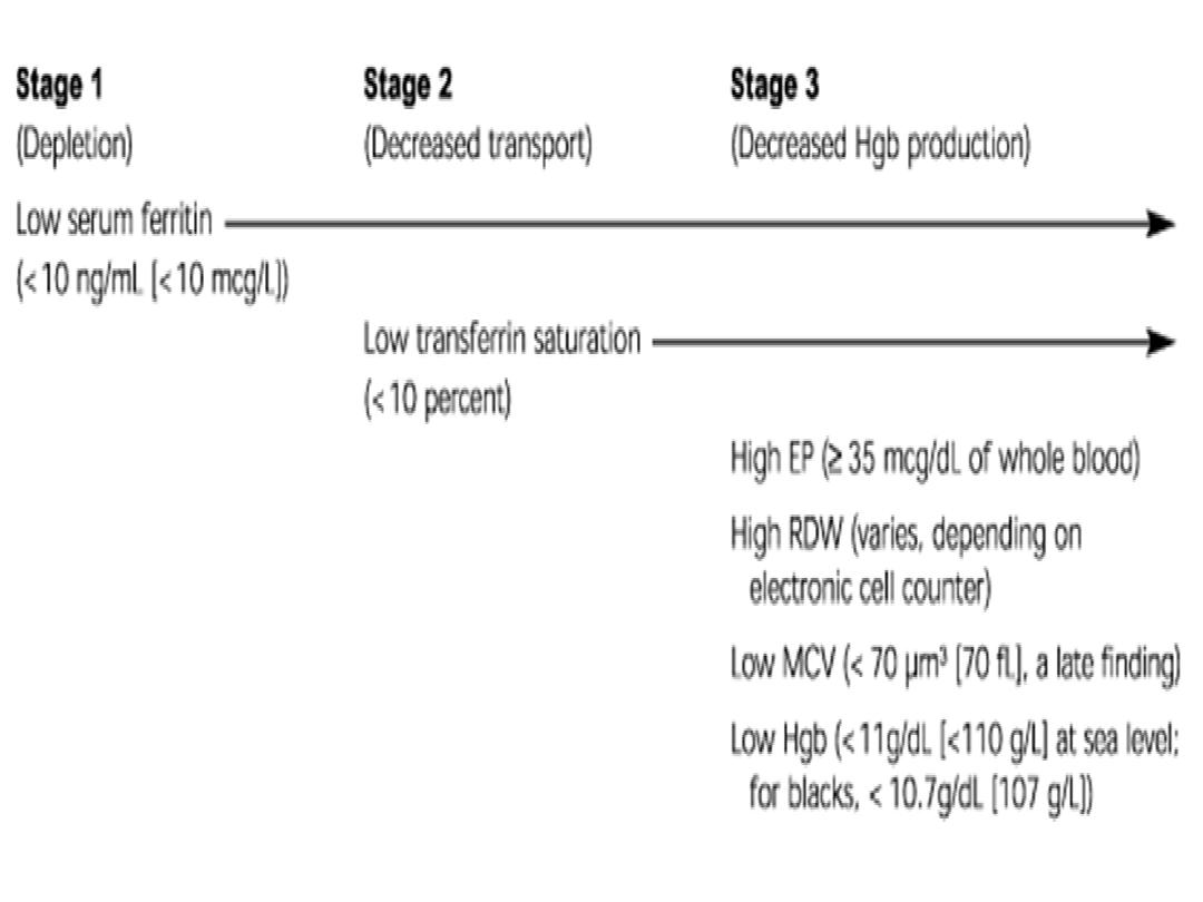
Iron Def Anemia
•
Is most common hematological dis. of infancy & childhood ,
its frequency related to certain basic aspects of iron
metabolism , resulting from lack of sufficient iron for
synthesis of Hb .
•
New born infants contains 0.5gm of iron, where in adult
contains 5gm of iron ( to make up this discrepancy needed 1
mg daily as o.8 absorbed )normal body need daily and 0.2
mg to replace shedding of blood cell
•
Note : only absorbed 10% of iron therefore needed daily10
mg 0f iron within food .
Etiology :---
1- inadequate iron intake .
2- blood loss as in peptic ulcer, polyp, IBD, occult blood, milk
allergy , meckles diverticulum.
3- defect in iron absorption as in malabsorption syndrome .

C\F:----
•
1- pallor .
•
2- pagophagia .
•
3-if Hb bellow 5gm\dl developed irritability,
anoreoxia , tachycardia & cardiac dilatation .
•
4- may associated with impaired altertness &
learning

Lab finding :-
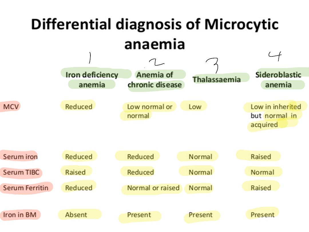
1-decrease s. iron , increase IBC(iron binding
capacity), decrease s. feritin
2- hypochromic microcytic in blood film.
3- WBC is normal , platelets count is increased (
however , platelets may be decreased in sever IDA).
D/D:-
1- other causes of hypochromic microcytic
2-anemia ofchronic dis.
Treatment :-
1- treatment of ppt. factors of IDA.
2-oral iron therapy ( 4-6mg\kg of elemental iron for
8wk)

Response to Iron therapy
•
12—24 hr mood change( decrease irritability)
•
36-48 hr bone marrow response, erythroid
hyperplasia
•
48-72 hr reticulocytosis(peaking at 5-7days )
•
4-30 days increase Hb level
•
1-3 months repletion of stores
3- blood transfusion :-- is indicated :-
a- Hb of less than 4 gm/ dl
b-superimposed infection which interfere with
response .