Fifth Stage
Internal Medicine
Dr. Abbas / Lec . 2
1
TESTS OF FUNCTION (CLINICAL NEUROPHYSIOLOGY)
Recording of electrical activity over the brain and assessment of nerve and muscle
function are essential in certain conditions. The major tests are electroencephalography
(EEG), evoked potentials (EPs) and nerve conduction studies/electromyography
(NCS/EMG
Electroencephalography
Electrical activity arising in the cerebral cortex can be detected using electrodes
placed on the scalp, although this is estimated to detect only 0.1-1% of the brain's
electrical activity at any one time.. When the eyes are shut, the most obvious frequency
over the occipital cortex is 8-13/s;
this is known as alpha rhythm, and
disappears when the eyes are
opened. Other frequency bands seen
over different parts of the brain in
different circumstances are beta
(faster than 13/s), theta (4-8/s) and
delta (slower than 4/s). Lower
frequencies predominate in the very
young and during sleep .
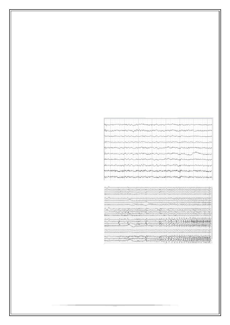
EEG
(Electroencephalography) may
support the clinical diagnosis of
epilepsy (by demonstrating
paroxysmal abnormalities containing
spikes or sharp waves), may provide
a guide to prognosis, and may help
classify the seizure disorder .
Advantages of EEG:
1- epilepsy is used to aid in diagnosis and classification and prognosis and localized
epileptic area when the surgery indicated.
Demonstration of sharp and spike wave which may be generalized or focal
2- in herpes simplex encephalitis
3- hepatic encephalopathy
4- Creutzfeldt-Jakop disease
5- focal brain lesion such abcessor tumour

2
Other diffuse brain disorders:
Recognizable slow-wave EEG abnormalities appear in encephalitis, prion
(Creutzfeldt-Jakob) disease and metabolic states (e.g. hypoglycaemia and hepatic
coma).
In Brainstem death: The EEG is isoelectric (flat), but is no longer necessary to
confirm brain death
Electromyography[E.M.G]
Is the recording and study of the insertional, spontaneous and voluntary electrical
activity of muscle; this allow evaluation of anterior horn cell, peripheral nerve and
muscle…
Indications:
•
Evaluate patient with weakness
•
To differentiate acute denervation from chronic denervation
•
To differentiate myopathy from neuropathy
•
To differentiate acute[active] inflamatory myopathy from chronic myopathy
•
Myotnia disorder like myotonia dystrophica.
•
Singe fiber EMG for neuromuscular junction transmission
Nerve Conduction Study
Is recording and measurement of the compound nerve and muscle action
potential elecited in response to an electrical stimulus, motor nerve conduction study
and sensory nerve conduction study
Study of electrical activity of nerve after electrical stimulation of examined nerve [
record the action potential and conduction velocity]
Abnoamality of N.C.s
•
Reduce amplitude[axonal neuropathy]
•
Slow coduction velocity[demylinating neuropathy]
•
Conduction block[compressive neuropathy]
•
Prolog terminal latency[demylinting neuropathy]
Clinical values of NCs
1- Diagnosis of neuropathy, asses the severity and distribution, distal, proximal or
diffuse
2- Whether Sensory or motor
3- Pathologic process: axonal or demyelinating
4- Compressive neuropathy like carpal tunnel syndrom

3
Evoked potentials
The spinal and cerebral potentials evoked by noninvasive stimulation of specific
afferent pathways are an important mean of monitoring the functional integrity of these
pathways
The cortical response to visual, auditory or electrical stimulation can be measured on
an EEG as an evoked potential [EP]
Assessing the latency [time delay] and amplitude can information about integrity of
the relevant pathway.
Indications:
1- Visual evoked potential – (multiple sclerosis):
Visual evoked potential studies are commonly used in the evaluation of suspected
multiple sclerosis demyelination in the optic nerve or central optic pathways.
2- Auditory evoked potential – (cerebello-pontine angle tumor and deafness in
children):
Brain stem auditory evoked potential studies in the diagnosis of diseases cranial
nerve VIII or its central projections. Lesions at the cerebellopontine angle and the
brain stem. Brain stem auditory evoked potentials the diagnosis of deafness in
infants.
3- Somatosensory visual evoked – (spinal cord disease):
Somatosensory evoked potentials are used to identify slowing of central sensory
conduction that results. They are also used to evaluate spinal cord-mediated
sensory abnormalities .
Routine blood tests
Many systemic conditions can affect the nervous system can identified by simple
blood tests. [full blood count ,ESR,C-reactive protein, biochemical screening] may
provide clue.
Lumbar puncture
It is a procedure that often performed in emergency department to obtain
information about the CSF
Procedure:
1- Local anesthetic injection.
2- Needle inserted between lumber spine processes [usually between L3 and L4]
through the Dura and into the spinal canal.
3- Intracranial pressure recorded and CSF send for analysis.

4
Normal CSF:
•
The color is clear and colorless
•
CSF pressure is 50-250 mm water
•
White cell count is 0-4 and lymphocyte
•
Red cell count is 0-4
•
Glucose is > 50-60 % of blood level
•
Protein is < 0.45 gm\L
•
Microbiology is sterile
•
Oligoclonal band negative
Tests usually performed on CSF include centrifuging to determine the colour of the
supernatant (yellow, or xanthochromic, some hours after subarachnoid haemorrhage),
biochemistry (glucose, total protein, and protein electrophoresis to detect oligoclonal
bands), microbiology (e.g. polymerase chain reaction (PCR) for herpes simplex or
tuberculosis), immunology (e.g. paraneoplastic antibodies) and cytology (to detect
malignant cells).
Indications:
1- In the CNS infection [meningitis and encephalitis]
2- Subarachniod hemorrhage
3- Inflamatory condition [multiple sclerosis, sarciodosis and cerebral lupus]
4- Some neurological malignancies (e.g. carcinomatous meningitis, lymphoma and
leukaemia)
5- Measure CSF pressure for diagnosis, therapeutic and monitoring of idiopathic
intracranial hypertension
6- And can be used in therapeutic procedures, either to lower CSF pressure or to
administer drugs .
Contraindication:
1- Mass in the brain or spinal cord
2- Raised intracranial pressure
3- Altered level of consciousness
4- Bleeding tendency
5- Infection at site of LP
6- Meningiocele and spina bifida
Biopsy
1- Nerve biopsy---peripheral neuropathy
-Target nerve is sural or radial nerve
2- Muscle biopsy to differentiate myositis from myopathy
-quidricepes
3- Brain biopsy—dementia and some brain tumor
It should only be consider when the diagnosis is elusive

5
Immunological test and genetic test
1-screen for antibodies in neuromuscular disorder
2-Screen for antibodies in channel disoreder and paraneoplastic dissorder
Thank you,,,