
1
College of Medicine
Dept. of Physio &Medical Physics
Electricity within the body
*****************************************************************
Physical phenomena involving electricity and magnetism have been observed since
ancient times.
There are two aspects of electricity and magnetism in medicine: -
1. Electrical and magnetic effects generated inside the body.
2. Applications of electricity and magnetism to the surface of the body.
The electricity generated inside the body serves for the control and operation of
nerves, muscles, and organs.
The nervous system plays a fundamental role in nearly every body function.
Basically, a central computer (the brain) receives internal and external signals and
(usually) makes the proper response.
The nervous system and the neuron
The nervous system can be divided structurally into two parts: -
1. The central nervous system.
2. The peripheral nervous system.
On the other hand, the nervous system can be divided functionally into two parts: -
1. The somatic nervous system (SNS) [Voluntary].
2. The autonomic nervous system (ANS) [Involuntary]
The autonomic nervous system itself reasonably can be classified to:
1. Sympathetic nervous system
2. Parasympathetic nervous system
Time: 2 Hours
May 4, 2020
Course: MPH2
Dr Hadi Al-Sagu
r

2
The autonomic nervous system controls various internal organs such as the heart,
intestines, and glands. The control of the autonomic nervous system is essentially
involuntary.
The Peripheral Nervous System (PNS) consist mainly of nerves that connect the CNS
to every other part of the body.
Therefore nerves can be divided based on the directionality of the carried signal:
Afferent neurons are sensory neurons that carry nerve impulses from sensory stimuli
(organs) towards the brain or spinal cord (CNS), while Efferent neurons are motor
neurons that carry neural impulses away from the brain or spinal cord (CNS) towards
muscles (organs) to cause movement.
Neuron
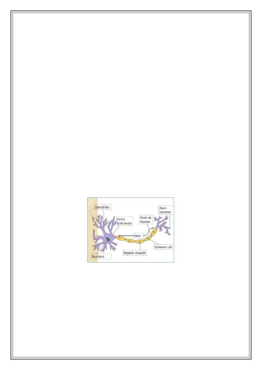
The dendrites are the parts of the neuron specialized for receiving information from
stimuli or from other cells. If the stimulus is strong enough, the neuron transmits an
electrical signal outward along a fiber called an axon. The axon, or nerve fiber, which
may be as long as 1m, carries the electrical signal to muscles, glands, or other
neurons.
The basic structural unit of the nervous system is
the neuron; a nerve cell specialized for reception,
interpretation, and transmission of electrical
messages. There are many types of neurons.
Basically, a neuron consists of a cell body that
receives electrical messages from other neurons
through contacts called synapses located on the
dendrites or on the cell body.
The diagram is the neuron
3
Examination of the axons of various neurons with an electron microscope indicates
that there are two different types of nerve fibers: -
1. The membranes of some axons are covered with a fatty insulating layer
called myelin that has small uninsulated gaps called nodes of Ranvier
every few millimeters; these nerves are, referred to as myelinated
nerves. Myelin helps to prevent action potentials, which are the
electrical signals that travel along axons, from decaying due to the
electrical current leaking out through the axonal membrane. Myelinated
axons thus conduct action potentials more quickly and efficiently than
unmyelinated axons.
2. The axons of other nerves have no myelin sleeve (sheath), and these
nerves are called unmyelinated nerves. Like these sorts of nerves can be
marked at babies due to the lack of mature myelin sheaths. As a result,
babies’ movements are jerky or shaky and uncoordinated. Their
movements become smoother and coordinated once the myelin sheaths
develop.
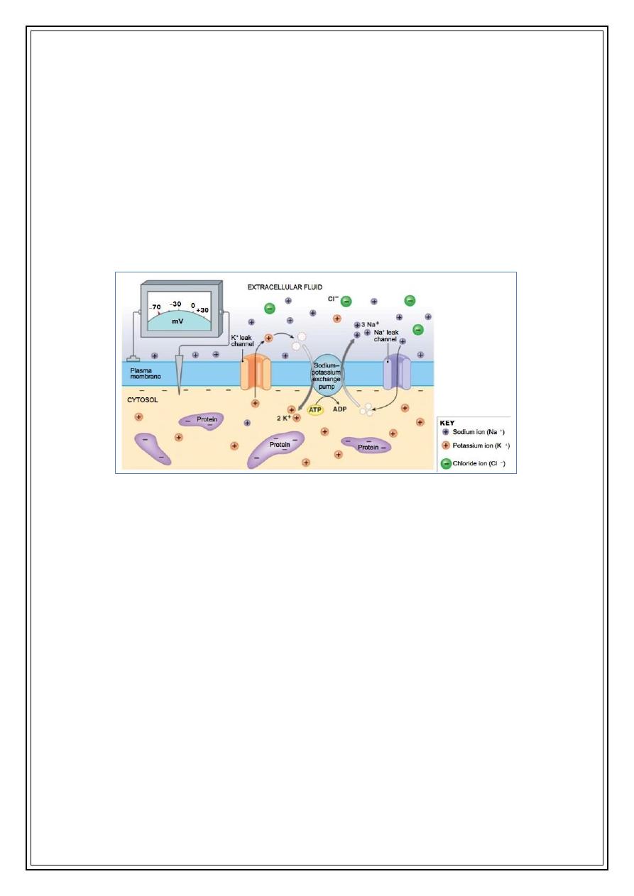
Electrical potentials of nerves
Across the surface or membrane of every neuron is an electrical potential
(voltage) difference due to the presence of more negative ions on the inside of the
membrane than on the outside. The neuron is said to be polarized. The inside of the
cell is typically -60 to -90 mV more negative than the outside. This potential difference
is called the resting potential of the neuron. When the neuron is stimulated, a large
The diagram is the neuron
4
momentary change in the resting potential occurs at the point of stimulation. This
potential change, called the action potential, propagates along the axon.
Electrical potentials of nerves
Why this potential difference exists?
The two basic reasons are:
1. The chemical compositions of the fluids inside and outside the cell are different.
2. The membrane allows some ions to entre and leave the cell more easily than others.
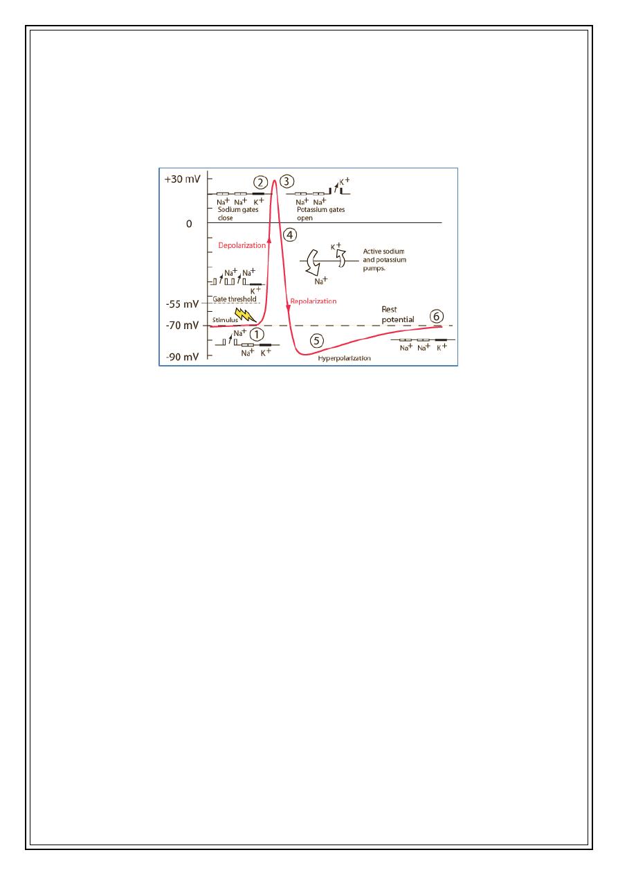
The action potential sequence is essential for neural communication and the process
involves several steps:
1. A stimulus is received by the dendrites of a nerve cell. This causes the
Na
+
channels to open. If the opening is sufficient to drive the interior potential
from -70 mV up to -55 mV, the process continues.
2. Having reached the action threshold, more Na
+
channels (sometimes called
voltage-gated channels) open. The Na
+
influx drives the interior of the cell
membrane up to about +30 mV. The process to this point is called
depolarization.
The figure explain the potential difference (-70) across the cell membrane
5
3. The Na+ channels close and the K+ channels open. Since the K+ channels are
much slower to open, the depolarization has time to be completed. Having both
Na+ and K+ channels open at the same time would drive the system toward
neutrality and prevent the creation of the action potential.
4. With the K+ channels open, the membrane begins to repolarize back toward its
rest potential.
5. The repolarization typically overshoots the rest potential to about -90 mV. This
is called hyperpolarization and would seem to be counterproductive, but it is
actually important in the transmission of information. Hyperpolarization
prevents the neuron from receiving another stimulus during this time, or at least
raises the threshold for any new stimulus. Part of the importance of
hyperpolarization is in preventing any stimulus already sent up an axon from
triggering another action potential in the opposite direction. In other words,
hyperpolarization assures that the signal is proceeding in one direction.
6. After hyperpolarization, the Na
+
/K
+
pumps eventually bring the membrane back
to its resting state of -70 mV.
Two primary factors affect the speed of propagation of the action potential are:
1. The resistance within the core of the membrane.
2. The capacitance (or the charge stored) across the membrane.
The diagram shows the six sequenced stages of the action potential