Injuries of the spine
Spinal injuries carry a double threat: damage to the vertebral column and damage to the neural tissues.Structural elements of the spine:
The vertical lines show Denis’ classification of the structural elements of the spine. The three elements are: the posterior complex, the middle component and the anterior column. This concept is particularly useful in assessing the stability of lumbar injuries.Types of spinal injuries:
Stable injury :is one in which the vertebral components will not be displaced by normal movements. The neural elements are undamaged there is little risk of them becoming damaged.
Unstable injury:
is one in which there is a significant risk of displacement and consequent damage – or further damage – to the neural tissues.Mechanism of injury:
There are three basic mechanisms of injury:Traction injury : In the lumbar spine resisted muscle effort may avulse transverse processes; in the cervical spine the seventh spinous process can be avulsed (‘clayshoveller’s racture’).
Direct injury :Penetrating injuries to the spine, particularly from firearms and knives, are becoming increasingly common.
Indirect injury : This is the most common cause of significant spinal damage; it occurs most typically in a fall from a height.
DIAGNOSIS:
History :
Every patient with a blunt injury above the clavicle, a head injury or loss of consciousness should be considered to have a cervical spine injury until proven otherwise.
Every patient who is involved in a fall from a height or a high-speed deceleration accident should similarly be considered to have a thoracolumbar injury.
Examination :
Local examination: the head , face , neck and back are thoroughly inspected for bruises ,deformity, or penetrating injury , and the bones and soft tissues of the neck and the back are gently palpated for tenderness.General examination( Shock) :
Three types of shock may be encountered in patients with spinal injury:
Hypovolaemic shock : is suggested by tachycardia, peripheral shutdown and, in later stages, hypotension.
Neurogenic : shock reflects loss of the sympathetic pathways in the spinal cord; The combination of paralysis, warm and well-perfused peripheral areas, bradycardia and hypotension with a low diastolic blood pressure suggests neurogenic shock.
Spinal shock: occurs when the spinal cord fails temporarily following injury. Below the level of the injury, the muscles are flaccid, the reflexes absent and sensation is lost. This rarely lasts for more than 48 hours and during this period it is difficult to tell whether the neurological lesion is complete or incomplete.
Neurological examination:
A full neurological examination is carried out in every case; this may have to be repeated several times during the first few days. Each dermatome, myotome and reflex is tested.Radiological examination:
X-ray : X-ray examination of the spine is mandatory for all accident victims complaining of pain or stiffness in the neck or back or peripheral paraesthesiae.CT scan : CT is ideal for showing structural damage to individual vertebrae and displacement of bone fragments into the vertebral canal.
MRI : MRI is the method of choice for displaying the intervertebral discs, ligamentum flavum and neural structures, and is indicated for all patients with neurological signs and those who are considered for surgery.
PRINCIPLES OF DEFINITIVE TREATMENT
Patients with no neurological injury:Stable injuries : the patient is treated by supporting the spine in a position that will cause no further strain; a firm collar or lumbar brace will usually suffice, but the patient may need to rest in bed until pain and muscle spasm subside.
Unstable injuries:
The spine should be held secure until the tissues heal and the spine becomes stable.In the cervical spine this should be done as soon as possible by traction or by internal fixation.
In the thoracolumbar spine internal fixation can be carried out.
b. Patients with a neurological injury
Stable spinal injuries with neurological injury (rare) the patient can be treated conservatively and rehabilitated as soon as possible.Unstable spinal injuries with neurological injury can be treated either
Conservatively by 2-hourly turning routines, skin toilet, bladder care and specialized physiotherapy and occupational therapy.
Early operative stabilization.
The indications for urgent surgical stabilization are:
(a) an unstable fracture with progressive neurological deficit and MRI signs of likely further neurological deterioration.(b) controversially an unstable fracture in a patient with multiple injuries.
Regional spinal injuries
Upper cervical spine injury:
C1 ring fracture(Jefferson’s fracture):Sudden severe load on the top of the head may cause a ‘bursting’ force which fractures the ring of the atlas . There is no neurological damage.The fracture is seen on the open-mouth view . A CT scan is particularly helpful in defining the fracture.
Treatment:
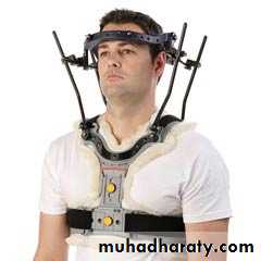
If the fracture is undisplaced and stable the patient can be treated by wears a semi-rigid collar until the fracture unites.If the fracture is displaced and unstable should be treated bya halo-vest for several weeks. If there is persisting instability on x-ray, a posterior C1/2 fixation and fusion is needed.
C2 pars interarticularis fractures‘hangman’s fracture’
There are bilateral fractures of the pars interarticularis of C2 and the C2/3 disc is torn; the mechanism is extension with distraction.
Fracture of C2 ‘Hangman’s fracture’ – fracture of
the pars interarticularis of C2.Treatment:
Undisplaced fractures which are shown to be stable on supervised flexion–extension viewscan be treated in a semi-rigid orthosis until united (usually 6–12 weeks).Displaced fractures but no kyphotic angulation may need reduction and the neck is held in a halo-vest until union occurs.
C2 Odontoid process fracture
They usually occur as flexion injuries in young adults after high velocity accidents or severe falls. However, they also occur in elderly, osteoporotic people as a result of low-energy trauma in which the neck is forced into hyperextension, e.g. a fall onto the face or forehead.
Classification :
Type I – An avulsion fracture of the tip of the odontoid process.
Type II – A fracture at the junction of the odontoid process and the body of the axis.Type III – A fracture through the body of the axis.
Treatment :
Type I fractures: treated by immobilization in a rigid collar until discomfort subsides.
Type II fractures:
Undisplaced fractures can be held by fitting a halo-vest or – in elderly patients – a rigid collar.Displaced fractures should be reduced by traction and can then be held by operative posterior C1/2 fusion;
Type III fractures:
undisplaced, these are treated in a halo-vest for 8–12 weeks.displaced, attempts should be made at reducing the fracture by halo traction, the neck is then immobilized in a halo-vest for 8–12 weeks.
Lower cervical spine injury:
Wedge compression fractureA pure flexion injury results in a wedge compression fracture of the vertebral body . The middle and posterior elements remain intact and the injury is stable. All that is needed is a comfortable collar for 6–12 weeks.
Burst and compression-flexion (‘teardrop’) fractures
These severe injuries are due to axial compression of the cervical spine, usually in diving or athletic accidents.
If the vertebral body is crushed in neutral position of the neck the result is a ‘burst fracture’, and this due to axial compression of the cervical spine,
With combined axial compression and flexion, an antero-inferior fragment of the vertebral body is sheared off, producing the eponymous ‘tear-drop’on the lateral x-ray.
Treatment:
If there is no neurological deficit, the patient can be treated surgically or by confinement to bed and traction for 2–4 weeks, followed by a further period of immobilization in a halo-vest for 6–8 weeks.If there is any deterioration of neurological status and the fracture is unstable should be treated by urgent anterior decompression.
Hyperextension injury
Hyperextension strains of soft-tissue structures are common and may be caused by comparatively mild acceleration forces. Bone and joint disruptions, however, are rare.Avulsion injury of the spinous process
Fracture of the C7 spinous process may occur with severe voluntary contraction of the muscles at the back of the neck; it is known as the clay-shoveller’s fracture. The injury is painful but harmless. No treatment is required.THORACOLUMBAR INJURIES
Most injuries of the thoracolumbar spine occur in the transitional area – T11 to L2 – between the somewhat rigid upper and middle thoracic column and the flexiblelumbar spine.The common mechanisms of injury are:
Flexion–compression – failure of the anterior column and wedge-compression of the vertebral body. Usually stable, but greater than 50 per cent loss of anterior height suggests some disruption of the posterior ligamentous structures.
•Lateral compression – lateral wedging of the vertebral body resulting in a localized ‘scoliotic’ deformity.
Axial compression – failure of anterior and middle columns causing a ‘burst’ fracture and the danger of retropulsion of a posterior fragment into the spinal canal. Often unstable.
Flexion–rotation – failure of all three columns and a risk of displacement or dislocation. Usually unstable.
Flexion–distraction – the so-called ‘jack-knife’ injury causing failure of the posterior and middle columns and sometimes also anterior compression.
Extension – tensile failure of the anterior column and compression failure of the posterior column. Unstable.