1
Areas of lower limb
stage
st
1
Dr.Kalid Ali Zayer
Femoral triangle
Contents
1. Borders
2. Contents
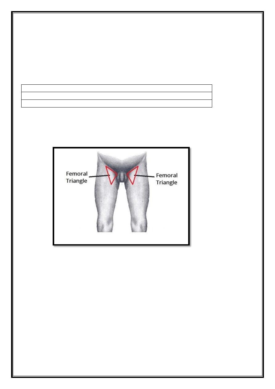
The is a hollow area in the anterior thigh. Many large neurovascular structures pass
through this area, and can be accessed relatively easily. Thus, it is an area of both
anatomical and clinical importance.
Fig 1– Surface anatomy of the femoral triangle.
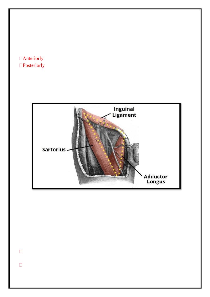
Borders
As this area is a triangle, it has three borders:
Superior border – Formed by the inguinal ligament, a ligament that runs
from the anterior superior iliac spine to the pubic tubercle.
Lateral border – Formed by the medial border of the sartorius muscle.
Medial border – Formed by the medial border of the adductor longus
muscle. The rest of this muscle forms part of the floor of the triangle.
2
o Note: Some sources consider the lateral border of the adductor longus to be the
medial border of the femoral triangle. However, the majority state that it is the
medial border of the adductor longus – and this is definition we have gone with.
It also has a floor and a roof:
, the roof of the femoral triangle is formed by the fascia lata.
, the base of the femoral triangle is formed by the pectineus,
iliopsoas and adductor longus muscles.
The inguinal ligament acts as a flexor retinaculum, supporting the contents of the
femoral triangle during flexion at the hip.
Fig 2 – The borders of the right femoral triangle.
Contents
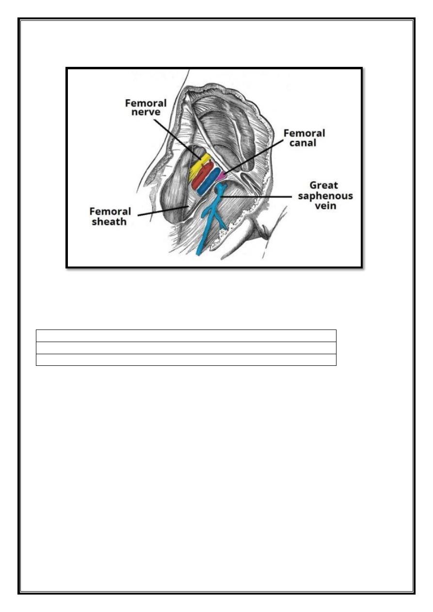
The femoral triangle contains some of the major neurovascular structures of the
lower limb.
Its contents (lateral to medial) are:
Femoral nerve
– Innervates the anterior compartment of the thigh, and
provides sensory branches for the leg and foot.
Femoral artery
– Responsible for the majority of the arterial supply to the
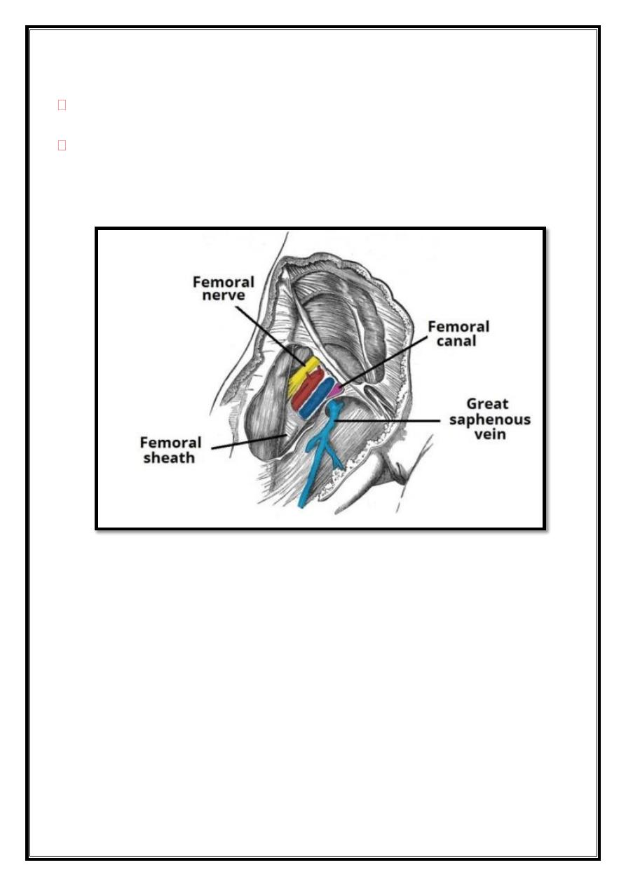
3
lower limb.
Femoral vein
– The great saphenous vein drains into the femoral vein within
the triangle.
Femoral canal
– A structure which contains deep lymph nodes and vessels.
The femoral artery, vein and canal are contained within a fascial compartment –
known as the femoral sheath
Fig 3 – The contents of the femoral triangle.
A good way of remembering the contents is using the acronym NAVEL:
N: Nerve.
A: Artery.
V: Vein.
E: Empty space
(this is important as it allows the veins and lymph vessels to
distend, so they can cope with different levels of flow).
L: Lymph canal.
The femoral canal is an anatomical compartment located in the anterior thigh. It
is the smallest and most medial part of the femoral sheath. It is approximately
1.3cm long.
Borders
The femoral canal is located in the anterior thigh within the femoral triangle. It
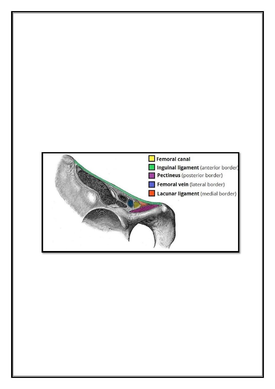
4
can be thought of as a rectangular shaped compartment with four borders and an
opening:
Medial border – lacunar ligament.
Lateral border – femoral vein.
Anterior border – inguinal ligament.
Posterior border – pectineal ligament, superior ramus of the pubic
bone, and the pectineus muscle
The opening to the femoral canal is located at its superior border, known as the
femoral ring.
The femoral ring is closed by a connective tissue layer – the femoral septum. This
septum is pierced by the lymphatic vessels exiting the canal.
Fig 4 – Borders of the femoral canal.
Contents
The femoral canal contains:
Lymphatic vessels – draining the deep inguinal lymph
nodes.
Deep lymph node – the lacunar node.
Empty space.
Loose connective tissue.
The empty space allows distension of the adjacent femoral vein, so it can cope
with increased venous return, or increased intra-abdominal pressure.
5
Fig 5 – The contents of the femoral triangle.
Adductor canal
Contents
1. Borders
2. Contents
The adductor canal (Hunter’s canal, subsartorial canal) is a narrow conical tunnel
located in the thigh. It is 15cm long, extending from the apex of the femoral
triangle to the adductor hiatus of the adductor magnus. The canal serves as a
passageway from structures moving between the anterior thigh and posterior leg.
Borders
The
adductor
canal
is
bordered
by
muscular
structures:
Anterior: Sartorius.
Lateral: Vastus medialis.
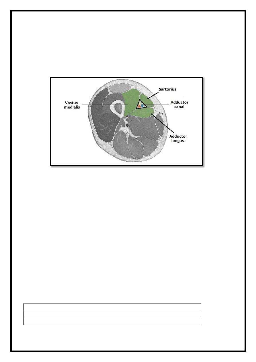
6
Posterior: Adductor longus and adductor magnus.
The apex of the adductor canal is marked by the adductor hiatus – a gap between
the adductor and hamstring attachments of the adductor magnus.
Fig 6 – Cross-section of the thigh, showing the borders of the
adductor canal. Note: the adductor magnus is not visible in this
illustration.
Contents
The adductor canal serves as a passageway for structures moving between the
anterior thigh and posterior leg. It contains the femoral artery, femoral vein,
nerve to the vastus medialis and the saphenous nerve (the largest cutaneous
branch of the femoral nerve).
As the femoral artery and vein exit the canal, they become the popliteal artery
and vein respectively.
Popliteal fossa
Contents
1. Borders
2. Contents
7
The popliteal fossa is a diamond shaped area found on the posterior side of the
knee. It is the main path in which structures move from the thigh to the leg. In any
anatomical area such as this, it is important to look at the borders, contents, and
any clinical relevance.
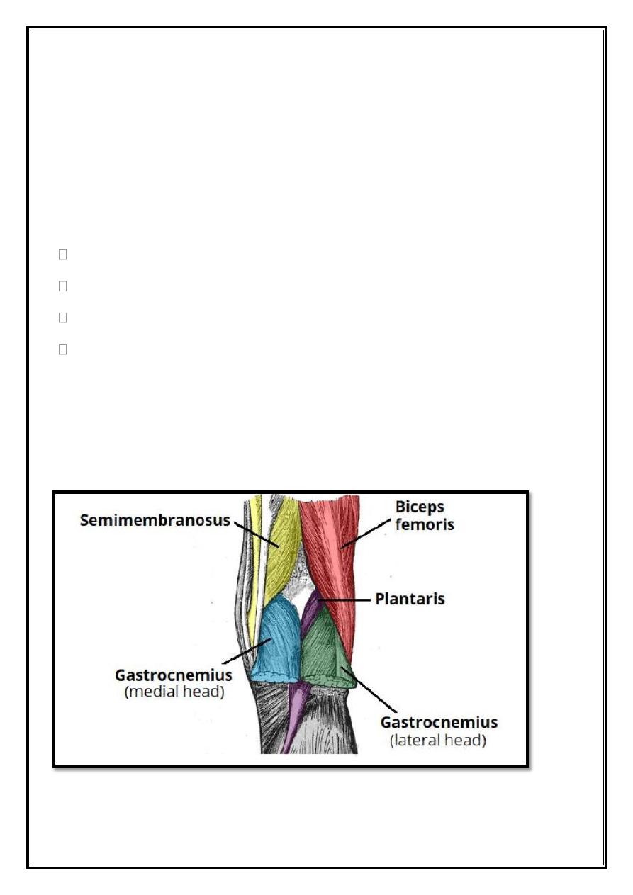
Borders
The popliteal fossa is diamond shaped, with four borders. These borders are
formed by the muscles in the posterior compartment of the leg and thigh:
Superomedial border – semimembranosus.
Superolateral border – biceps femoris.
Inferomedial border – medial head of the gastrocnemius.
Inferolateral border – lateral head of the gastrocnemius and plantaris.
The popliteal fossa also has a floor and a roof. The floor of the popliteal fossa is
formed by the posterior surface of the knee joint capsule, and by the posterior
surface of the femur.
The roof is made of up two layers; popliteal fascia and skin. The popliteal fascia
is continuous with the fascia lata of the leg.
8
Fig 7 – The borders of the popliteal fossa are formed by the muscles
of the thigh and leg.
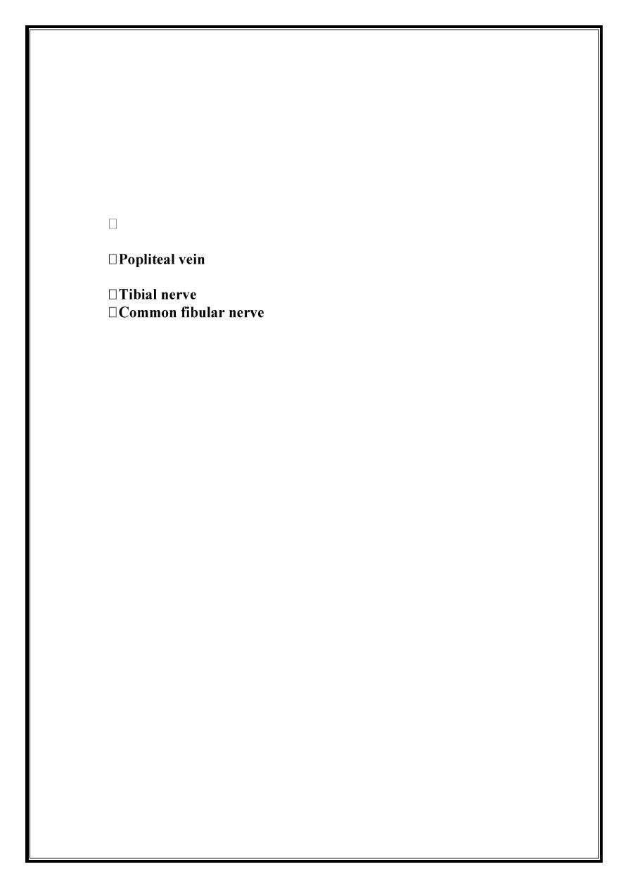
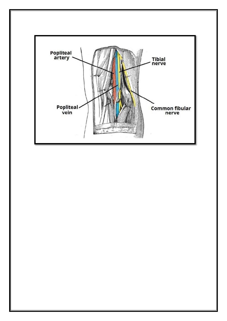
Contents
The popliteal fossa is the main conduit for neurovascular structures entering and
leaving the leg. Its contents are (medial to lateral):
Popliteal artery
The tibial and common fibular nerves are the most superficial of the contents of
the popliteal fossa. They are both branches of the sciatic nerve. The common
fibular nerve follows the biceps femoris tendon, running along the lateral margin
of the popliteal fossa.
The small saphenous vein pierces the popliteal fascia of the popliteal fossa to
enter
the
diamond,
and
empty
into
the
popliteal
vein.
In the popliteal fossa, the deepest structure is the popliteal artery. It is a
continuation of the femoral artery, and travels into the leg to supply it with blood.
9
Fig 8 – The contents of the popliteal fossa.