Thi -Qar University Human genetic
Medical College Lec 3
Microbiology Department Dr Dhafer A. Alghezi
1
Chromosomes
A chromosome is a structure that occurs within cells and contains the cell's
genetic material. The genetic material, which determines how an organism develops,
is a molecule of deoxyribonucleic acid (DNA). In prokaryotes, or cells without a
nucleus, the chromosome is merely a circle of DNA. In eukaryotes, or cells with a
distinct nucleus, chromosomes are much more complex in structure.
The structure of chromosomes
A chromosome is an organized structure of DNA and protein that is found in cells.
It is a single piece of coiled DNA containing many genes, regulatory elements and
other nucleotide sequences. Chromosomes also contain DNA-bound proteins, which
serve to package the DNA and control its functions. Chromosomes vary widely
between different organisms. The DNA molecule may be circular or linear, and can
be composed of 10,000 to 1,000,000,000 nucleotides in a long chain. Two
chromosomal regions have special importance in the formation of chromosome
aberrations: the centromeres and the telomeres.
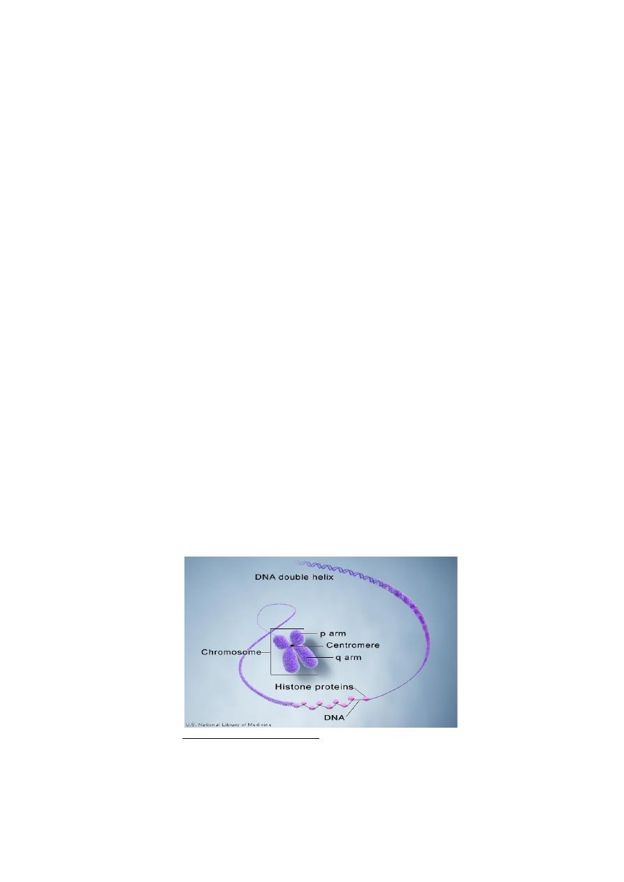
The centromere is primary constriction of chromosomes where sister chromatids
are connected, situated in strictly imposed position of the chromosome. It divides the
chromosome into two sections, or “arms.” The short arm of the chromosome is
labeled the “p arm.” The long arm of the chromosome is labeled the “q arm.” The
location of the centromere on each chromosome gives the chromosome its
characteristic shape, and can be used to help describe the location of specific genes.
The chromosome ends, the telomeres are rich in TTAGGG repetitive sequences,
and ensure the integrity and stability of the chromosome structure and play a role in
cell aging, in tumorigenicity and the formation of structural chromosome aberrations,
since in the absence of a telomere the chromosome structure becomes unstable and
fragments without telomeres easily adhere, opening the way for a wide variety of
disorders (Figure 1).
Figure 1: chromosome structure.
Humans have 46 chromosomes that occur in 23 pairs. Twenty-two of these pairs
are called autosomes. All of these chromosomes are found in both males and females.
One pair of chromosomes is called sex chromosomes because this pair contains the
Thi -Qar University Human genetic
Medical College Lec 3
Microbiology Department Dr Dhafer A. Alghezi
2
genes that control gender. Males have the sex chromosomes X and Y, and females
have two X chromosomes.
Any cell in the body except red blood cells, which lack a nucleus, can be a source
of chromosomes for examination. In adults, it is easiest to obtain and use white blood
cells separated from a blood sample for the purpose of looking at the chromosomes.
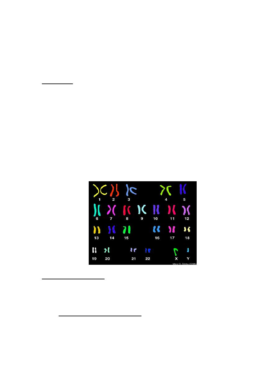
A karyotype:
A karyotype is the characteristic chromosome complement of a eukaryote species.
It describes the number of chromosomes, and what they look like under a light
microscope (Figure 2). The term is also used for the complete set of chromosomes in
a species, or an individual organism. The study of whole sets of chromosomes is
sometimes known as karyology. The preparation and study of karyotypes is part of
Cytogenetics which represents a field of genetics dealing with species or cell specific
number of chromosomes, and their structure and characteristic segments, their
functional roles, and all the differences - namely the chromosomal mutations - related
to them.
Karyotypes can be used for many purposes; such as, to study chromosomal
aberrations, cellular function, taxonomic relationships, and to gather information
about past evolutionary events.
Figure 2: Human karyotypes
Chromosome Inheritance:
Normally, an individual receives 22 pairs of autosomes and two sex
chromosomes. Each pair of autosomes carries alleles for particular traits. The alleles
can be different, as when one calls for freckles and one does not.
A. Changes in chromosomes number:
Sometimes individuals are born with either too many or too few autosomes or sex
chromosomes, most likely due to non-disjunction during meiosis.
Non-disjunction is a failure of the homologous chromosomes, or daughter
chromosomes, to separate correctly during meiosis I and meiosis II, respectively.

Thi -Qar University Human genetic
Medical College Lec 3
Microbiology Department Dr Dhafer A. Alghezi
3
Non-disjunction may occur during meiosis I, when both members of a homologous
pair go into the same daughter cell. It can also occur during meiosis II, when the sister
chromatids fail to separate and both daughter chromosomes go into the same gamete.
Some abnormal eggs have 24 chromosomes, whereas others have only 22
chromosomes. If an egg with 24 chromosomes is fertilized with a normal sperm, the
result is a trisomy, so called because one type of chromosome is present in three
copies. If an egg with 22 chromosomes is fertilized with a normal sperm, the result is
a monosomy, so called because one type of chromosome is present in a single copy.
An abnormal number of autosomes causes a developmental abnormality. Monosomy
of all but the X chromosome is fatal. The affected infant rarely develops to full term.
Trisomy is usually fatal, though there are some exceptions. Among autosomal
trisomies, only trisomy 21 (Down syndrome) has a reasonable chance of survival after
birth. The chances of survival are greater when trisomy or monosomy involves the sex
chromosomes. In normal XX females, one of the X chromosomes becomes a darkly
staining mass of chromatin called a Barr body (named after the person who
discovered it). A Barr body is an inactive X chromosome. Beyond a single one
become Barr body; this explains why poly-X females and XXY males are seen fairly
frequently.
Down Syndrome: An Autosomal Trisomy:
The most common autosomal trisomy seen among humans is Down syndrome,
also called trisomy 21.
Persons with Down syndrome usually have three copies of chromosome 21
because the egg had two copies instead of one. The chances of woman having a Down
syndrome child increase rapidly with age, starting at about age 40. The reasons for
this are still being investigated. Although an older woman is more likely to have a
Down syndrome child, most babies with Down syndrome are born to women younger
than age 40 because this is the age group having the most babies. Karyotyping can
detect a Down syndrome child.
Down syndrome is easily recognized by these common characteristics: short
stature; an eyelid fold; a flat face; stubby fingers; a wide gap between the first and
second toes; a large, fissured tongue; a round head; and a palm crease, the so-called
simian line. Unfortunately, mental retardation, which can vary in intensity, is also a
characteristic. The genes that cause Down syndrome are located on the bottom third
of chromosome 21.
B. Changes in Sex chromosome number:
An abnormal sex chromosome number is a result of inheriting too many or too
few X or Y chromosomes. Turner syndrome (XO) and Klinefelter syndrome (XXY)
diseases are two examples for this changing.
Turner Syndrome:
It is a rare chromosomal condition that affects development in females. It is caused by
partial or complete loss (monosomy) of the second sex chromosome. From birth, a
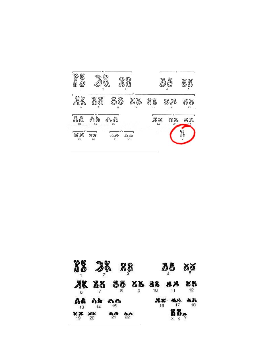
Thi -Qar University Human genetic
Medical College Lec 3
Microbiology Department Dr Dhafer A. Alghezi
4
girl with Turner syndrome has only one sex chromosome, an X (Figure 3). the
Turner syndrome female is a short, with a broad chest and folds of skin on the
back of the neck. The ovaries, oviducts, and uterus are very small and
underdeveloped. Turner females do not undergo puberty or menstruation, and their
breasts do not develop.
Figure 3: Monosomy (Turner syndrome) karyotype
Klinefelter Syndrome:
It is a chromosomal variation in males in which one extra X chromosome is present,
resulting in a XXY sex chromosome karyotype (Figure 4). 1: 650 of males are born
with two X chromosomes and one Y chromosome. The symptoms of this condition
(referred to as “47, XXY”) are often so subtle that only 25% are ever diagnosed, and
those are usually not diagnosed until after age 15. Disorder occurring due to
nondisjunction of the X chromosome.
The sperm containing both X and Y combines with an egg containing X,
results in a male child.
The egg may contribute the extra X chromosome.
Males with some development of breast tissue normally seen in females.
Little body hair is present, and such person are typically tall, have small testes.
Infertility results from absent sperm.
Evidence of mental retardation may or may not be present.
Figure 4: Klinefelter Syndrome karyotype
Thi -Qar University Human genetic
Medical College Lec 3
Microbiology Department Dr Dhafer A. Alghezi
5
Poly-X females:
A poly-X female has more than two X chromosomes and extra Barr bodies in the
nucleus. Females with three X chromosomes have no distinctive phenotype. Females
with more than three X chromosomes occur rarely.
Jacobs Syndrome:
XYY males with Jacobs syndrome can only result from nondisjunction during
spermatogenesis. Affected males are usually taller than average, suffer from persistent
acne, and tend to have speech and reading problems.
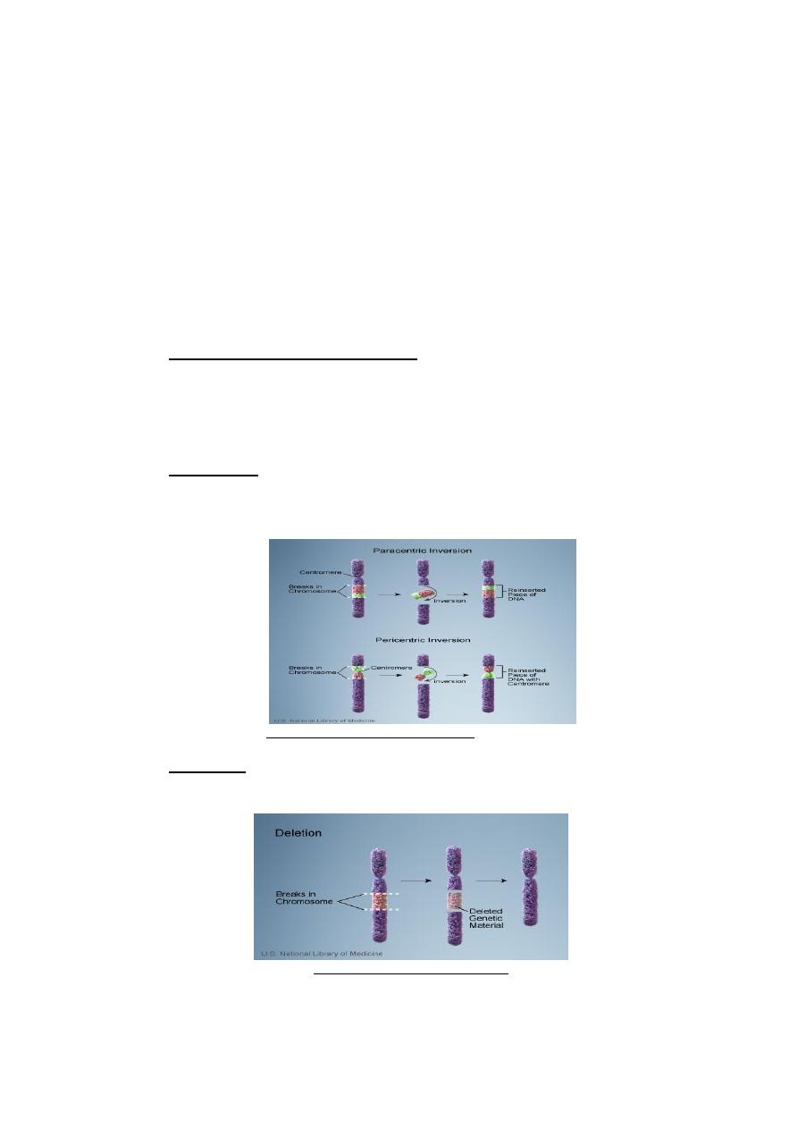
C. Changes in Chromosome Structure:
There are various agents in the environment, such as radiation, certain organic
chemicals or even viruses, that cause chromosome to break. Changes in chromosome
structure include deletions, translocations, duplications, and inversions of
chromosome segments.
1. An inversion: occurs when a segment of a chromosome is turned around 180
c (Figure 5)°, you might think this is not a problem because the same genes are
present, but the new position might lead to altered gene activity.
Figure 5: An inversion of chromosome
2. A deletion: occurs when an end of a chromosome breaks off or when two
simultaneous breaks lead to the loss of an internal segment (Figure 6).
Figure 6: A deletion of chromosome
Thi -Qar University Human genetic
Medical College Lec 3
Microbiology Department Dr Dhafer A. Alghezi
6
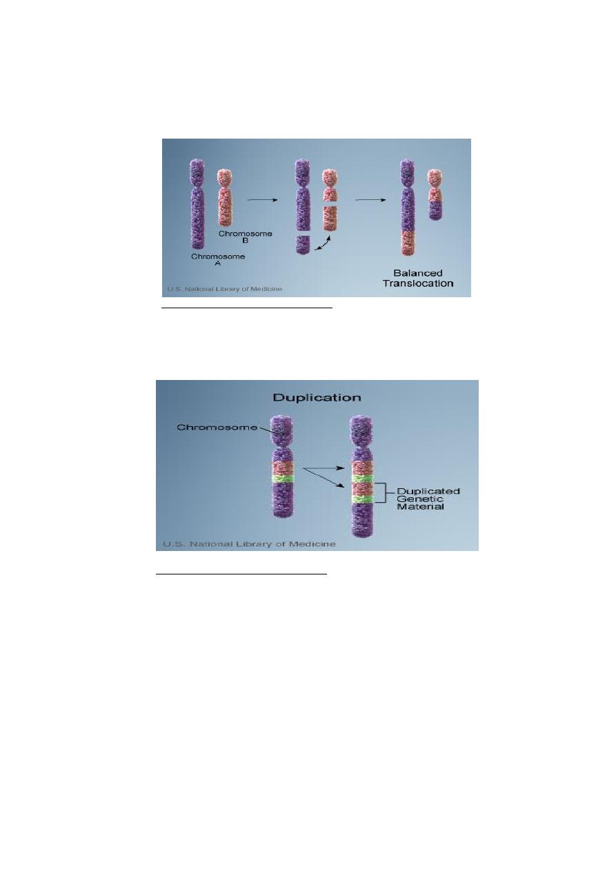
3. A translocation: is the movement of a chromosome segment from one
chromosome to another non homologous chromosome (Figure 7) .
Translocation heterozygotes usually have reduced fertility due to production
of abnormal gametes.
Figure 7: A translocation of chromosome
4. A duplication: is the doubling of chromosome segment (Figure 8) . There are
several ways a duplication can occur. A broken from one chromosome can
simply attach to its homologue, or unequal crossing- over may occur leading
to a duplication and deletion.
Figure 7: A translocation of chromosome